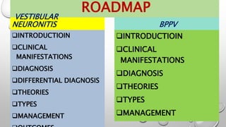
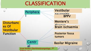
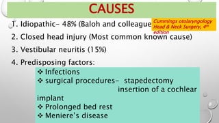
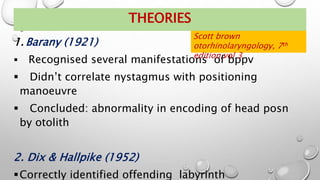
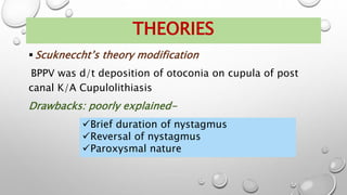
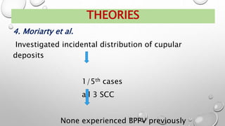
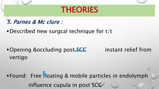
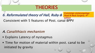
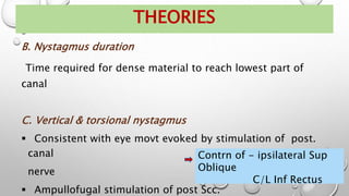
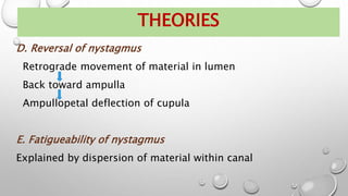
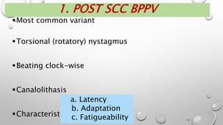
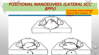
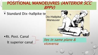
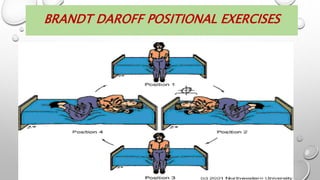
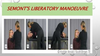
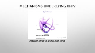
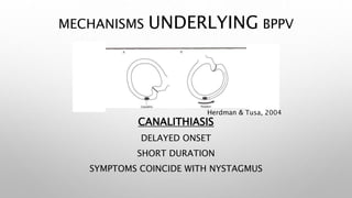
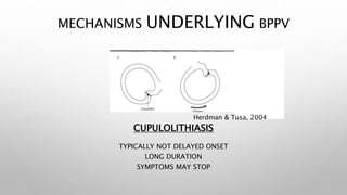
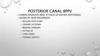
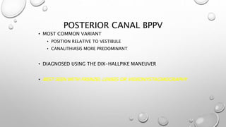
This document provides an overview of vestibular neuronitis and benign paroxysmal positional vertigo (BPPV). It discusses the introduction, clinical manifestations, diagnosis, theories, types, and management of each condition. Vestibular neuronitis is characterized by sudden onset of vertigo and imbalance due to loss of vestibular input from one inner ear. BPPV causes brief attacks of vertigo triggered by certain head movements, and is most commonly caused by debris in the posterior semicircular canal. Both conditions are diagnosed using bedside tests like Dix-Hallpike, and typically managed initially with repositioning maneuvers, vestibular rehabilitation, and sometimes medications.
![INTRODUCTION
Disorder in which there is sudden, spontaneous, isolated,
total or subtotal loss of afferent vestibular input from one
labyrinth resulting in severe vertigo
[ Scott brown otorhinolaryngology, 7th edition,vol
3]
2nd most common cause of vertigo of peripheral origin
[Cummings otolaryngology Head & Neck Surgery,](https://image.slidesharecdn.com/zlflytsnrrkkey4tdbkz-vestibular-neuronitis-bppv-240404125037-6bc407ef/85/VESTIBULAR_NEURONITIS__bppv-ent-hns-pptx-5-320.jpg)
![ Synonyms:
INTRODUCTION
Vestibular neuritis
Acute unilateral peripheral
neuropathy
Labyrinthitis
Neurolabyrinthitis
[ Scott brown otorhinolaryngology, 7th
edition,vol 3]
No gender bias
Middle aged people](https://image.slidesharecdn.com/zlflytsnrrkkey4tdbkz-vestibular-neuronitis-bppv-240404125037-6bc407ef/85/VESTIBULAR_NEURONITIS__bppv-ent-hns-pptx-6-320.jpg)
![C. HEARING LOSS
TINNITUS
Typically
absent
b. Nystagmus
Spontaneous, horizontal-torsional
Strictly unidirectional
Quick phase towards unaffected side
Magnifying frenzel lenses may be reqd
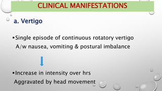
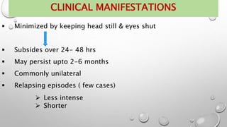
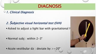
CLINICAL MANIFESTATIONS
[ Scott brown otorhinolaryngology, 7th
edition,vol 3]](https://image.slidesharecdn.com/zlflytsnrrkkey4tdbkz-vestibular-neuronitis-bppv-240404125037-6bc407ef/85/VESTIBULAR_NEURONITIS__bppv-ent-hns-pptx-9-320.jpg)
![D. Head impulse test
CLINICAL MANIFESTATIONS
[ Scott brown otorhinolaryngology, 7th
edition,vol 3]](https://image.slidesharecdn.com/zlflytsnrrkkey4tdbkz-vestibular-neuronitis-bppv-240404125037-6bc407ef/85/VESTIBULAR_NEURONITIS__bppv-ent-hns-pptx-10-320.jpg)
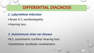
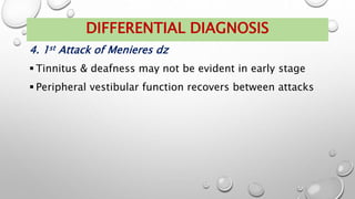
![DIFFERENTIAL DIAGNOSIS
1. Cerebellar infarction
Head impulse test –ve
Nystagmus: Bidirectional
Not suppressed by visual fixation
Can’t stand without support even with eyes open
[ Scott brown otorhinolaryngology, 7th
edition,vol 3]](https://image.slidesharecdn.com/zlflytsnrrkkey4tdbkz-vestibular-neuronitis-bppv-240404125037-6bc407ef/85/VESTIBULAR_NEURONITIS__bppv-ent-hns-pptx-13-320.jpg)


![Hypothalamus short ppt by Dr. Neha [PT].pptx](https://cdn.slidesharecdn.com/ss_thumbnails/hypothalamusbydr-260124145759-b9f94a93-thumbnail.jpg?width=640&height=640&fit=bounds)