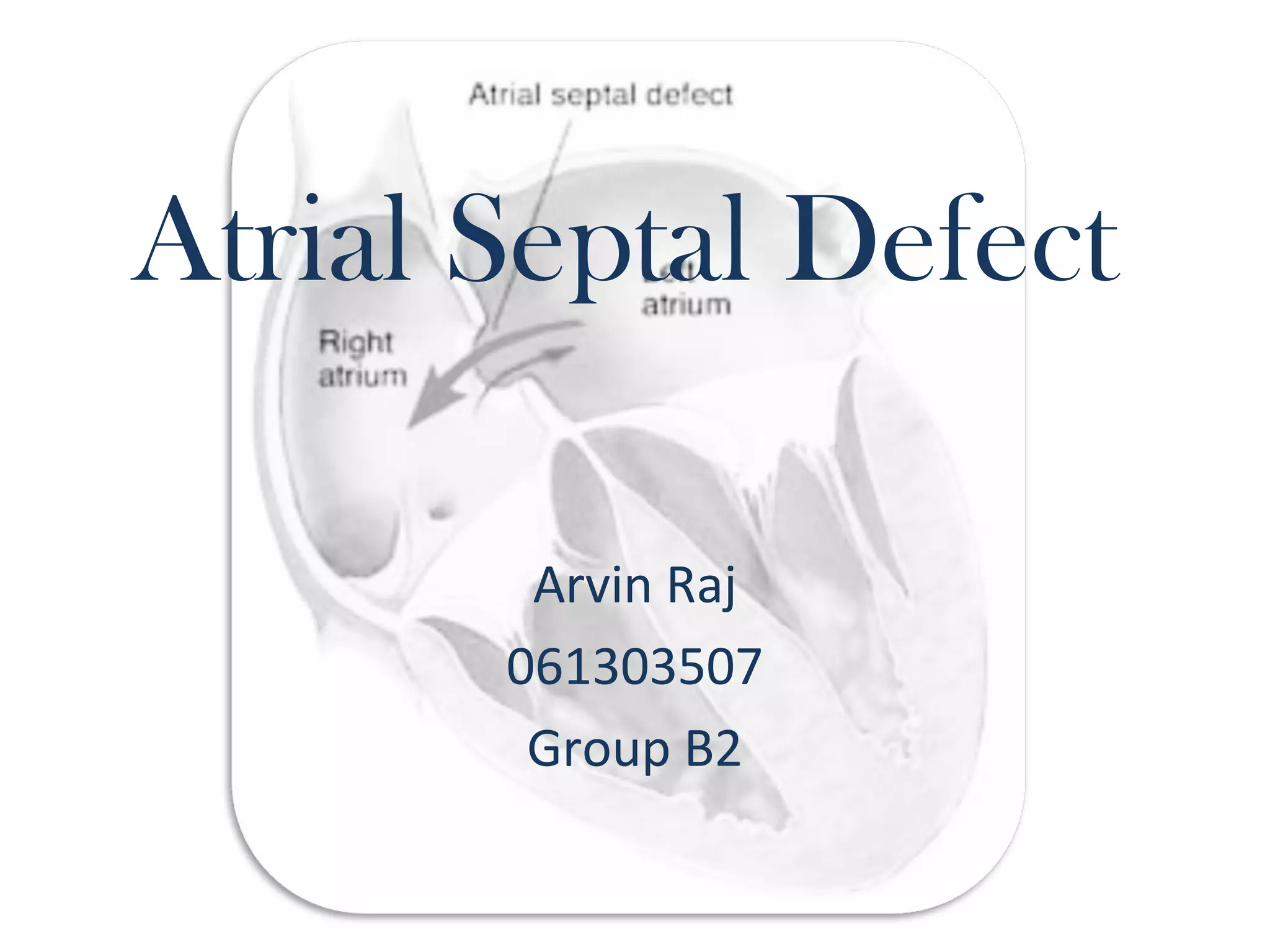
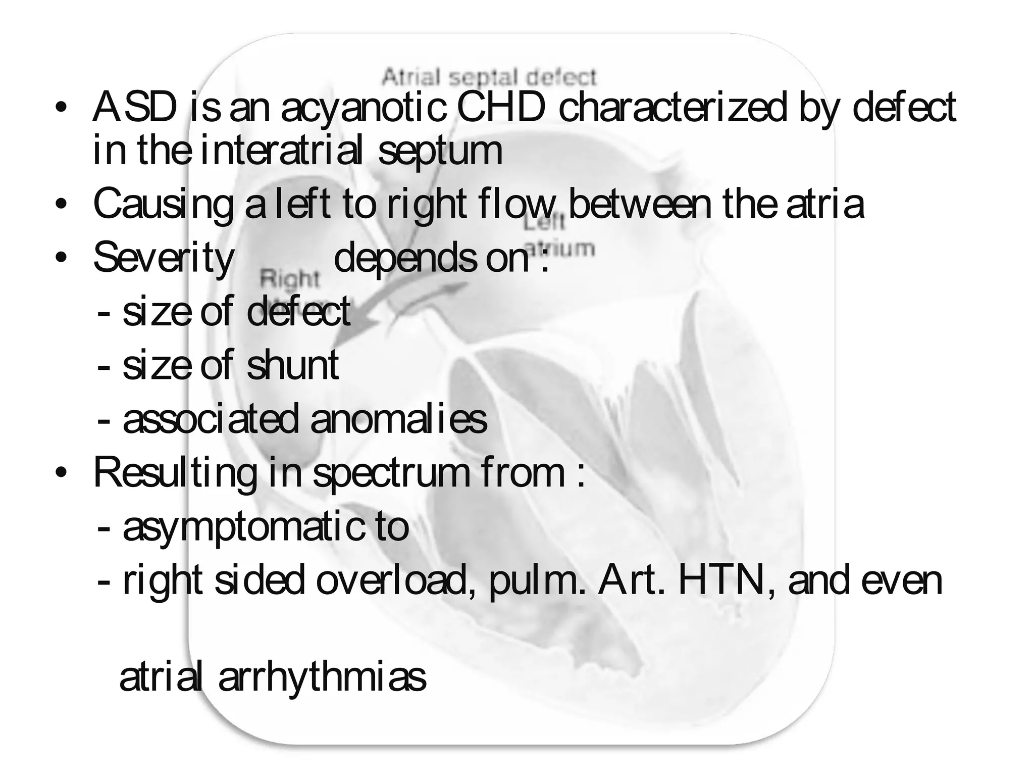
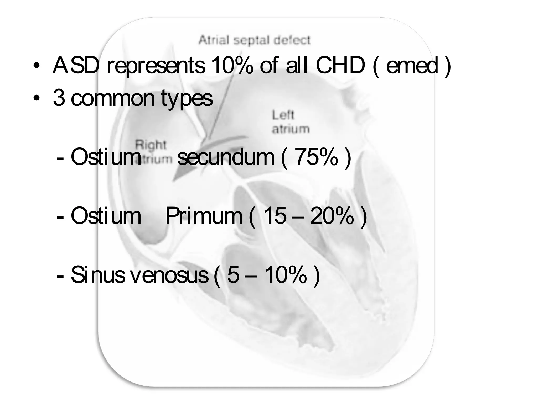
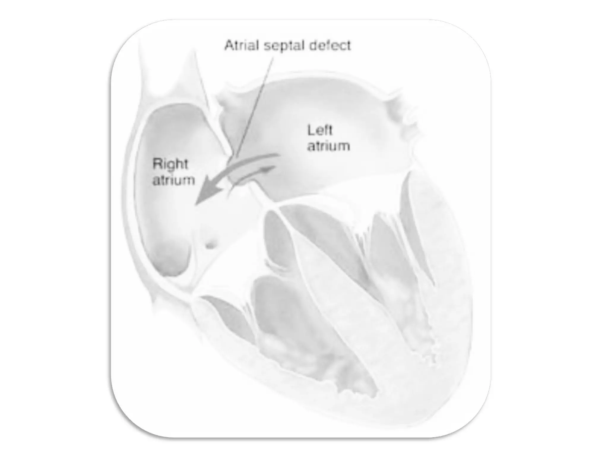
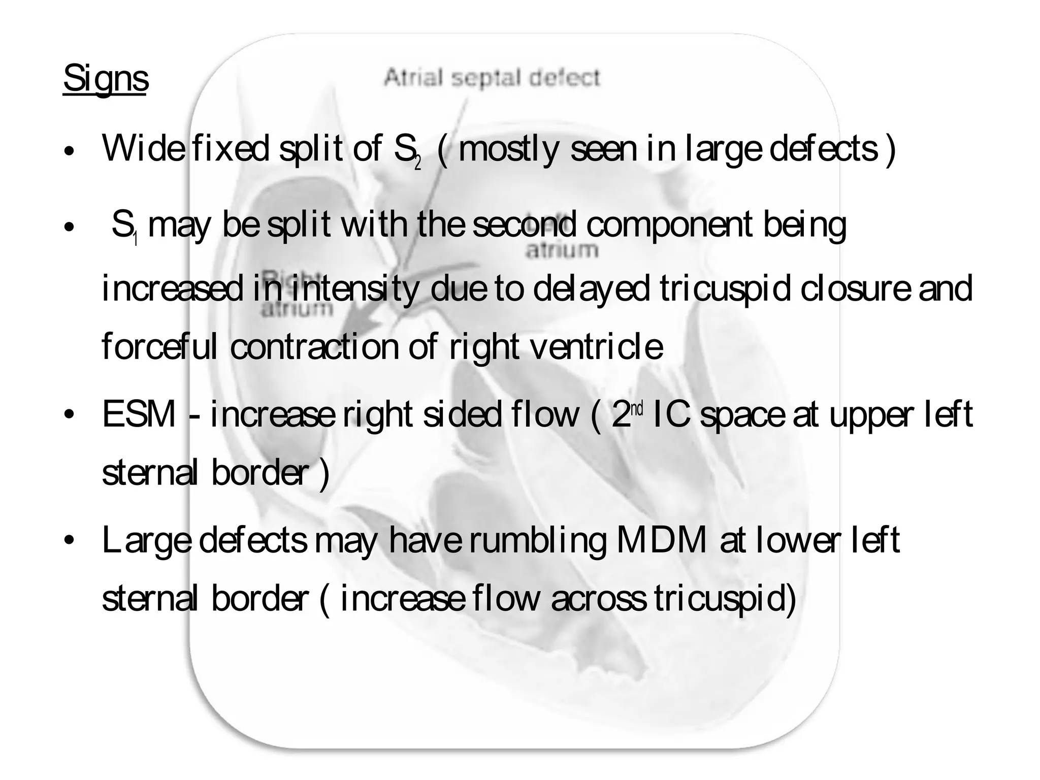
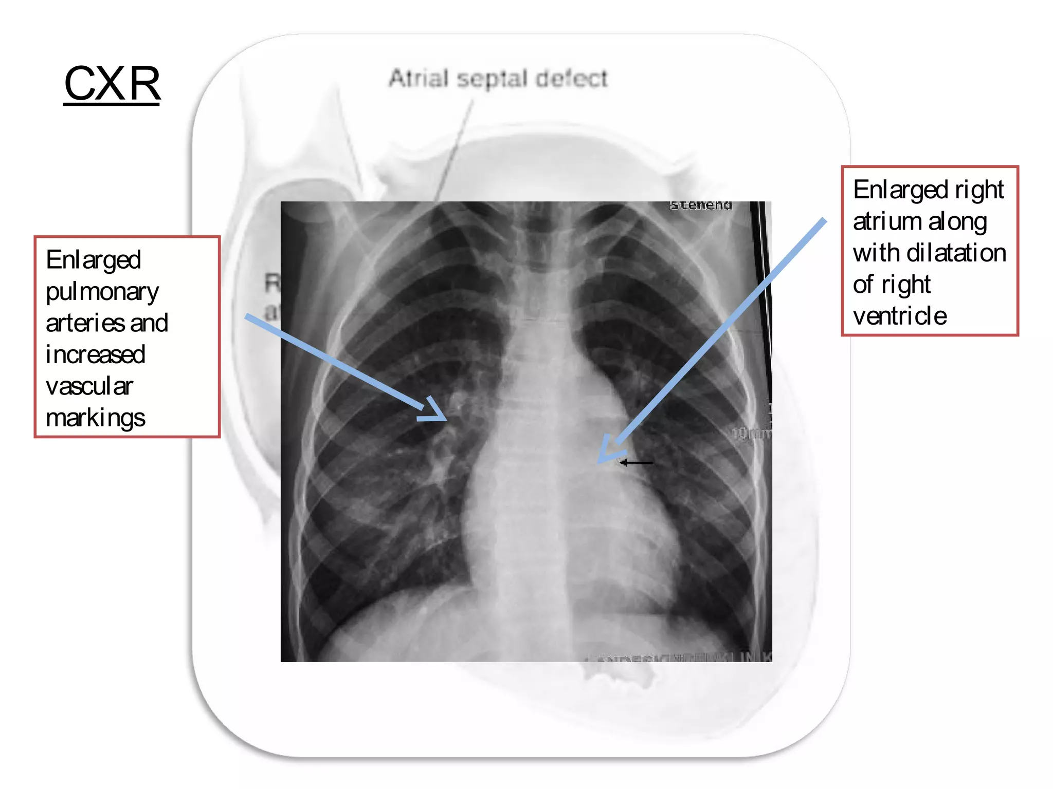
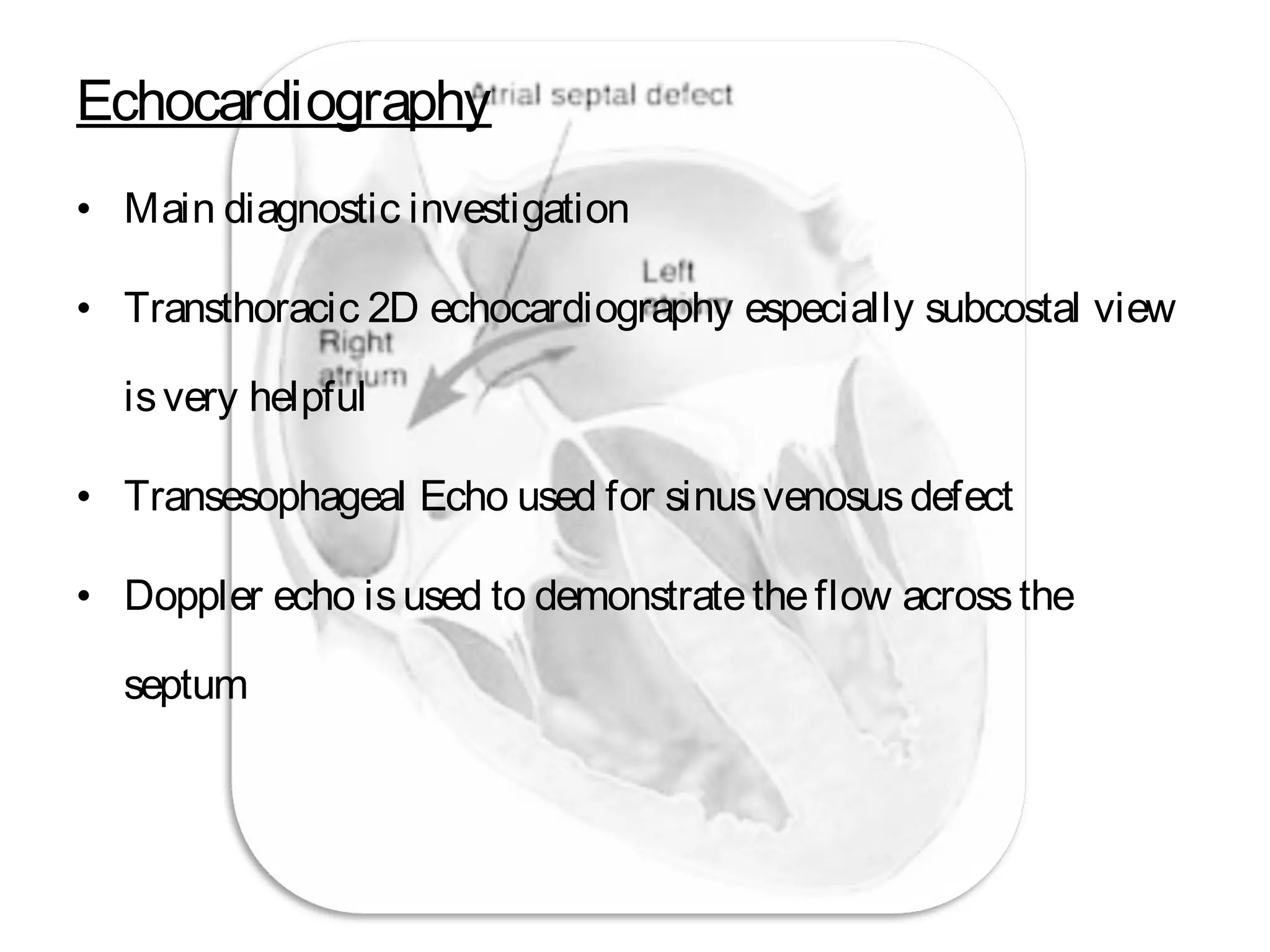
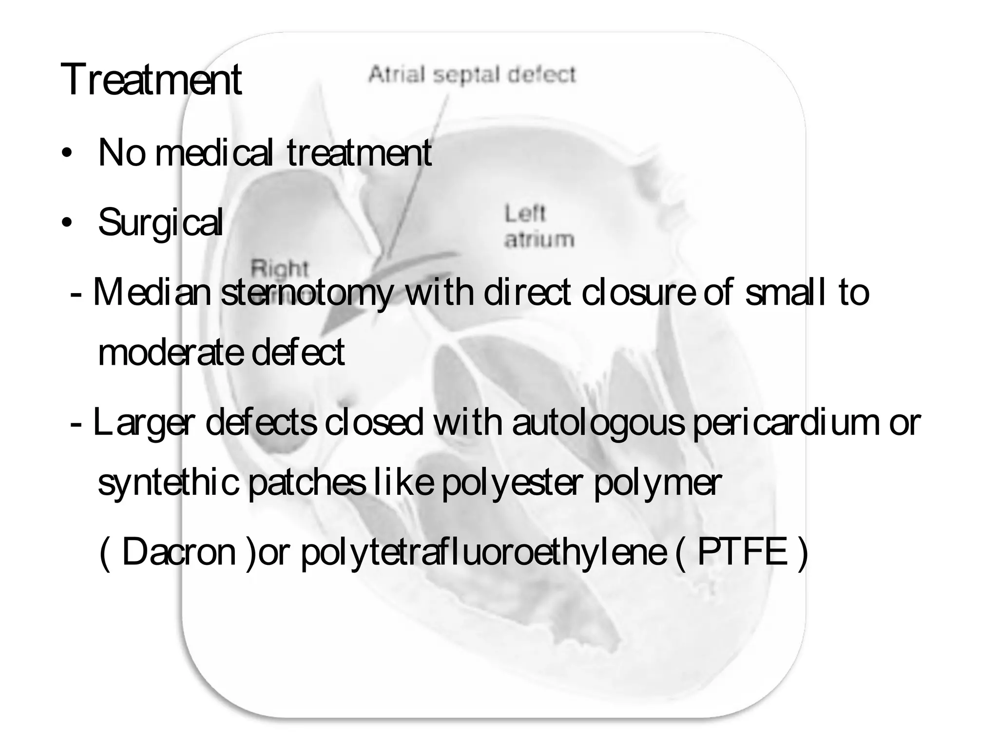
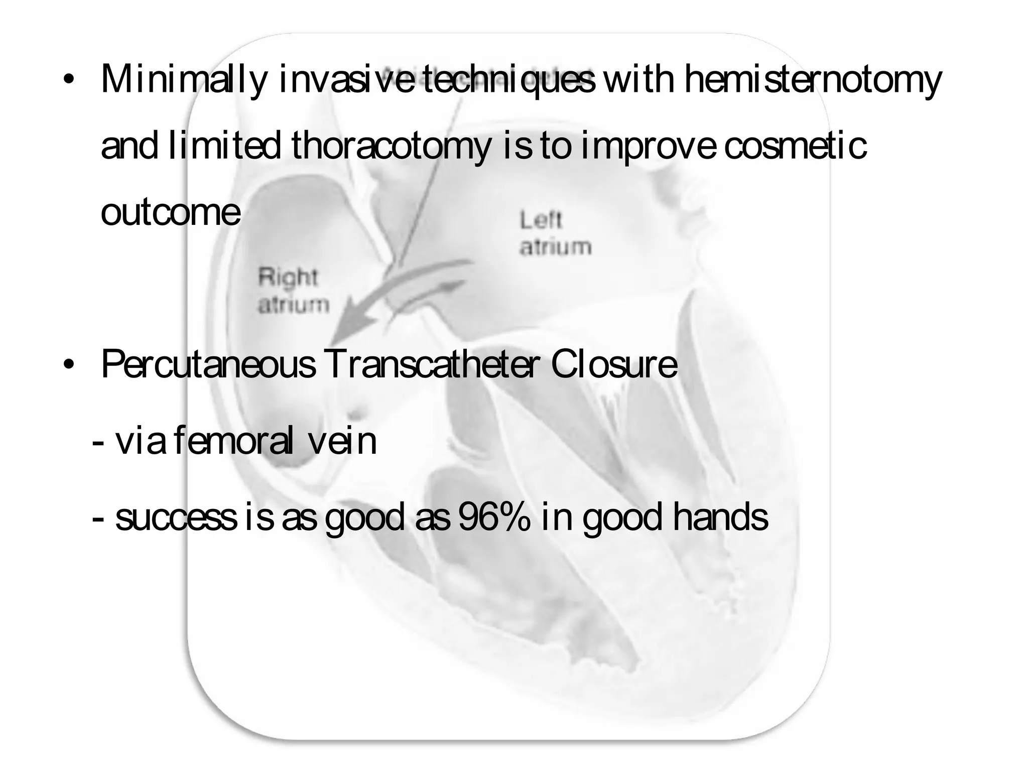
Atrial Septal Defect
Arvin Raj
061303507
Group B2
• ASD isan acyanotic CHD characterized by defect
in theinteratrial septum
• Causing aleft to right flow between theatria
• Severity dependson :
- sizeof defect
- sizeof shunt
- associated anomalies
• Resulting in spectrum from :
- asymptomatic to
- right sided overload, pulm. Art. HTN, and even
atrial arrhythmias
• ASD represents10% of all CHD ( emed )
• 3 common types
- Ostium secundum ( 75% )
- Ostium Primum ( 15 – 20% )
- Sinusvenosus( 5 – 10% )
• Male: female= 1:2
• Most infant and children areasymptomatic,
but thisagain dependson severity of defect
• Symptomsaremoreprevalent aspatient ages,
usually around ageof 40
• Magnitudeof L – R shunt dependson :
- Defect size
- Complianceof ventricles
- Relativeresistancein both pulmonary and
systemic circulation
• Shunting occursduring latevent systoleand
early diastole
• Thevolumeoverload isusually well tolerated
in children
• Even though thepulmonary flow may bemore
than twice
• However if left untreated… reversal of shunt
can eventually occur at alater age.
Presentation
Symptoms
• Often asymptomatic
• Easy fatigability
• Recurrent chest infection
• Exertional dyspnoea
• Palpitationsrelated to arryhthmias
Signs
• Widefixed split of S2 ( mostly seen in largedefects)
• S1 may besplit with thesecond component being
increased in intensity dueto delayed tricuspid closureand
forceful contraction of right ventricle
• ESM - increaseright sided flow ( 2nd
IC spaceat upper left
sternal border )
• Largedefectsmay haverumbling MDM at lower left
sternal border ( increaseflow acrosstricuspid)
CXR
Enlarged
pulmonary
arteriesand
increased
vascular
markings
Enlarged right
atrium along
with dilatation
of right
ventricle
ECG
Enlarged ‘p’
wave
indicating
Right atrial
hypertrophy
rSR’ seen and tall R wave
Indicating RBBB and
RVH
Also notethat theaVF is
predominantly upwardsas
compared to Lead I
indicating Right Axis
Deviation
LAD with rSR’ in V1 issuggestive
of Ostium primum defect
Echocardiography
• Main diagnostic investigation
• Transthoracic 2D echocardiography especially subcostal view
isvery helpful
• Transesophageal Echo used for sinusvenosusdefect
• Doppler echo isused to demonstratetheflow acrossthe
septum
MRI
• Can beuseto identify sizeand location of defect
• A major advantageof MRI istheability to quantify
right ventricular size, volume, and function along
with theability to identify thesystemic and
pulmonary venousreturn.
Treatment
• No medical treatment
• Surgical
- Median sternotomy with direct closureof small to
moderatedefect
- Larger defectsclosed with autologouspericardium or
syntethic patcheslikepolyester polymer
( Dacron )or polytetrafluoroethylene( PTFE )
• Minimally invasivetechniqueswith hemisternotomy
and limited thoracotomy isto improvecosmetic
outcome
• PercutaneousTranscatheter Closure
- viafemoral vein
- successisasgood as96% in good hands
Atrial septal defect

Atrial septal defect

  • 1.
    Atrial Septal Defect ArvinRaj 061303507 Group B2
  • 2.
    • ASD isanacyanotic CHD characterized by defect in theinteratrial septum • Causing aleft to right flow between theatria • Severity dependson : - sizeof defect - sizeof shunt - associated anomalies • Resulting in spectrum from : - asymptomatic to - right sided overload, pulm. Art. HTN, and even atrial arrhythmias
  • 3.
    • ASD represents10%of all CHD ( emed ) • 3 common types - Ostium secundum ( 75% ) - Ostium Primum ( 15 – 20% ) - Sinusvenosus( 5 – 10% )
  • 5.
    • Male: female=1:2 • Most infant and children areasymptomatic, but thisagain dependson severity of defect • Symptomsaremoreprevalent aspatient ages, usually around ageof 40
  • 6.
    • Magnitudeof L– R shunt dependson : - Defect size - Complianceof ventricles - Relativeresistancein both pulmonary and systemic circulation • Shunting occursduring latevent systoleand early diastole
  • 7.
    • Thevolumeoverload isusuallywell tolerated in children • Even though thepulmonary flow may bemore than twice • However if left untreated… reversal of shunt can eventually occur at alater age.
  • 8.
    Presentation Symptoms • Often asymptomatic •Easy fatigability • Recurrent chest infection • Exertional dyspnoea • Palpitationsrelated to arryhthmias
  • 9.
    Signs • Widefixed splitof S2 ( mostly seen in largedefects) • S1 may besplit with thesecond component being increased in intensity dueto delayed tricuspid closureand forceful contraction of right ventricle • ESM - increaseright sided flow ( 2nd IC spaceat upper left sternal border ) • Largedefectsmay haverumbling MDM at lower left sternal border ( increaseflow acrosstricuspid)
  • 10.
  • 11.
    ECG Enlarged ‘p’ wave indicating Right atrial hypertrophy rSR’seen and tall R wave Indicating RBBB and RVH Also notethat theaVF is predominantly upwardsas compared to Lead I indicating Right Axis Deviation LAD with rSR’ in V1 issuggestive of Ostium primum defect
  • 12.
    Echocardiography • Main diagnosticinvestigation • Transthoracic 2D echocardiography especially subcostal view isvery helpful • Transesophageal Echo used for sinusvenosusdefect • Doppler echo isused to demonstratetheflow acrossthe septum
  • 13.
    MRI • Can beusetoidentify sizeand location of defect • A major advantageof MRI istheability to quantify right ventricular size, volume, and function along with theability to identify thesystemic and pulmonary venousreturn.
  • 14.
    Treatment • No medicaltreatment • Surgical - Median sternotomy with direct closureof small to moderatedefect - Larger defectsclosed with autologouspericardium or syntethic patcheslikepolyester polymer ( Dacron )or polytetrafluoroethylene( PTFE )
  • 15.
    • Minimally invasivetechniqueswithhemisternotomy and limited thoracotomy isto improvecosmetic outcome • PercutaneousTranscatheter Closure - viafemoral vein - successisasgood as96% in good hands