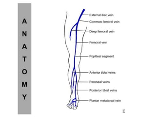
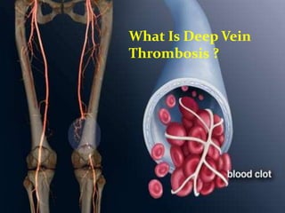
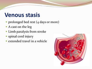
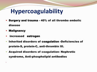
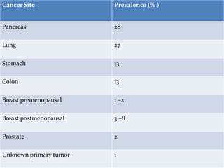
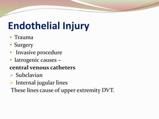
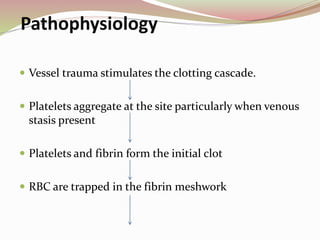
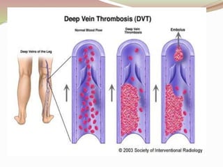
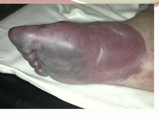
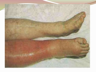
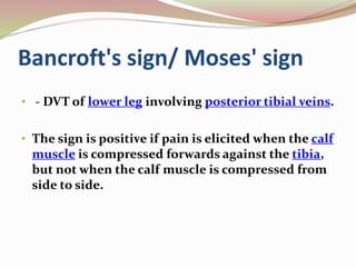
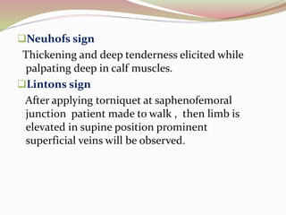
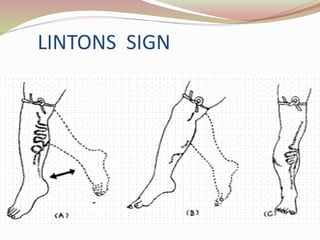
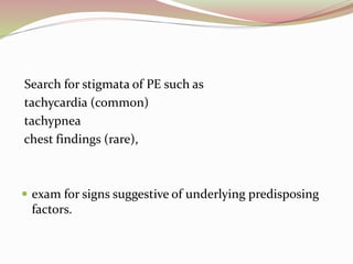
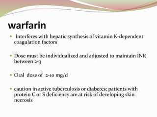
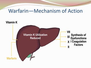
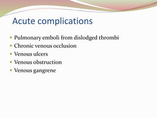
DEEP VEIN THROMBOSIS
What Is Deep Vein
Thrombosis ?
Definition
Deep vein thrombosis is the
formation of a blood clot in one of
the deep veins of the body, usually
in the leg.
It is like iceberg disease
Symptomatic deep vein thrombosis is
"tip of the iceberg"
Epidemiology
Venous ThromboEmbolism related deaths
3,00,000/anum
7% diagnosed and treated
34% sudden pulmonary embolism
59% as undected
Incidence
 An annual incidence of symptomatic Venous
ThromboEmbolism as 117 per 100,000 persons .
 Venous ThromboEmbolism in hospitalized patients
has increased from 0.8% to 1.3% over a period of 20
years (reported in 2005).
Without prophylaxis the incidence of deep vein
thrombosis is about –
 14% in gynaecological surgery
22% in neurosurgery
26% in abdominal surgery
45%-60% in patients undergoing hip and knee
surgeries.
15% to 40% Urologic surgery.
ETIOLOGY
Starts in
Lower
extremity
calf veins
progressing
proximally
to involve
Popliteal,
Femoral,
iliac system
Virchow triad
 More than 100 years ago, Rudolf Virchow described a
triad of factors
Venous stasis
Endothelial
damage
Hypercoagulable
state
Venous stasis
 prolonged bed rest (4 days or more)
 A cast on the leg
 Limb paralysis from stroke
 spinal cord injury
 extended travel in a vehicle
Hypercoagulability
 Surgery and trauma - 40% of all thrombo embolic
disease
 Malignancy
 increased estrogen
 Inherited disorders of coagulation -Deficiencies of
protein-S, protein-C, anti-thrombin III.
 Acquired disorders of coagulation- Nephrotic
syndrome, Anti-phospholipid antibodies

Hypercoagulable State of
Malignancy
 Up to 15% of cancer patients presents with VTE
VTE is not equally common in all types of cancer.
 The highest incidence is found in mucin-producing
adenocarcinomas, pancreas and gastrointestinal
tract, lung cancer, and ovarian cancer.
Cancer Site Prevalence (% )
Pancreas 28
Lung 27
Stomach 13
Colon 13
Breast premenopausal 1 –2
Breast postmenopausal 3 –8
Prostate 2
Unknown primary tumor 1
Endothelial Injury
 Trauma
 Surgery
 Invasive procedure
 Iatrogenic causes –
central venous catheters
 Subclavian
 Internal jugular lines
These lines cause of upper extremity DVT.
Pathophysiology
 Vessel trauma stimulates the clotting cascade.
 Platelets aggregate at the site particularly when venous
stasis present
 Platelets and fibrin form the initial clot
 RBC are trapped in the fibrin meshwork
 The thrombus propagates in the direction of the
blood flow.
 Inflammation is triggered, causing tenderness,
swelling, and erythema.
 Pieces of thrombus may break loose and travel
through circulation- emboli.
 Fibroblasts eventually invade the thrombus,
scarring vein wall and destroying valves. Patency
may be restored valve damage is permanent,
affecting directional flow.
40-60% also have capillary involvement irreversible
venous gangrene
Hydrostatic pressure in arterial and venous
capillaries exceeds the oncotic pressure
Fluid sequestration in the interstitium
Circulatory shock, and arterial insufficiency
which causes gangrene.
RISK FACTORS
 Endothelial damage
 Trauma
 Surgery
 Pacing wires
 Central venous catheters
 Dialysis access catheters
 Local vein damage
 Previous history
 Prolonged rest
Risk factors....
 Repetitive motion injury
 Venous stasis
 Bed rest or immobilization
 Obesity
 History of varicosities
 Spinal cord injury
 Age (>40 yr)
Risk factors...
 Coagulopathy
 Cancer
 Pregnancy
 Oral contraceptive use
 Presence of congenital proteins C and S
 Presence of anticardiolipin antibody
 Antithrombin III deficiency
 Polycythemia
 Septicemia
Presentation and Physical Examination
 Calf pain or tenderness, or both
 Swelling with pitting oedema
 Increased skin temperature and fever
 Superficial venous dilatation
 Cyanosis can occur with severe obstruction
Less frequent manifestations of venous thrombosis
include
 Phlegmasia alba dolens,
 Phlegmasia cerulea dolens, and
 Venous gangrene.
These are clinical spectrum of the same disorder.
PHLEGMASIA ALBA DOLENS
Thrombosis in only major deep venous channels
sparing collateral veins
Causing painful congestion and oedema
of leg, with lymphangitis
Which further increases
Oedema
PHLEGMASIA CERULEA DOLENS
Thrombosis extends to collateral veins.
congestions, massive fluid sequestration,
edema
CLINICAL MANIFESTATIONS
 sudden severe pain , swelling,
cyanosis and edema of the affected limb.
 There is a high risk of massive pulmonary
embolism, even under anticoagulation.
Foot gangrene may also occur.
 An underlying malignancy is found in 50% of
cases. Usually, it occurs in those afflicted by a life-
threatening illness.
Clinical examination
 Palpate distal pulses and evaluate capillary refill to
assess limb perfusion.
 Move and palpate all joints to detect acute arthritis or
other joint pathology.
 Neurologic evaluation may detect nerve root irritation;
sensory, motor, and reflex deficits should be noted
 Homans sign: pain in the posterior calf or knee with
forced dorsiflexion of the foot.
Bancroft's sign/ Moses' sign
• - DVT of lower leg involving posterior tibial veins.
• The sign is positive if pain is elicited when the calf
muscle is compressed forwards against the tibia,
but not when the calf muscle is compressed from
side to side.
Neuhofs sign
Thickening and deep tenderness elicited while
palpating deep in calf muscles.
Lintons sign
After applying torniquet at saphenofemoral
junction patient made to walk , then limb is
elevated in supine position prominent
superficial veins will be observed.
LINTONS SIGN
Search for stigmata of PE such as
tachycardia (common)
tachypnea
chest findings (rare),
 exam for signs suggestive of underlying predisposing
factors.
WELLS CLINICAL
PREDICTION SCORE
Interpretation
 High probability: ≥ 3 (Prevalence of DVT - 53%)
 Moderate probability: 1-2 (Prevalence of DVT - 17%)
 Low probability: ≤ 0 (Prevalence of DVT - 5%)
Limitations of Wells score
It useful in secondary and tertiary care centers,
has not been properly validated for use in
primary care centers patients with the
suspicion of DVT.
 The performance of Wells score was decreased
when evaluating elderly patients or those with a
prior DVT or having those having other
comorbidities, which might be equivalent to
what is found in a primary care setting.
Diagnostic Studies
 Clinical examination alone is able to confirm
only 20-30% of cases of DVT
 Blood Tests
The D-dimer
 Imaging Studies
D-dimer
It specific degradation product of cross-linked fibrin.
Because concurrent production and breakdown of clot
characterize thrombosis, patients with
thromboembolic disease have elevated levels of D-
dimer.
Three major approaches for measuring D-dimer
ELISA
latex agglutination
blood agglutination test
False-positive D-dimers occur in
patients with
 recent (within 10 days) surgery or trauma,
 recent myocardial infarction or stroke,
 acute infection,
 disseminated intravascular coagulation,
 pregnancy or recent delivery,
active collagen vascular disease, or metastatic cancer
Algorithm for diagnostic imaging
Assess
clinical
likelihood
Low D
dimer
Normal No DVT
High Imaging test
needed
High Imaging test
needed
Imaging Studies
 Invasive
 venography,
radiolabeled fibrinogen
 noninvasive
ultrasound,
plethysmography,
MRI techniques
Venography -Popliteal Vein Thrombosis
Venography
 It detects thrombi in both calf and thigh
 It can conclude and exclude the diagnosis of DVT
when other objective testings are not conclusive.
 Advantages
 It is useful if the patient has a high clinical probability
of thrombosis and a negative ultrasound.
 It is also valuable in symptomatic patients with a
history of prior thrombosis in whom the ultrasound is
non-diagnostic.
Disadvantage
 It can primary cause of DVT in 3% of patients who
undergo this diagnostic procedure.
 An invasive and expensive.
 Although Venography was once considered the gold
standard for diagnosis of DVT, today it is more
commonly used in research environments and less
frequently utilized in clinical practice.
Nuclear Medicine Studies
Because the radioactive isotope incorporates into a
growing thrombus, this test can distinguish new clot
from an old clot.
Nuclear medicine studies done with I125-labeled
fibrinogen .
More commonly used in research.
Plethysmography
 Plethysmography measures change in lower extremity
volume in response to certain stimuli.
Impedance plethysmography
 Principle- Blood volume changes in the leg lead to
changes in electrical resistance.
 Venous return in the lower extremity is occluded by
inflation of a thigh cuff, and then the cuff is released,
resulting in a decrease in calf blood volume. Any
obstruction of the proximal veins diminishes the
volume change, which is detected by measuring
changes in electrical resistance (impedance) over the
calf.
Ultrasonography
 color-flow Duplex scanning is the imaging test of
choice for patients with suspected DVT
 inexpensive,
 noninvasive,
 widely available
 Ultrasound can also distinguish other causes of leg
swelling, such as tumor, popliteal cyst, abscess,
aneurysm, or hematoma.
clinical limitations
 Reader dependent
 Duplex scans are less likely to detect non-occluding
thrombi.
 During the second half of pregnancy, ultrasound
becomes less specific, because the gravid uterus
compresses the inferior vena cava, thereby changing
Doppler flow in the lower extremities.
 An inability to distinguish old clots from a
newly forming clot
 Lack of accuracy in detecting DVT in the pelvis or the
small vessels of the calf
 Lack of accuracy in detecting DVT in the presence of
obesity or significant edema
Magnetic Resonance Imaging
It detects leg, pelvis, and pulmonary thrombi and is
97% sensitive and 95% specific for DVT.
 It distinguishes a mature from an immature clot.
 MRI is safe in all stages of pregnancy.
 Test may not be appropriate for patients with
pacemakers or other metallic implants, it can be an
effective diagnostic option for some patients.
DIFFERENTIAL DIAGNOSIS
o Cellulitis
Thrombophlebitis
o Arthritis
Asymmetric peripheral edema secondary to CHF,
liver disease, renal failure, or nephrotic syndrome
lymphangitis
Extrinsic compression of iliac vein secondary to
tumor, hematoma, or abscess
Hematoma
Lymphedema
Muscle or soft tissue injury
Neurogenic pain
Postphlebitic syndrome
Ruptured Baker cyst
Stress fractures or other bony lesions
Superficial thrombophlebitis
Some Tips to Avoid DVT
Do not sit for long periods of time
 Elevate legs if you are sitting for moderate periods of time
 Keep hydrated-drink six glasses of water a day
 Do not wear constricting garments around thighs or legs
 Application of elastic compression stockings, the use of intermittent
pneumatic compression devices
 special body positioning and exercise
 subcutaneous unfractionated or low molecular weight heparin.
Management
 Using the pretest probability score calculated from the
Wells Clinical Prediction rule, patients are stratified
into 3 risk groups—high, moderate, or low.
 The results from duplex ultrasound are incorporated
as follows:
 If the patient is high or moderate risk and the duplex
ultrasound study is positive, treat for DVT.
 If the duplex study is negative and the patient is low
risk, DVT has been ruled out.
• When discordance exists between the pretest
probability and the duplex study result, further
evaluation is required.
 If the patient is high risk but the ultrasound study was
negative, the patient still has a significant probability of
DVT
 a venogram to rule out a calf vein DVT
 surveillance with repeat clinical evaluation and
ultrasound in 1 week.
 results of a D-dimer assay to guide management
 If the patient is low risk but the ultrasound study is
positive, some authors recommend a second
confirmatory study such as a venogram before treating
for DVT
EMERGENCY DEPARTMANT CARE
The primary objectives of the treatment of DVT
are to -
 prevent pulmonary embolism,
 reduce morbidity, and
 prevent or minimize the risk of developing the
postphlebitic syndrome.
General Therapeutic Measures :
 Bed rest .
 Encourage the patient to perform gentle foot & leg
exercises every hour.
 Increase fluid intake upto 2 l/day unless
contraindicated.
 Avoid deep palpation .
SPECIFIC TREATMENT :
 Anticoagulation
 Thrombolytic therapy for DVT
 Surgery for DVT
 Filters for DVT
 Compression stockings
 Initial treatment of DVT is with low-molecular-
weight heparin or unfractionated heparin for at
least 5 days, followed by warfarin (target INR, 2.0–
3.0) for at least 3 months.
Anticoagulation
 Heparin prevents extension of the thrombus
 It is a heterogeneous mixture of polysaccharide
fragments with varying molecular weights but with
similar biological activity.
Mechanism of action
 Heparin's anticoagulant effect is related directly to its
activation of antithrombin III.
 Antithrombin III, the body's primary anticoagulant,
inactivates thrombin and inhibits the activity of
activated factor X, factor IX in the coagulation
process.
Heparin: Mechanism of Action
Accelerates antithrombin III activity
Prothrombin Thrombin
Factor Xa
Factor Va
Factor X
Factor IXa
Factor VIIIa
Ca2+, PL
Ca2+, PL
Antithrombin III
(Heparin)
Side effects
• Bleeding
• Osteoporosis
• Thrombocytopenia
• Skins lesions- urticaria, papules, necrosis
• Hypoaldosteronism, hyperkalemia
CONTRAINDICATIONS-
• Bleeding disorders,
• Severe hypertension, threatened abortion, piles,
• large malignancies, tuberculosis’
• Ocular surgery and neurosurgery,
• Chronic alcoholics, cirrhosis, renal failure
Dose
 IV bolus dose of 5,000 to 10,000 units
followed by an infusion of 1,000 units per hour.
Other method of initiating therapy is to begin with
 Loading dose of 50-100 units/kg of heparin followed
by a constant infusion of 15-25 units/kg/hr.
Low molecular weight heparin
 Selectively inhibit factor Xa .
 Superior bioavailability
 Superior or equivalent safety and efficacy
 Subcutaneous once- or twice-daily dosing
 No laboratory monitoring
 Less phlebotomy (no monitoring/no intravenous line)
 Less thrombocytopenia
 The optimal regimen for the treatment of DVT is
anticoagulation with heparin or an LMWH followed
by full anticoagulation with oral warfarin for 3-6
months
 Warfarin therapy is overlapped with heparin for 4-5
days until the INR is therapeutically elevated to
between 2-3.
warfarin
 Interferes with hepatic synthesis of vitamin K-dependent
coagulation factors
 Dose must be individualized and adjusted to maintain INR
between 2-3
 Oral dose of 2-10 mg/d
 caution in active tuberculosis or diabetes; patients with
protein C or S deficiency are at risk of developing skin
necrosis
Warfarin—Mechanism of Action
Vitamin K
Warfarin
Synthesis of
Dysfunctiona
l Coagulation
Factors
VII
IX
X
II
Vitamin K Utilization
Reduced
Drawbacks of warfarin therapy
 Delayed onset and offset of action.
 Frequent blood test monitoring required:
- the dose response is unpredictable,
- has a narrow therapeutic range
 Reversibility of anticoagulant affect is slow.
 Requires labor-intensive follow up, Expert dose
management, Frequent patient communication.
Thrombolytic therapy for DVT
Advantages include
 Prompt resolution of symptoms,
 Prevention of pulmonary embolism,
 Restoration of normal venous circulation,
 Preservation of venous valvular function,
 Prevention of postphlebitic syndrome.
Disadvantage
Thrombolytic therapy does not prevent
 clot propagation,
 rethrombosis, or
 subsequent embolization.
 Heparin therapy and oral anticoagulant therapy
always must followed after a course of thrombolysis.
 Thrombolytic therapy is also not effective once the
thrombus is adherent and begins to organize
 The hemorrhagic complications of thrombolytic
therapy are about 3 times higher, including the small
but potentially fatal risk of intra-cerebral hemorrhage.
 At present, therefore, thrombo-lysis should be
reserved for exceptional circumstances, such as
patients with limb-threatening ischemia caused by
phlegmasia cerulea dolens.
CONTRAINDICATIONS TO
ANTICOAGULATION THERAPY
 Lack of patient cooperation
 Bleeding from the following systems:Gastrointestinal
,Genitourinary systems
 Hemorrhagic blood dyscrasias
 Aneurysms
 Severe trauma
 Alcoholism
Contraindications (contd....)
 Recent / impending surgery of: Eye , Spinal cord, Brain
 Severe hepatic or renal disease
 Recent cerebrovascular hemorrhage
 Infections
 Open ulcerative wounds
 Occupations that involve a significant hazard for
injury
 Recent delivery of a baby

Surgery for DVT
Indications
 when anticoagulant therapy is ineffective
 unsafe,
 contraindicated.
 The major surgical procedures for DVT are clot
removal and partial interruption of the inferior vena
cava to prevent pulmonary embolism.
 These pulmonary emboli removed at autopsy look like
casts of the deep veins of the leg where they
originated.

This patient underwent a thrombectomy. The thrombus has been laid over
the approximate location in the leg veins where it developed.
Catheter-Directed Thrombolysis
 Successful clot lysis in > 85%
 Better 1-yr patency,
 Long-term symptom resolution.
Filters for DVT
 Indications
 Contraindication to anticoagulation.
 Significant bleeding complication of anticoagulation
therapy.
 Pulmonary embolism with contraindication to
anticoagulation.
 Recurrent thrombo-embolic complication despite
adequate anticoagulation therapy.
 Inferior vena cava filters reduce the rate of pulmonary
embolism but have no effect on the other
complications of deep vein thrombosis. Thrombolysis
should be considered in patients with major proximal
vein thrombosis and threatened venous infarction
PROPHYLAXIS
 Indicated in who underwent major abdominal trauma
or orthopaedic surgery or patient having prolonged
immobolization (> 3 days).
 Benefits of VTE Prophylaxis
 Improved patient outcomes
 Reduced costs
Methods of VTE Prophylaxis
 Mechanical:
 Graduated Compression Stockings (GCS)
 Intermittent Pneumatic Compression Devices
(IPC)
 Pharmacologic
Low molecular weight Heparin.(5000u sc 8hourly )
It inhibits factor Xa and IIA activity.
Acute complications
 Pulmonary emboli from dislodged thrombi
 Chronic venous occlusion
 Venous ulcers
 Venous obstruction
 Venous gangrene
CHRONIC COMPLICATIONS
 Valvular destruction
 Chronic venous insufficiency
 Increased venous pressure
 Varicosities
 Increased distal pressure
 Fluid stasis
 Edema
NURSING DIAGNOSIS
1. Pain related to inflammatory response in affected
vein
2. Anxiety related to unexpected hospitalization and
uncertainty about the seriousness of her illness
3. Ineffective tissue perfusion: Peripheral related to
decreased venous circulation in the right leg
4. Risk for impaired skin integrity related to pooling of
venous blood in the right leg
DISCUSSION
CONCLUSION
Bindhya dvt

Bindhya dvt

  • 2.
  • 5.
    What Is DeepVein Thrombosis ?
  • 6.
    Definition Deep vein thrombosisis the formation of a blood clot in one of the deep veins of the body, usually in the leg.
  • 7.
    It is likeiceberg disease Symptomatic deep vein thrombosis is "tip of the iceberg"
  • 8.
    Epidemiology Venous ThromboEmbolism relateddeaths 3,00,000/anum 7% diagnosed and treated 34% sudden pulmonary embolism 59% as undected
  • 9.
    Incidence  An annualincidence of symptomatic Venous ThromboEmbolism as 117 per 100,000 persons .  Venous ThromboEmbolism in hospitalized patients has increased from 0.8% to 1.3% over a period of 20 years (reported in 2005).
  • 10.
    Without prophylaxis theincidence of deep vein thrombosis is about –  14% in gynaecological surgery 22% in neurosurgery 26% in abdominal surgery 45%-60% in patients undergoing hip and knee surgeries. 15% to 40% Urologic surgery.
  • 12.
  • 13.
    Virchow triad  Morethan 100 years ago, Rudolf Virchow described a triad of factors
  • 14.
  • 15.
    Venous stasis  prolongedbed rest (4 days or more)  A cast on the leg  Limb paralysis from stroke  spinal cord injury  extended travel in a vehicle
  • 16.
    Hypercoagulability  Surgery andtrauma - 40% of all thrombo embolic disease  Malignancy  increased estrogen  Inherited disorders of coagulation -Deficiencies of protein-S, protein-C, anti-thrombin III.  Acquired disorders of coagulation- Nephrotic syndrome, Anti-phospholipid antibodies 
  • 17.
    Hypercoagulable State of Malignancy Up to 15% of cancer patients presents with VTE VTE is not equally common in all types of cancer.  The highest incidence is found in mucin-producing adenocarcinomas, pancreas and gastrointestinal tract, lung cancer, and ovarian cancer.
  • 18.
    Cancer Site Prevalence(% ) Pancreas 28 Lung 27 Stomach 13 Colon 13 Breast premenopausal 1 –2 Breast postmenopausal 3 –8 Prostate 2 Unknown primary tumor 1
  • 19.
    Endothelial Injury  Trauma Surgery  Invasive procedure  Iatrogenic causes – central venous catheters  Subclavian  Internal jugular lines These lines cause of upper extremity DVT.
  • 20.
    Pathophysiology  Vessel traumastimulates the clotting cascade.  Platelets aggregate at the site particularly when venous stasis present  Platelets and fibrin form the initial clot  RBC are trapped in the fibrin meshwork
  • 21.
     The thrombuspropagates in the direction of the blood flow.  Inflammation is triggered, causing tenderness, swelling, and erythema.  Pieces of thrombus may break loose and travel through circulation- emboli.  Fibroblasts eventually invade the thrombus, scarring vein wall and destroying valves. Patency may be restored valve damage is permanent, affecting directional flow.
  • 22.
    40-60% also havecapillary involvement irreversible venous gangrene Hydrostatic pressure in arterial and venous capillaries exceeds the oncotic pressure Fluid sequestration in the interstitium Circulatory shock, and arterial insufficiency which causes gangrene.
  • 24.
    RISK FACTORS  Endothelialdamage  Trauma  Surgery  Pacing wires  Central venous catheters  Dialysis access catheters  Local vein damage  Previous history  Prolonged rest
  • 25.
    Risk factors....  Repetitivemotion injury  Venous stasis  Bed rest or immobilization  Obesity  History of varicosities  Spinal cord injury  Age (>40 yr)
  • 26.
    Risk factors...  Coagulopathy Cancer  Pregnancy  Oral contraceptive use  Presence of congenital proteins C and S  Presence of anticardiolipin antibody  Antithrombin III deficiency  Polycythemia  Septicemia
  • 28.
    Presentation and PhysicalExamination  Calf pain or tenderness, or both  Swelling with pitting oedema  Increased skin temperature and fever  Superficial venous dilatation  Cyanosis can occur with severe obstruction
  • 29.
    Less frequent manifestationsof venous thrombosis include  Phlegmasia alba dolens,  Phlegmasia cerulea dolens, and  Venous gangrene. These are clinical spectrum of the same disorder.
  • 30.
    PHLEGMASIA ALBA DOLENS Thrombosisin only major deep venous channels sparing collateral veins Causing painful congestion and oedema of leg, with lymphangitis Which further increases Oedema
  • 31.
    PHLEGMASIA CERULEA DOLENS Thrombosisextends to collateral veins. congestions, massive fluid sequestration, edema
  • 34.
    CLINICAL MANIFESTATIONS  suddensevere pain , swelling, cyanosis and edema of the affected limb.  There is a high risk of massive pulmonary embolism, even under anticoagulation. Foot gangrene may also occur.  An underlying malignancy is found in 50% of cases. Usually, it occurs in those afflicted by a life- threatening illness.
  • 37.
    Clinical examination  Palpatedistal pulses and evaluate capillary refill to assess limb perfusion.  Move and palpate all joints to detect acute arthritis or other joint pathology.  Neurologic evaluation may detect nerve root irritation; sensory, motor, and reflex deficits should be noted
  • 38.
     Homans sign:pain in the posterior calf or knee with forced dorsiflexion of the foot.
  • 39.
    Bancroft's sign/ Moses'sign • - DVT of lower leg involving posterior tibial veins. • The sign is positive if pain is elicited when the calf muscle is compressed forwards against the tibia, but not when the calf muscle is compressed from side to side.
  • 40.
    Neuhofs sign Thickening anddeep tenderness elicited while palpating deep in calf muscles. Lintons sign After applying torniquet at saphenofemoral junction patient made to walk , then limb is elevated in supine position prominent superficial veins will be observed.
  • 41.
  • 42.
    Search for stigmataof PE such as tachycardia (common) tachypnea chest findings (rare),  exam for signs suggestive of underlying predisposing factors.
  • 43.
  • 45.
    Interpretation  High probability:≥ 3 (Prevalence of DVT - 53%)  Moderate probability: 1-2 (Prevalence of DVT - 17%)  Low probability: ≤ 0 (Prevalence of DVT - 5%)
  • 46.
    Limitations of Wellsscore It useful in secondary and tertiary care centers, has not been properly validated for use in primary care centers patients with the suspicion of DVT.  The performance of Wells score was decreased when evaluating elderly patients or those with a prior DVT or having those having other comorbidities, which might be equivalent to what is found in a primary care setting.
  • 47.
    Diagnostic Studies  Clinicalexamination alone is able to confirm only 20-30% of cases of DVT  Blood Tests The D-dimer  Imaging Studies
  • 48.
    D-dimer It specific degradationproduct of cross-linked fibrin. Because concurrent production and breakdown of clot characterize thrombosis, patients with thromboembolic disease have elevated levels of D- dimer. Three major approaches for measuring D-dimer ELISA latex agglutination blood agglutination test
  • 49.
    False-positive D-dimers occurin patients with  recent (within 10 days) surgery or trauma,  recent myocardial infarction or stroke,  acute infection,  disseminated intravascular coagulation,  pregnancy or recent delivery, active collagen vascular disease, or metastatic cancer
  • 51.
    Algorithm for diagnosticimaging Assess clinical likelihood Low D dimer Normal No DVT High Imaging test needed High Imaging test needed
  • 52.
    Imaging Studies  Invasive venography, radiolabeled fibrinogen  noninvasive ultrasound, plethysmography, MRI techniques
  • 53.
  • 54.
    Venography  It detectsthrombi in both calf and thigh  It can conclude and exclude the diagnosis of DVT when other objective testings are not conclusive.  Advantages  It is useful if the patient has a high clinical probability of thrombosis and a negative ultrasound.  It is also valuable in symptomatic patients with a history of prior thrombosis in whom the ultrasound is non-diagnostic.
  • 55.
    Disadvantage  It canprimary cause of DVT in 3% of patients who undergo this diagnostic procedure.  An invasive and expensive.  Although Venography was once considered the gold standard for diagnosis of DVT, today it is more commonly used in research environments and less frequently utilized in clinical practice.
  • 56.
  • 57.
    Nuclear Medicine Studies Becausethe radioactive isotope incorporates into a growing thrombus, this test can distinguish new clot from an old clot. Nuclear medicine studies done with I125-labeled fibrinogen . More commonly used in research.
  • 58.
    Plethysmography  Plethysmography measureschange in lower extremity volume in response to certain stimuli.
  • 59.
    Impedance plethysmography  Principle-Blood volume changes in the leg lead to changes in electrical resistance.  Venous return in the lower extremity is occluded by inflation of a thigh cuff, and then the cuff is released, resulting in a decrease in calf blood volume. Any obstruction of the proximal veins diminishes the volume change, which is detected by measuring changes in electrical resistance (impedance) over the calf.
  • 60.
    Ultrasonography  color-flow Duplexscanning is the imaging test of choice for patients with suspected DVT  inexpensive,  noninvasive,  widely available  Ultrasound can also distinguish other causes of leg swelling, such as tumor, popliteal cyst, abscess, aneurysm, or hematoma.
  • 61.
    clinical limitations  Readerdependent  Duplex scans are less likely to detect non-occluding thrombi.  During the second half of pregnancy, ultrasound becomes less specific, because the gravid uterus compresses the inferior vena cava, thereby changing Doppler flow in the lower extremities.  An inability to distinguish old clots from a newly forming clot
  • 62.
     Lack ofaccuracy in detecting DVT in the pelvis or the small vessels of the calf  Lack of accuracy in detecting DVT in the presence of obesity or significant edema
  • 63.
    Magnetic Resonance Imaging Itdetects leg, pelvis, and pulmonary thrombi and is 97% sensitive and 95% specific for DVT.  It distinguishes a mature from an immature clot.  MRI is safe in all stages of pregnancy.  Test may not be appropriate for patients with pacemakers or other metallic implants, it can be an effective diagnostic option for some patients.
  • 64.
    DIFFERENTIAL DIAGNOSIS o Cellulitis Thrombophlebitis oArthritis Asymmetric peripheral edema secondary to CHF, liver disease, renal failure, or nephrotic syndrome lymphangitis Extrinsic compression of iliac vein secondary to tumor, hematoma, or abscess Hematoma Lymphedema
  • 65.
    Muscle or softtissue injury Neurogenic pain Postphlebitic syndrome Ruptured Baker cyst Stress fractures or other bony lesions Superficial thrombophlebitis
  • 67.
    Some Tips toAvoid DVT Do not sit for long periods of time  Elevate legs if you are sitting for moderate periods of time  Keep hydrated-drink six glasses of water a day  Do not wear constricting garments around thighs or legs  Application of elastic compression stockings, the use of intermittent pneumatic compression devices  special body positioning and exercise  subcutaneous unfractionated or low molecular weight heparin.
  • 68.
    Management  Using thepretest probability score calculated from the Wells Clinical Prediction rule, patients are stratified into 3 risk groups—high, moderate, or low.  The results from duplex ultrasound are incorporated as follows:  If the patient is high or moderate risk and the duplex ultrasound study is positive, treat for DVT.
  • 69.
     If theduplex study is negative and the patient is low risk, DVT has been ruled out. • When discordance exists between the pretest probability and the duplex study result, further evaluation is required.  If the patient is high risk but the ultrasound study was negative, the patient still has a significant probability of DVT
  • 70.
     a venogramto rule out a calf vein DVT  surveillance with repeat clinical evaluation and ultrasound in 1 week.  results of a D-dimer assay to guide management  If the patient is low risk but the ultrasound study is positive, some authors recommend a second confirmatory study such as a venogram before treating for DVT
  • 71.
    EMERGENCY DEPARTMANT CARE Theprimary objectives of the treatment of DVT are to -  prevent pulmonary embolism,  reduce morbidity, and  prevent or minimize the risk of developing the postphlebitic syndrome.
  • 72.
    General Therapeutic Measures:  Bed rest .  Encourage the patient to perform gentle foot & leg exercises every hour.  Increase fluid intake upto 2 l/day unless contraindicated.  Avoid deep palpation .
  • 73.
    SPECIFIC TREATMENT : Anticoagulation  Thrombolytic therapy for DVT  Surgery for DVT  Filters for DVT  Compression stockings
  • 74.
     Initial treatmentof DVT is with low-molecular- weight heparin or unfractionated heparin for at least 5 days, followed by warfarin (target INR, 2.0– 3.0) for at least 3 months.
  • 75.
    Anticoagulation  Heparin preventsextension of the thrombus  It is a heterogeneous mixture of polysaccharide fragments with varying molecular weights but with similar biological activity.
  • 76.
    Mechanism of action Heparin's anticoagulant effect is related directly to its activation of antithrombin III.  Antithrombin III, the body's primary anticoagulant, inactivates thrombin and inhibits the activity of activated factor X, factor IX in the coagulation process.
  • 77.
    Heparin: Mechanism ofAction Accelerates antithrombin III activity Prothrombin Thrombin Factor Xa Factor Va Factor X Factor IXa Factor VIIIa Ca2+, PL Ca2+, PL Antithrombin III (Heparin)
  • 78.
    Side effects • Bleeding •Osteoporosis • Thrombocytopenia • Skins lesions- urticaria, papules, necrosis • Hypoaldosteronism, hyperkalemia CONTRAINDICATIONS- • Bleeding disorders, • Severe hypertension, threatened abortion, piles, • large malignancies, tuberculosis’ • Ocular surgery and neurosurgery, • Chronic alcoholics, cirrhosis, renal failure
  • 79.
    Dose  IV bolusdose of 5,000 to 10,000 units followed by an infusion of 1,000 units per hour. Other method of initiating therapy is to begin with  Loading dose of 50-100 units/kg of heparin followed by a constant infusion of 15-25 units/kg/hr.
  • 80.
    Low molecular weightheparin  Selectively inhibit factor Xa .  Superior bioavailability  Superior or equivalent safety and efficacy  Subcutaneous once- or twice-daily dosing  No laboratory monitoring  Less phlebotomy (no monitoring/no intravenous line)  Less thrombocytopenia
  • 81.
     The optimalregimen for the treatment of DVT is anticoagulation with heparin or an LMWH followed by full anticoagulation with oral warfarin for 3-6 months  Warfarin therapy is overlapped with heparin for 4-5 days until the INR is therapeutically elevated to between 2-3.
  • 82.
    warfarin  Interferes withhepatic synthesis of vitamin K-dependent coagulation factors  Dose must be individualized and adjusted to maintain INR between 2-3  Oral dose of 2-10 mg/d  caution in active tuberculosis or diabetes; patients with protein C or S deficiency are at risk of developing skin necrosis
  • 83.
    Warfarin—Mechanism of Action VitaminK Warfarin Synthesis of Dysfunctiona l Coagulation Factors VII IX X II Vitamin K Utilization Reduced
  • 84.
    Drawbacks of warfarintherapy  Delayed onset and offset of action.  Frequent blood test monitoring required: - the dose response is unpredictable, - has a narrow therapeutic range  Reversibility of anticoagulant affect is slow.  Requires labor-intensive follow up, Expert dose management, Frequent patient communication.
  • 85.
    Thrombolytic therapy forDVT Advantages include  Prompt resolution of symptoms,  Prevention of pulmonary embolism,  Restoration of normal venous circulation,  Preservation of venous valvular function,  Prevention of postphlebitic syndrome.
  • 86.
    Disadvantage Thrombolytic therapy doesnot prevent  clot propagation,  rethrombosis, or  subsequent embolization.  Heparin therapy and oral anticoagulant therapy always must followed after a course of thrombolysis.
  • 87.
     Thrombolytic therapyis also not effective once the thrombus is adherent and begins to organize  The hemorrhagic complications of thrombolytic therapy are about 3 times higher, including the small but potentially fatal risk of intra-cerebral hemorrhage.  At present, therefore, thrombo-lysis should be reserved for exceptional circumstances, such as patients with limb-threatening ischemia caused by phlegmasia cerulea dolens.
  • 88.
    CONTRAINDICATIONS TO ANTICOAGULATION THERAPY Lack of patient cooperation  Bleeding from the following systems:Gastrointestinal ,Genitourinary systems  Hemorrhagic blood dyscrasias  Aneurysms  Severe trauma  Alcoholism
  • 89.
    Contraindications (contd....)  Recent/ impending surgery of: Eye , Spinal cord, Brain  Severe hepatic or renal disease  Recent cerebrovascular hemorrhage  Infections  Open ulcerative wounds  Occupations that involve a significant hazard for injury  Recent delivery of a baby 
  • 90.
    Surgery for DVT Indications when anticoagulant therapy is ineffective  unsafe,  contraindicated.  The major surgical procedures for DVT are clot removal and partial interruption of the inferior vena cava to prevent pulmonary embolism.
  • 91.
     These pulmonaryemboli removed at autopsy look like casts of the deep veins of the leg where they originated. 
  • 92.
    This patient underwenta thrombectomy. The thrombus has been laid over the approximate location in the leg veins where it developed.
  • 93.
    Catheter-Directed Thrombolysis  Successfulclot lysis in > 85%  Better 1-yr patency,  Long-term symptom resolution.
  • 94.
    Filters for DVT Indications  Contraindication to anticoagulation.  Significant bleeding complication of anticoagulation therapy.  Pulmonary embolism with contraindication to anticoagulation.  Recurrent thrombo-embolic complication despite adequate anticoagulation therapy.
  • 95.
     Inferior venacava filters reduce the rate of pulmonary embolism but have no effect on the other complications of deep vein thrombosis. Thrombolysis should be considered in patients with major proximal vein thrombosis and threatened venous infarction
  • 97.
    PROPHYLAXIS  Indicated inwho underwent major abdominal trauma or orthopaedic surgery or patient having prolonged immobolization (> 3 days).  Benefits of VTE Prophylaxis  Improved patient outcomes  Reduced costs
  • 98.
    Methods of VTEProphylaxis  Mechanical:  Graduated Compression Stockings (GCS)  Intermittent Pneumatic Compression Devices (IPC)  Pharmacologic Low molecular weight Heparin.(5000u sc 8hourly ) It inhibits factor Xa and IIA activity.
  • 103.
    Acute complications  Pulmonaryemboli from dislodged thrombi  Chronic venous occlusion  Venous ulcers  Venous obstruction  Venous gangrene
  • 104.
    CHRONIC COMPLICATIONS  Valvulardestruction  Chronic venous insufficiency  Increased venous pressure  Varicosities  Increased distal pressure  Fluid stasis  Edema
  • 106.
    NURSING DIAGNOSIS 1. Painrelated to inflammatory response in affected vein 2. Anxiety related to unexpected hospitalization and uncertainty about the seriousness of her illness 3. Ineffective tissue perfusion: Peripheral related to decreased venous circulation in the right leg 4. Risk for impaired skin integrity related to pooling of venous blood in the right leg
  • 107.
  • 108.