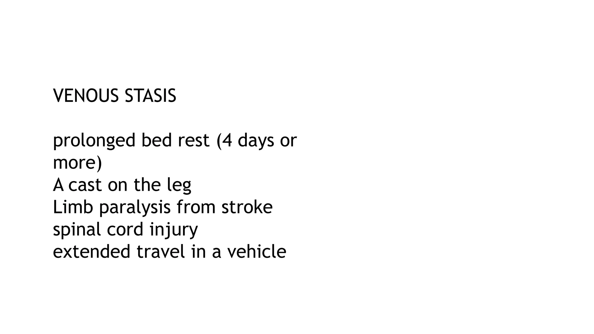
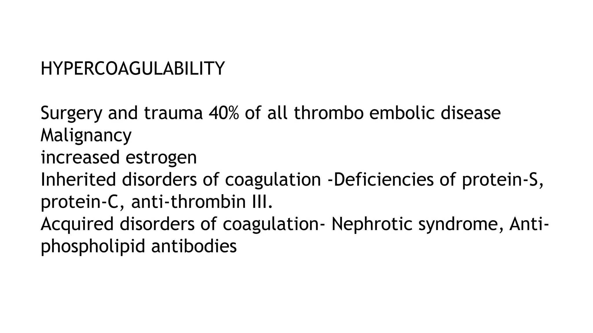
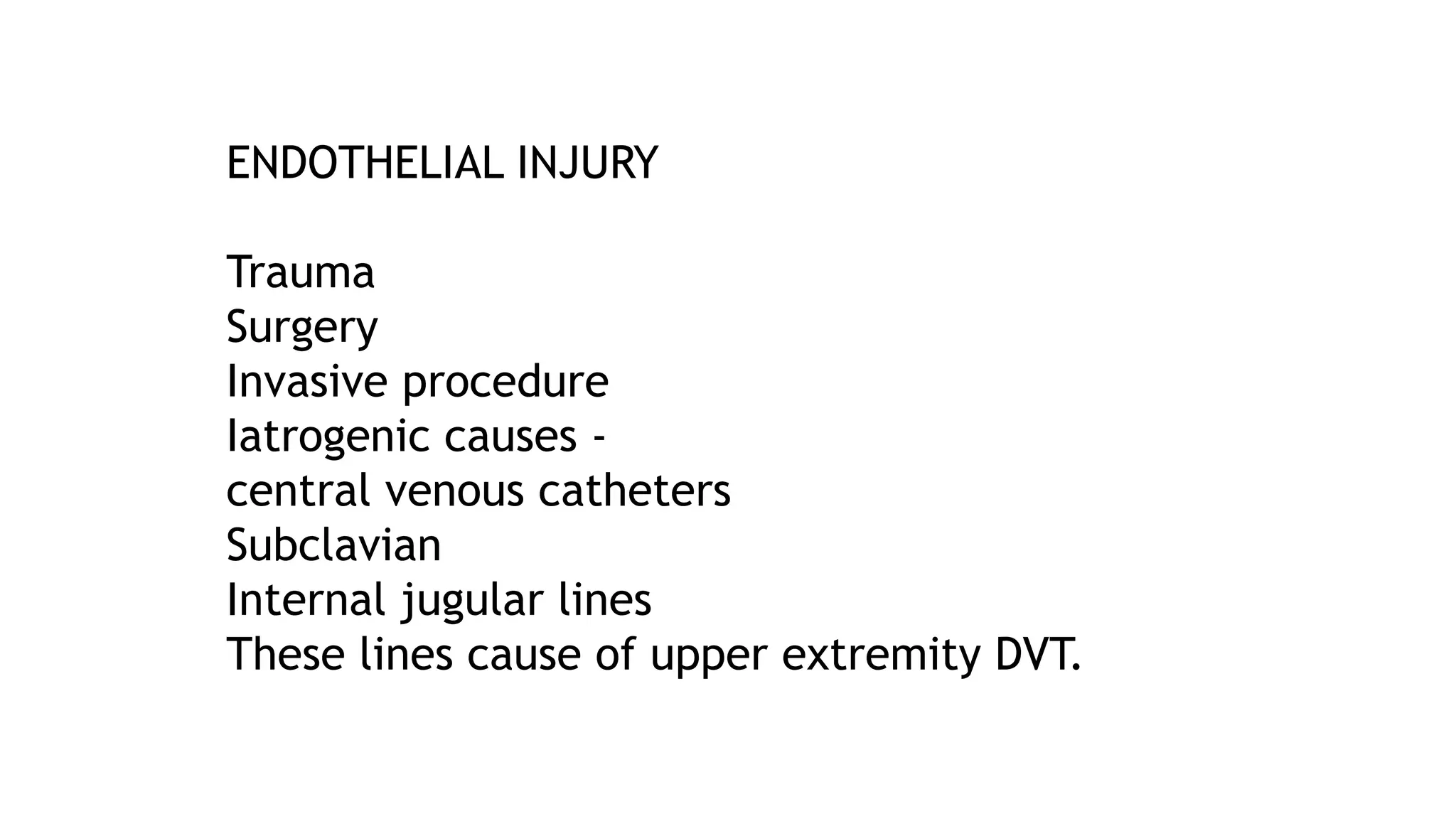
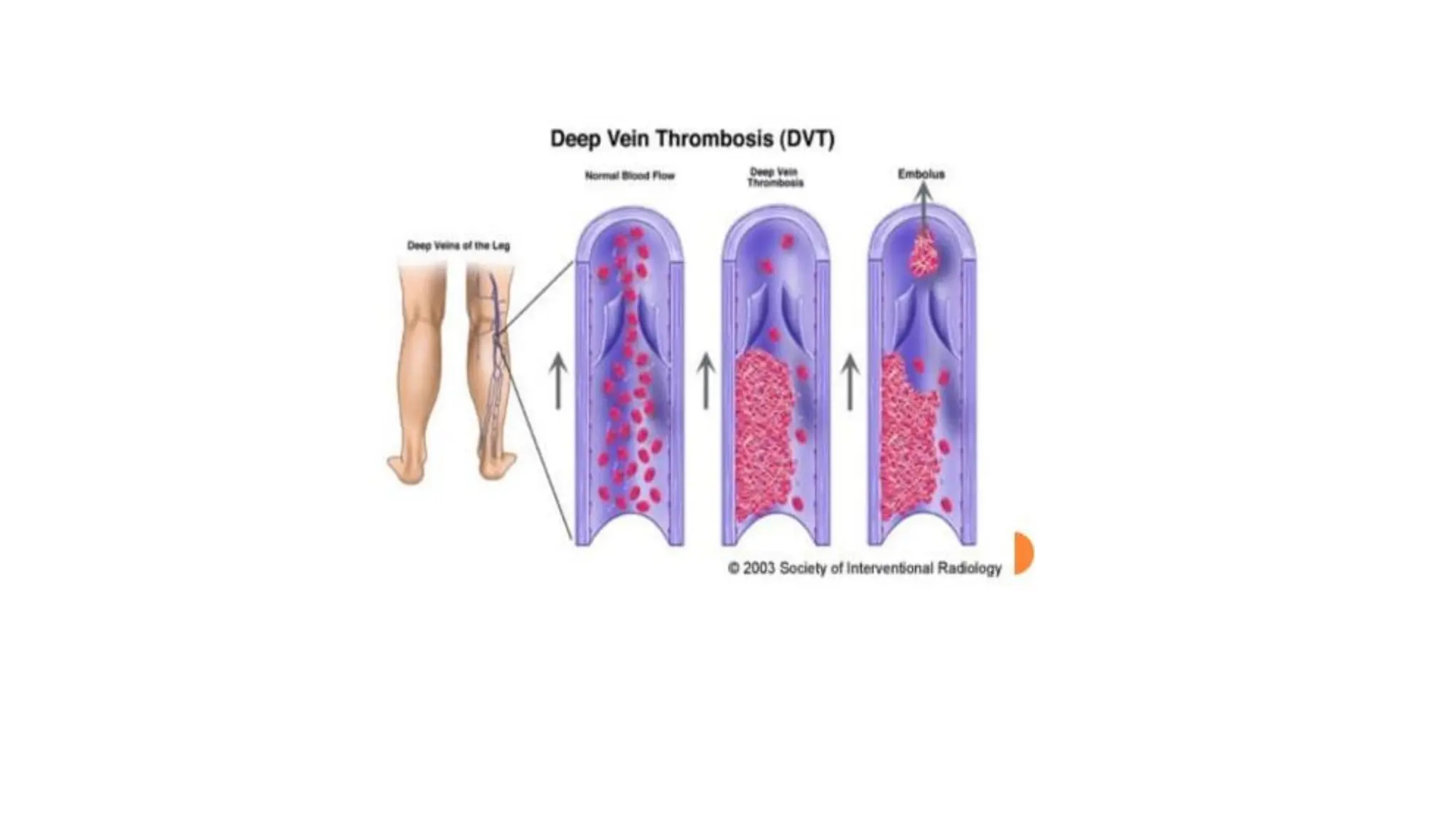
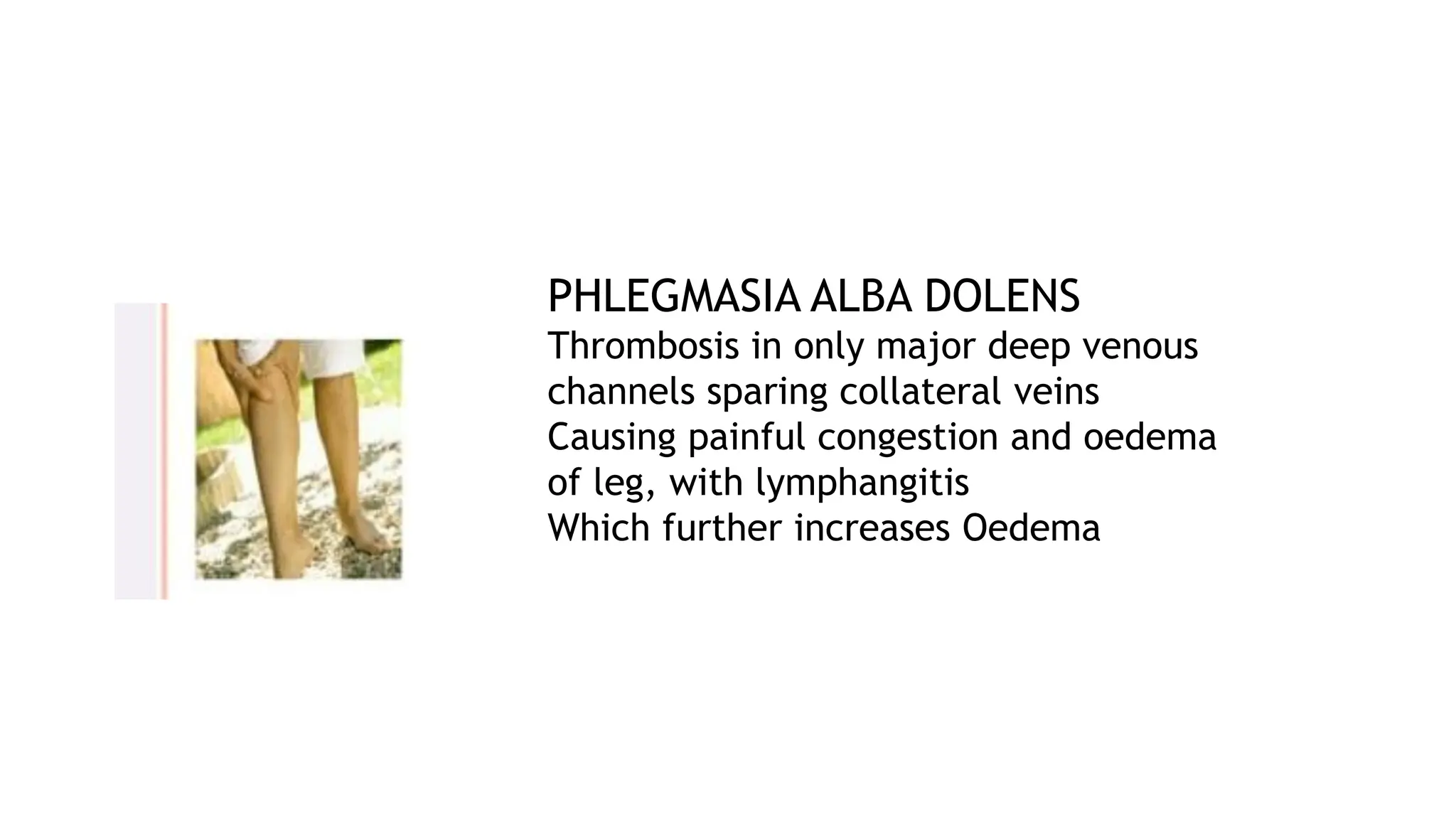
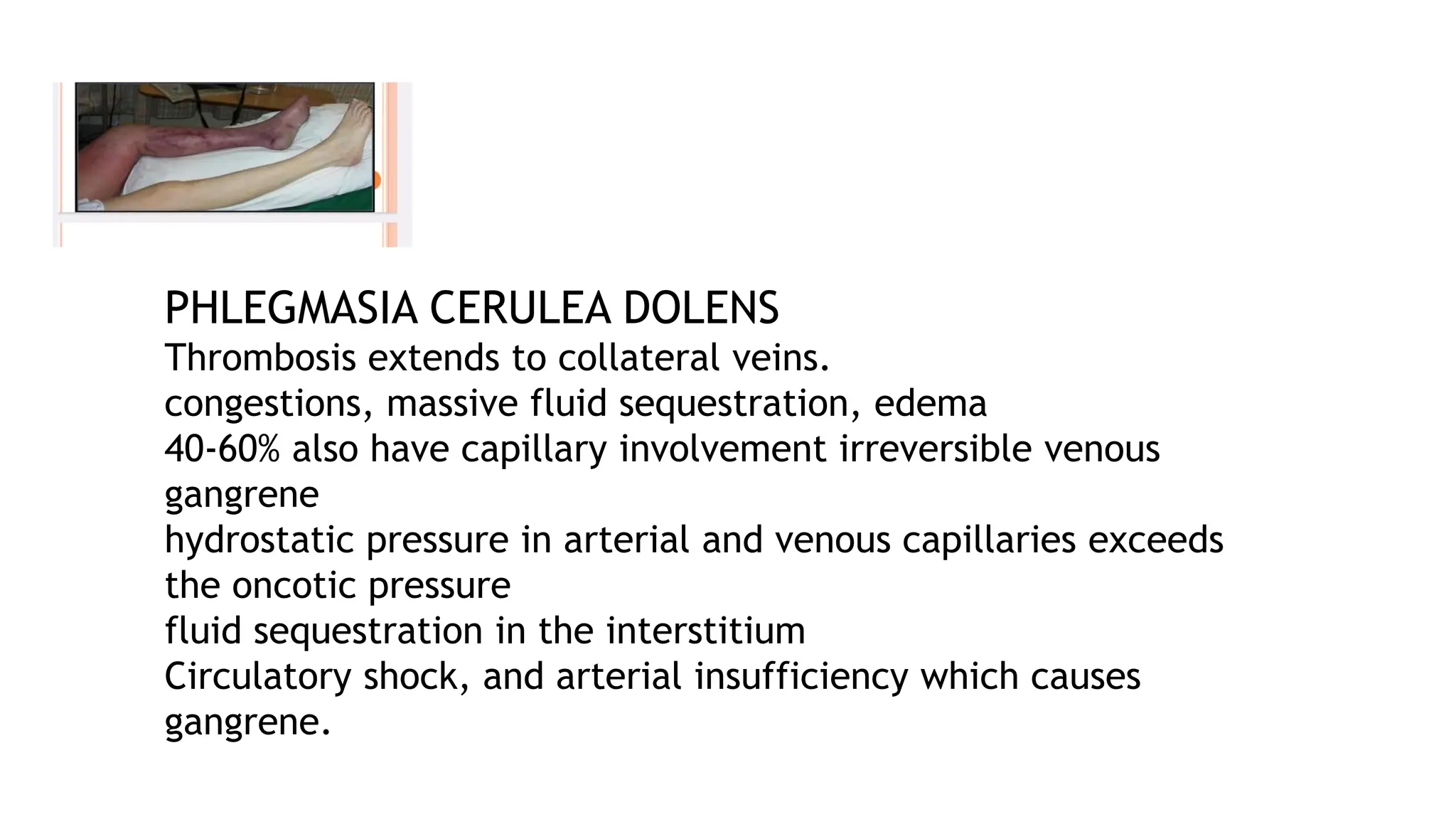
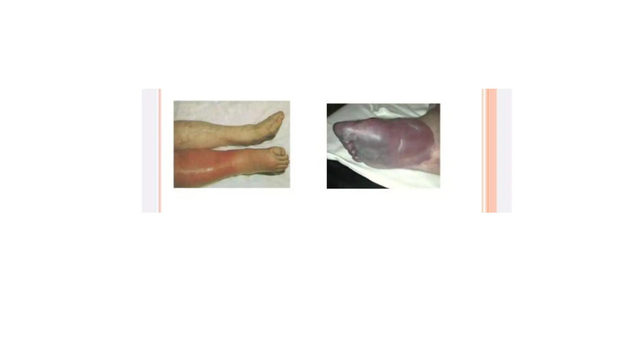
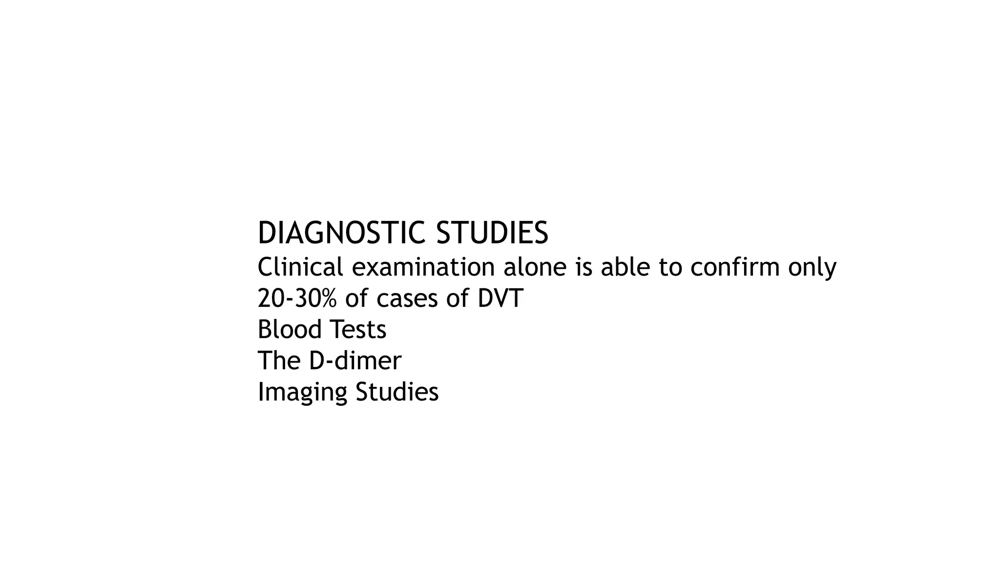
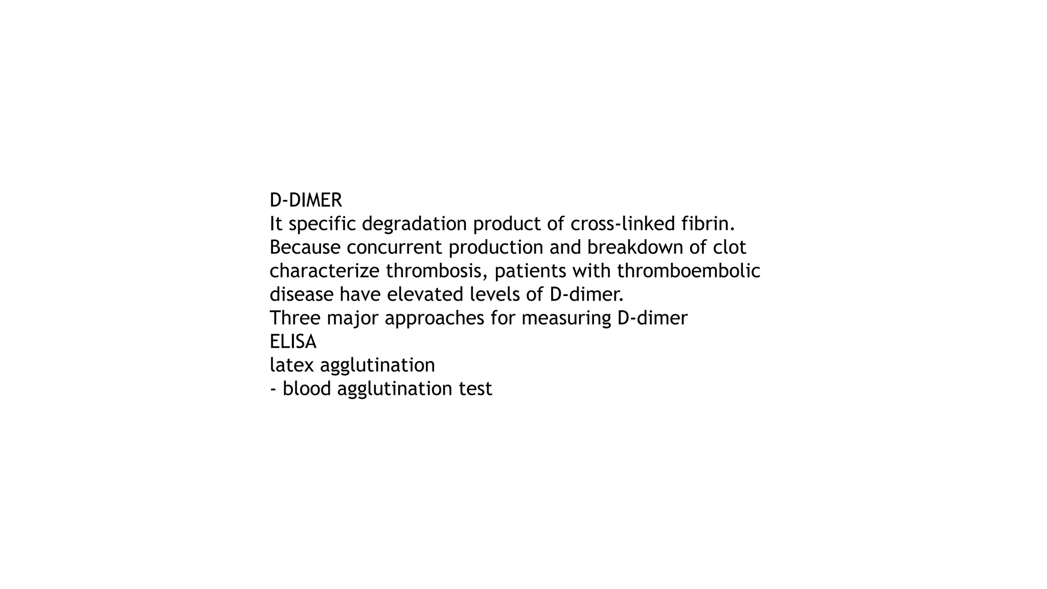
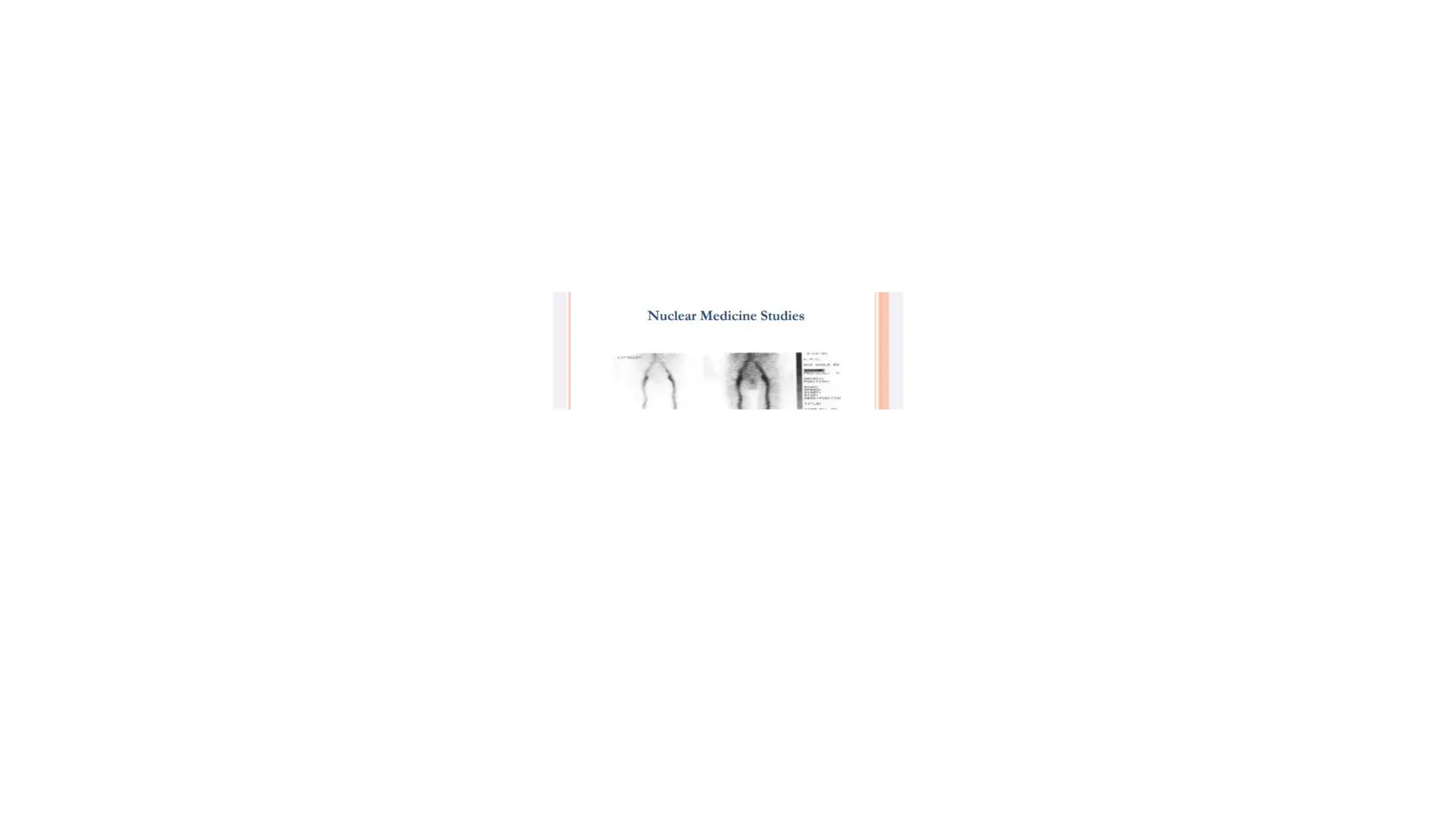
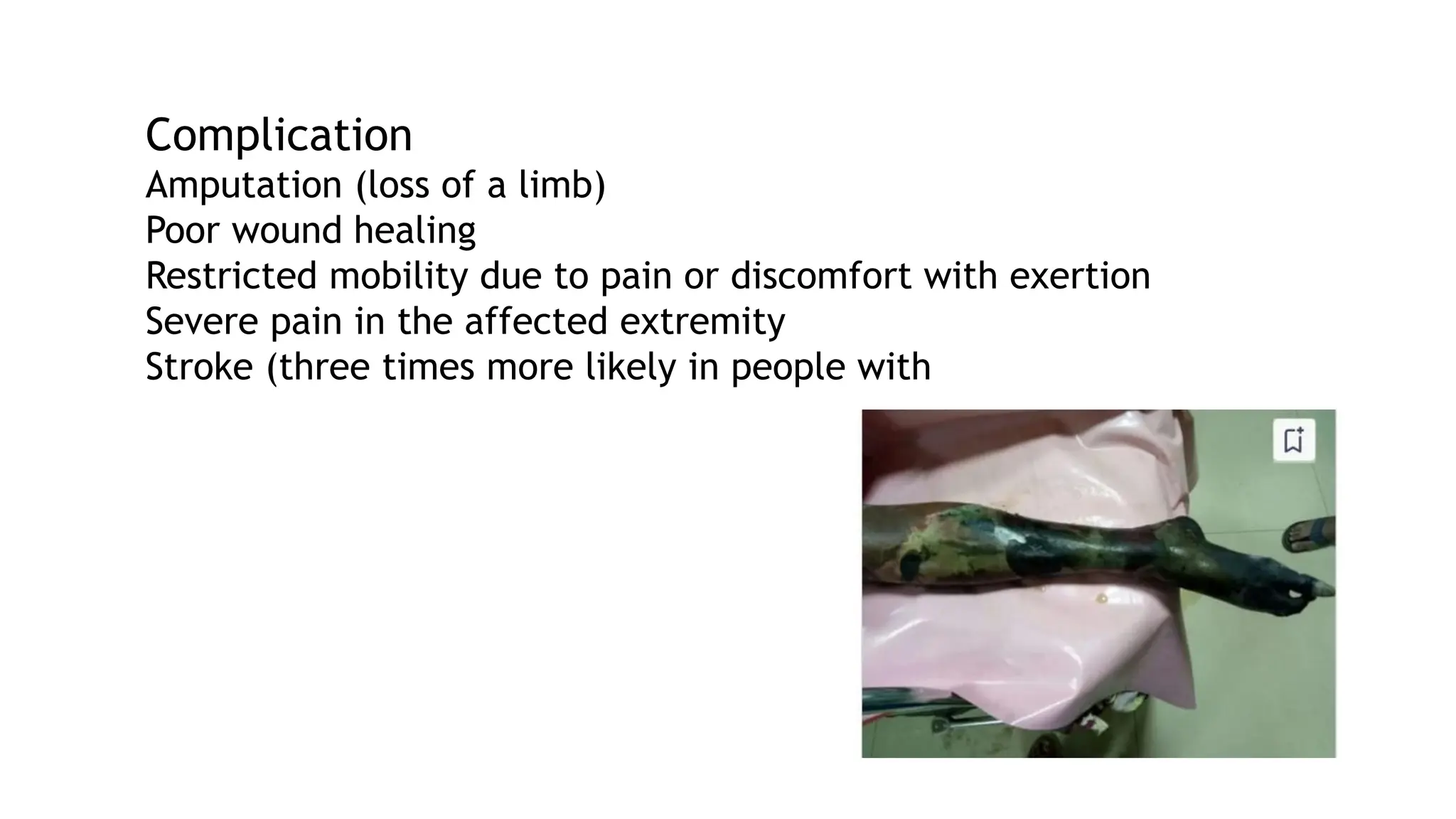
DEFINITION
Deep vein thrombosis is the formation of a blood clot in
one of the deep veins of the body, usually in the leg.
DEEP VEIN THROMBOSIS
ETIOLOGY
Starts in Lower
extremity calf veins
progressing proximally
to involve
Popliteal, Femoral, iliac
system
Virchow's triad
Describes three factors that are thought to
contribute to thrombosis
VIRCHOW TRIAD
More than 100 years ago, Rudolf Virchow
described a triad of factors of
Venous stasis
Hypercoagulable state
Endothelial damage
VENOUS STASIS
prolonged bed rest (4 days or
more)
A cast on the leg
Limb paralysis from stroke
spinal cord injury
extended travel in a vehicle
HYPERCOAGULABILITY
Surgery and trauma 40% of all thrombo embolic disease
Malignancy
increased estrogen
Inherited disorders of coagulation -Deficiencies of protein-S,
protein-C, anti-thrombin III.
Acquired disorders of coagulation- Nephrotic syndrome, Anti-
phospholipid antibodies
ENDOTHELIAL INJURY
Trauma
Surgery
Invasive procedure
Iatrogenic causes -
central venous catheters
Subclavian
Internal jugular lines
These lines cause of upper extremity DVT.
HYPERCOAGULABLE STATE OF MALIGNANCY
Up to 15% of cancer patients presents with VTE
VTE is not equally common in all types of cancer.
The highest incidence is found in mucin- producing
adenocarcinomas, pancreas and gastrointestinal tract,
lung cancer, and ovarian cancer.
PATHOPHYSIOLOGY
Vessel trauma stimulates the clotting cascade.
• Platelets aggregate at the site particularly when
venous stasis present
Platelets and fibrin form the initial clot
RBC are trapped in the fibrin meshwork
The thrombus propagates in the direction of the blood
flow.
Inflammation is triggered, causing tenderness, swelling,
and erythema.
Pieces of thrombus may break loose and travel through
circulation- emboli.
Fibroblasts eventually invade the thrombus, scarring vein
wall and destroying valves. Patency may be restored valve
damage is permanent, affecting directional flow.
• Thrombophlebitis a thrombus accompanied by inflammation of the
vein (phlebitis).
• Phlebothrombosis refers to a thrombus with minimal
inflammation.
Dislodgment and migration of a thrombus are known as
thromboembolism.
Which is common in phlebothrombosis.
PRESENTATION AND PHYSICAL EXAMINATION
Calf pain or tenderness, or both
Swelling with pitting oedema
Increased skin temperature and fever
• Superficial venous dilatation
Cyanosis can occur with severe obstruction
Less frequent manifestations of venous thrombosis include
Phlegmasia alba dolens,
Phlegmasia cerulea dolens, and
Venous gangrene.
These are clinical spectrum of the same disorder.
PHLEGMASIA ALBA DOLENS
Thrombosis in only major deep venous
channels sparing collateral veins
Causing painful congestion and oedema
of leg, with lymphangitis
Which further increases Oedema
PHLEGMASIA CERULEA DOLENS
Thrombosis extends to collateral veins.
congestions, massive fluid sequestration, edema
40-60% also have capillary involvement irreversible venous
gangrene
hydrostatic pressure in arterial and venous capillaries exceeds
the oncotic pressure
fluid sequestration in the interstitium
Circulatory shock, and arterial insufficiency which causes
gangrene.
c/f
sudden severe pain, swelling, cyanosis and edema of the
affected limb.
There is a high risk of massive pulmonary embolism, even
under anticoagulation.
Foot gangrene may also occur.
An underlying malignancy is found in 50% of cases. Usually, it
occurs in those
CLINICAL EXAMINATION
Palpate distal pulses and evaluate capillary refill to assess
limb perfusion.
Move and palpate all joints to detect acute
arthritis or other joint pathology.
• Neurologic evaluation may detect nerve root irritation;
sensory, motor, and reflex deficits should be noted
Homans sign: pain in the posterior calf or
knee with forced dorsiflexion of the
foot.
Moses sign
Gentle squeezing of the lower part of the calf from side to side.
Neuhofs sign
Thickening and deep tenderness elicited while palpating deep in
calf muscles.
Lintons sign
After applying torniquet at saphenofemoral junction patient
made to walk, then limb is elevated in supine posation
prominent superficial veins will be observed
Search for stigmata of PE such as tachycardia (common)
tachypnea chest findings (rare),
exam for signs suggestive of underlying predisposing
factors.
WELLS CLINICAL PREDICTION GUIDE
It pre-test probability score
Helps in early risk stratification and appropriate use of
laboratory tests and imaging modalities.
wells criteria is an additional tool to diagnosis rather than
being a stand-alone test.
Variable
Interpretation
High probability: ≥ 3 (Prevalence of DVT-53%)
Moderate probability: 1-2 (Prevalence of DVT- 17%)
• Low probability: ≤0 (Prevalence of DVT-5%)
DIAGNOSTIC STUDIES
Clinical examination alone is able to confirm only
20-30% of cases of DVT
Blood Tests
The D-dimer
Imaging Studies
D-DIMER
It specific degradation product of cross-linked fibrin.
Because concurrent production and breakdown of clot
characterize thrombosis, patients with thromboembolic
disease have elevated levels of D-dimer.
Three major approaches for measuring D-dimer
ELISA
latex agglutination
- blood agglutination test
PLASMIN CLEAVES FACTOR XIIIA-CROSS- LINKED FIBRIN
INTO AN ARRAY OF INTERMEDIA TE FORMS. THE D-DIMER
AND E FRAGMENTS ARE THE RESULT OF TERMINAL FIBRIN
DEGRADATIO N
IMAGING STUDIES Invasive venography, radiolabeled
fibrinogen noninvasive - ultrasound, plethysmography, -
MRI techniques
Venography
VENOGRAM:
POPLITEAL VEIN
THROMBOSIS
VENOGRAPHY
It detects thrombi in both calf and thigh
It can conclude and exclude the diagnosis of DVT when
other objective testings are not conclusive.
Advantages
It is useful if the patient has a high clinical probability of
thrombosis and a negative ultrasound.
It is also valuable in symptomatic patients with a history
of prior thrombosis in whom the ultrasound is non-
diagnostic.
DISADVANTAGE
It can primary cause of DVT in 3% of patients who
undergo this diagnostic procedure.
An invasive and expensive.
Although Venography was once considered the gold
standard for diagnosis of DVT, today it is more commonly
used in research environments and less frequently
utilized in clinical practice.
NUCLEAR MEDICINE STUDIES
Because the radioactive isotope incorporates into a
growing thrombus, this test can distinguish new clot from
an old clot.
Nuclear medicine studies done with P-labeled fibrinogen.
More commonly used in research.
PLETHYSMOGRAPHY Plethysmography measures
change in lower extremity volume in response to certain
stimuli.
IMPEDANCE PLETHYSMOGRAPHY
Principle- Blood volume changes in the leg lead to
changes in electrical resistance.
• Venous return in the lower extremity is occluded by
inflation of a thigh cuff, and then the cuff is released,
resulting in a decrease in calf blood volume. Any
obstruction of the proximal veins diminishes the volume
change, which is detected by measuring changes in
electrical resistance (impedance) over the calf.
IMPEDANCE PLETHYSMOGRAPHY
Principle- Blood volume changes in the leg lead to
changes in electrical resistance.
Venous return in the lower extremity is occluded by
inflation of a thigh cuff, and then the cuff is released,
resulting in a decrease in calf blood volume. Any
obstruction of the proximal veins diminishes the volume
change, which is detected by measuring changes in
electrical resistance (impedance) over the calf.
ULTRASONOGRAPHY
color-flow Duplex scanning is the imaging test of choice
for patients with suspected DVT
• inexpensive,
noninvasive,
widely available
Ultrasound can also distinguish other causes of leg
swelling, such as tumor, popliteal cyst, abscess,
aneurysm, or hematoma.
CLINICAL LIMITATIONS
Reader dependent
Duplex scans are less likely to detect non- occluding thrombi.
During the second half of pregnancy, ultrasound becomes less specific,
because the gravid uterus compresses the inferior vena cava, thereby
changing Doppler flow in the lower extremities.
An inability to distinguish old clots from a newly forming clot
MAGNETIC RESONANCE IMAGING
It detects leg, pelvis, and pulmonary thrombi and is 97% sensitive and 95%
specific for DVT.
It distinguishes a mature from an immature
clot.
MRI is safe in all stages of pregnancy.
Test may not be appropriate for patients with pacemakers or other metallic
implants, it can be an effective diagnostic option for some patients.
DIFFERENTIAL DIAGNOSIS
• Cellulitis
Thrombophlebitis
• Arthritis
Asymmetric peripheral edema secondary to CHF, liver disease,
renal failure, or nephrotic syndrome
lymphangitis
Extrinsic compression of iliac vein secondary to tumor,
hematoma, or abscess
Hematoma
Lymphedema
Muscle or soft tissue injury
Neurogenic pain
Postphlebitic syndrome
Ruptured Baker cyst
Stress fractures or other bony lesions
Superficial thrombophlebitis
MANAGEMENT
Using the pretest probability score calculated from the Wells
Clinical Prediction rule, patients are stratified into 3 risk
groups-high, moderate, or low.
The results from duplex ultrasound are incorporated as follows:
If the patient is high or moderate risk and the duplex ultrasound
study is positive, treat for DVT.
If the duplex study is negative and the patient is low
risk. DVT has been ruled out.
When discordance exists between the pretest
probability and the duplex study result, further
evaluation is required.
If the patient is high risk but the ultrasound study was
negative, the patient still has a significant probability
of DVT
a venogram to rule out a calf vein DVT surveillance with
repeat clinical evaluation and ultrasound in 1 week.
results of a D-dimer assay to guide management
If the patient is low risk but the ultrasound study is
positive, some authors recommend a second
confirmatory study such as a venogram before treating
for DVT
EMERGENCY DEPARTMANT CARE
The primary objectives of the treatment of DVT are to-
prevent pulmonary embolism,
reduce morbidity, and
• prevent or minimize the risk of developing the postphlebitic
syndrome.
GENERAL THERAPEUTIC MEASURES:
Bed rest.
Encourage the patient to perform gentle foot & leg exercises every hour.
Increase fluid intake upto 2 l/day unless contraindicated.
Avoid deep palpation..
SPECIFIC TREATMENT:
Anticoagulation
Thrombolytic therapy for DVT
Surgery for DVT
Filters for DVT
Compression stockings
Initial treatment of DVT is with low- molecular-weight
heparin or unfractionated heparin for at least 5 days,
followed by warfarin (target INR, 2.0-3.0) for at least 3
months.
ANTICOAGULATION
Heparin prevents extension of the thrombus
It is a heterogeneous mixture of polysaccharide fragments
with varying molecular weights but with similar biological
activity.
MECHANISM OF ACTION
Heparin's anticoagulant effect is related directly to its
activation of antithrombin III.
Antithrombin III, the body's primary anticoagulant,
inactivates thrombin and inhibits the activity of activated
factor X, factor IX in the coagulation process.
Side effects
Bleeding
Osteoporosis
Thrombocytopenia
Skins lesions- urticaria, papules, necrosis
Hypoaldosteronism, hyperkalemia
CONTRAINDICATIONS-
Bleeding disorders,
Severe hypertension, threatened abortion, piles,
large malignancies, tuberculosis'
Ocular surgery and neurosurgery,
Chronic alcoholics, cirrhosis, renal failure
DOSE
IV bolus dose of 5,000 to 10,000 units
followed by an infusion of 1,000 units per hour.
Other method of initiating therapy is to begin with
Loading dose of 50-100 units/kg of heparin followed by a
constant infusion of 15-25 units/kg/hr.
THROMBOLYTIC THERAPY FOR DVT
Advantages include
Prompt resolution of symptoms,
Prevention of pulmonary embolism,
Restoration of normal venous circulation,
• Preservation of venous valvular function,
• Prevention of postphlebitic syndrome.
DISADVANTAGE
Thrombolytic therapy does not prevent
clot propagation,
rethrombosis, or
subsequent embolization.
• Heparin therapy and oral anticoagulant therapy always
must followed after a course of thrombolysis.
Thrombolytic therapy is also not effective once the thrombus is
adherent and begins to organize
The hemorrhagic complications of thrombolytic therapy are about 3
times higher, including the small but potentially fatal risk of intra-
cerebral hemorrhage.
At present, therefore, thrombo-lysis should be reserved for
exceptional circumstances, such as patients with limb-threatening
ischemia caused by phlegmasia cerulea dolens.
SURGERY FOR DVT
Indications
when anticoagulant therapy is ineffective
unsafe,
contraindicated.
The major surgical procedures for DVT are clot removal
and partial interruption of the inferior vena cava to
prevent pulmonary embolism.
FILTERS FOR DVT
Indications
Contraindication to anticoagulation.
Significant bleeding complication of anticoagulation
therapy.
Pulmonary embolism with contraindication to
anticoagulation.
Recurrent thrombo-embolic complication despite
adequate anticoagulation therapy.
Inferior vena cava filters reduce the rate of pulmonary
embolism but have no effect on the other complications of deep
vein thrombosis. Thrombolysis should be considered in patients
with major proximal vein thrombosis and threatened venous
infarction
PROPHYLAXIS
Indicated in who underwent major abdominal trauma or
orthopaedic surgery or patient having prolonged
immobolization (> 3 days).
Benefits of VTE Prophylaxis
Improved patient outcomes
• Reduced costs
METHODS OF VTE PROPHYLAXIS
Mechanical:
• Graduated Compression Stockings (GCS)
• Intermittent Pneumatic Compression Devices (IPC)
Pharmacologic
Low molecular weight Heparin.(5000u se Shourly) It
inhibits factor Xa and IIA activity.
PERIPHERAL VASCULAR DISEASE
Shemil
Clinical instructor DM WIMS
Definition
share
Peripheral vascular disease (PVD) is a circulation disorder
that causes narrowing of blood vessels to parts of the
body other than the brain and heart.
There are two types of PVD
Functional PVDs don't involve defects in blood vessels' structure. (The blood
vessels aren't physically damaged.) These diseases often have symptoms related
to "spasm" that may come and go.(Raynaud disease)
Organic PVDs are caused by structural changes in the blood vessels. Examples
could include inflammation and tissue damage. (peripheral artery disease)
Causes
1. Atherosclerosis
2. Blood clots
3. Diabetes
4. Inflammation of the arteries or arteritis
5. Infection(salmonellosis and syphilis)
6. Structural defects
7. Injury
Risk factors
Family history of heart disease, high blood pressure, high cholesterol,
or stroke
Older than 50 years
Overweight or obesity
Inactive lifestyle
Smoking
Diabetes
High blood pressure
High cholesterol or LDL plus high triglycerides and low HDL
Symptoms
Dull, cramping pain in one or both calves, thighs, or hips
when walking, called intermittent claudication
Buttock pain,
Numbness or tingling in the legs,
Weakness, burning or aching pain in the feet or toes
while resting.
A sore on a leg or a foot that will not heal,
One or both legs or feet feel cold or change color (pale,
bluish, dark reddish),
Hair loss on the legs, and
Impotence
1. Edinburgh claudication questionnaire.
2. Ankle brachial index(ABI).
3. Treadmill exercise test.
4. Angiography, or arteriography
5. Doppler ultrasound flow studies
6. Magnetic resonance imaging
Ankle/brachial index (ABI)
This test compares the blood pressure in the arm (brachial)
with the blood pressure in the legs.
In a person with healthy blood vessels, the pressure should
be higher in the legs than in the arms.
An ABI above 0.90 is normal; 0.71-0.90 indicates mild PVD;
0.41-0.70 indicates moderate disease; and less than 0.40
indicates severe PVD.
Treadmill exercise test
If necessary, the ABI will be followed by a treadmill exercise
test.
Blood pressures in your arms and legs will be taken before and
after exercise (walking on a treadmill, usually until you have
symptoms).
A significant drop in leg blood pressures and ABIs after exercise
suggests PVD
Angiography, or arteriography
Doppler ultrasound flow studies
Magnetic resonance imaging
Lifestyle changes
Stopping smoking (smokers are 4 times more likely to get
PAD and have symptoms of PAD than nonsmokers.)
• Controlling diabetes
Controlling blood pressure
• Being physically active
• Eating a diet low in saturated fats
Medicines
Antiplatelet agents to prevent blood clots
• Cholesterol-lowering medicine.
Anti hypertensive.
Vascular surgery
A bypass graft using a blood vessel from another part of the body or a
tube made of synthetic material is placed in the area of the blocked or
narrowed artery to reroute the blood flow
Angioplasty
Balloon angioplasty (a small balloon is inflated inside the blocked
artery to open the blocked area).
Atherectomy (the blocked area inside the artery is "shaved" away by a
tiny device on the end of a catheter)
Laser angioplasty (a laser is used to "vaporize" the blockage in the
artery)
Stent (a tiny coil is expanded inside the blocked artery to open the
blocked area and is left in place to keep the artery open)
Complication
Amputation (loss of a limb)
Poor wound healing
Restricted mobility due to pain or discomfort with exertion
Severe pain in the affected extremity
Stroke (three times more likely in people with
Prevention
Smoking cessation, including avoidance of second hand smoke and use
of any tobacco products.
Dietary changes including reduced fat, cholesterol, and simple
carbohydrates (such as sweets), and increased amounts of fruits and
vegetables,
Weight reduction
Exercise plan of a minimum of 30 minutes daily
Control of diabetes
Control of high blood pressure

This is a presentation of the topic on deep vain thrabolisis

  • 1.
    DEFINITION Deep vein thrombosisis the formation of a blood clot in one of the deep veins of the body, usually in the leg. DEEP VEIN THROMBOSIS
  • 2.
    ETIOLOGY Starts in Lower extremitycalf veins progressing proximally to involve Popliteal, Femoral, iliac system Virchow's triad Describes three factors that are thought to contribute to thrombosis VIRCHOW TRIAD More than 100 years ago, Rudolf Virchow described a triad of factors of
  • 3.
  • 4.
    VENOUS STASIS prolonged bedrest (4 days or more) A cast on the leg Limb paralysis from stroke spinal cord injury extended travel in a vehicle
  • 5.
    HYPERCOAGULABILITY Surgery and trauma40% of all thrombo embolic disease Malignancy increased estrogen Inherited disorders of coagulation -Deficiencies of protein-S, protein-C, anti-thrombin III. Acquired disorders of coagulation- Nephrotic syndrome, Anti- phospholipid antibodies
  • 6.
    ENDOTHELIAL INJURY Trauma Surgery Invasive procedure Iatrogeniccauses - central venous catheters Subclavian Internal jugular lines These lines cause of upper extremity DVT.
  • 7.
    HYPERCOAGULABLE STATE OFMALIGNANCY Up to 15% of cancer patients presents with VTE VTE is not equally common in all types of cancer. The highest incidence is found in mucin- producing adenocarcinomas, pancreas and gastrointestinal tract, lung cancer, and ovarian cancer.
  • 9.
    PATHOPHYSIOLOGY Vessel trauma stimulatesthe clotting cascade. • Platelets aggregate at the site particularly when venous stasis present Platelets and fibrin form the initial clot RBC are trapped in the fibrin meshwork
  • 10.
    The thrombus propagatesin the direction of the blood flow. Inflammation is triggered, causing tenderness, swelling, and erythema. Pieces of thrombus may break loose and travel through circulation- emboli. Fibroblasts eventually invade the thrombus, scarring vein wall and destroying valves. Patency may be restored valve damage is permanent, affecting directional flow.
  • 12.
    • Thrombophlebitis athrombus accompanied by inflammation of the vein (phlebitis). • Phlebothrombosis refers to a thrombus with minimal inflammation. Dislodgment and migration of a thrombus are known as thromboembolism. Which is common in phlebothrombosis.
  • 13.
    PRESENTATION AND PHYSICALEXAMINATION Calf pain or tenderness, or both Swelling with pitting oedema Increased skin temperature and fever • Superficial venous dilatation Cyanosis can occur with severe obstruction
  • 14.
    Less frequent manifestationsof venous thrombosis include Phlegmasia alba dolens, Phlegmasia cerulea dolens, and Venous gangrene. These are clinical spectrum of the same disorder.
  • 15.
    PHLEGMASIA ALBA DOLENS Thrombosisin only major deep venous channels sparing collateral veins Causing painful congestion and oedema of leg, with lymphangitis Which further increases Oedema
  • 16.
    PHLEGMASIA CERULEA DOLENS Thrombosisextends to collateral veins. congestions, massive fluid sequestration, edema 40-60% also have capillary involvement irreversible venous gangrene hydrostatic pressure in arterial and venous capillaries exceeds the oncotic pressure fluid sequestration in the interstitium Circulatory shock, and arterial insufficiency which causes gangrene.
  • 17.
    c/f sudden severe pain,swelling, cyanosis and edema of the affected limb. There is a high risk of massive pulmonary embolism, even under anticoagulation. Foot gangrene may also occur. An underlying malignancy is found in 50% of cases. Usually, it occurs in those
  • 19.
    CLINICAL EXAMINATION Palpate distalpulses and evaluate capillary refill to assess limb perfusion. Move and palpate all joints to detect acute arthritis or other joint pathology. • Neurologic evaluation may detect nerve root irritation; sensory, motor, and reflex deficits should be noted
  • 20.
    Homans sign: painin the posterior calf or knee with forced dorsiflexion of the foot.
  • 21.
    Moses sign Gentle squeezingof the lower part of the calf from side to side. Neuhofs sign Thickening and deep tenderness elicited while palpating deep in calf muscles. Lintons sign After applying torniquet at saphenofemoral junction patient made to walk, then limb is elevated in supine posation prominent superficial veins will be observed
  • 22.
    Search for stigmataof PE such as tachycardia (common) tachypnea chest findings (rare), exam for signs suggestive of underlying predisposing factors.
  • 23.
    WELLS CLINICAL PREDICTIONGUIDE It pre-test probability score Helps in early risk stratification and appropriate use of laboratory tests and imaging modalities. wells criteria is an additional tool to diagnosis rather than being a stand-alone test. Variable
  • 25.
    Interpretation High probability: ≥3 (Prevalence of DVT-53%) Moderate probability: 1-2 (Prevalence of DVT- 17%) • Low probability: ≤0 (Prevalence of DVT-5%)
  • 26.
    DIAGNOSTIC STUDIES Clinical examinationalone is able to confirm only 20-30% of cases of DVT Blood Tests The D-dimer Imaging Studies
  • 27.
    D-DIMER It specific degradationproduct of cross-linked fibrin. Because concurrent production and breakdown of clot characterize thrombosis, patients with thromboembolic disease have elevated levels of D-dimer. Three major approaches for measuring D-dimer ELISA latex agglutination - blood agglutination test
  • 28.
    PLASMIN CLEAVES FACTORXIIIA-CROSS- LINKED FIBRIN INTO AN ARRAY OF INTERMEDIA TE FORMS. THE D-DIMER AND E FRAGMENTS ARE THE RESULT OF TERMINAL FIBRIN DEGRADATIO N
  • 31.
    IMAGING STUDIES Invasivevenography, radiolabeled fibrinogen noninvasive - ultrasound, plethysmography, - MRI techniques
  • 32.
  • 34.
    VENOGRAPHY It detects thrombiin both calf and thigh It can conclude and exclude the diagnosis of DVT when other objective testings are not conclusive. Advantages It is useful if the patient has a high clinical probability of thrombosis and a negative ultrasound. It is also valuable in symptomatic patients with a history of prior thrombosis in whom the ultrasound is non- diagnostic.
  • 35.
    DISADVANTAGE It can primarycause of DVT in 3% of patients who undergo this diagnostic procedure. An invasive and expensive. Although Venography was once considered the gold standard for diagnosis of DVT, today it is more commonly used in research environments and less frequently utilized in clinical practice.
  • 37.
    NUCLEAR MEDICINE STUDIES Becausethe radioactive isotope incorporates into a growing thrombus, this test can distinguish new clot from an old clot. Nuclear medicine studies done with P-labeled fibrinogen. More commonly used in research.
  • 38.
    PLETHYSMOGRAPHY Plethysmography measures changein lower extremity volume in response to certain stimuli.
  • 39.
    IMPEDANCE PLETHYSMOGRAPHY Principle- Bloodvolume changes in the leg lead to changes in electrical resistance. • Venous return in the lower extremity is occluded by inflation of a thigh cuff, and then the cuff is released, resulting in a decrease in calf blood volume. Any obstruction of the proximal veins diminishes the volume change, which is detected by measuring changes in electrical resistance (impedance) over the calf.
  • 40.
    IMPEDANCE PLETHYSMOGRAPHY Principle- Bloodvolume changes in the leg lead to changes in electrical resistance. Venous return in the lower extremity is occluded by inflation of a thigh cuff, and then the cuff is released, resulting in a decrease in calf blood volume. Any obstruction of the proximal veins diminishes the volume change, which is detected by measuring changes in electrical resistance (impedance) over the calf.
  • 41.
    ULTRASONOGRAPHY color-flow Duplex scanningis the imaging test of choice for patients with suspected DVT • inexpensive, noninvasive, widely available Ultrasound can also distinguish other causes of leg swelling, such as tumor, popliteal cyst, abscess, aneurysm, or hematoma.
  • 42.
    CLINICAL LIMITATIONS Reader dependent Duplexscans are less likely to detect non- occluding thrombi. During the second half of pregnancy, ultrasound becomes less specific, because the gravid uterus compresses the inferior vena cava, thereby changing Doppler flow in the lower extremities. An inability to distinguish old clots from a newly forming clot
  • 43.
    MAGNETIC RESONANCE IMAGING Itdetects leg, pelvis, and pulmonary thrombi and is 97% sensitive and 95% specific for DVT. It distinguishes a mature from an immature clot. MRI is safe in all stages of pregnancy. Test may not be appropriate for patients with pacemakers or other metallic implants, it can be an effective diagnostic option for some patients.
  • 44.
    DIFFERENTIAL DIAGNOSIS • Cellulitis Thrombophlebitis •Arthritis Asymmetric peripheral edema secondary to CHF, liver disease, renal failure, or nephrotic syndrome lymphangitis Extrinsic compression of iliac vein secondary to tumor, hematoma, or abscess Hematoma Lymphedema
  • 45.
    Muscle or softtissue injury Neurogenic pain Postphlebitic syndrome Ruptured Baker cyst Stress fractures or other bony lesions Superficial thrombophlebitis
  • 46.
    MANAGEMENT Using the pretestprobability score calculated from the Wells Clinical Prediction rule, patients are stratified into 3 risk groups-high, moderate, or low. The results from duplex ultrasound are incorporated as follows: If the patient is high or moderate risk and the duplex ultrasound study is positive, treat for DVT.
  • 47.
    If the duplexstudy is negative and the patient is low risk. DVT has been ruled out. When discordance exists between the pretest probability and the duplex study result, further evaluation is required. If the patient is high risk but the ultrasound study was negative, the patient still has a significant probability of DVT
  • 48.
    a venogram torule out a calf vein DVT surveillance with repeat clinical evaluation and ultrasound in 1 week. results of a D-dimer assay to guide management If the patient is low risk but the ultrasound study is positive, some authors recommend a second confirmatory study such as a venogram before treating for DVT
  • 49.
    EMERGENCY DEPARTMANT CARE Theprimary objectives of the treatment of DVT are to- prevent pulmonary embolism, reduce morbidity, and • prevent or minimize the risk of developing the postphlebitic syndrome.
  • 50.
    GENERAL THERAPEUTIC MEASURES: Bedrest. Encourage the patient to perform gentle foot & leg exercises every hour. Increase fluid intake upto 2 l/day unless contraindicated. Avoid deep palpation..
  • 51.
    SPECIFIC TREATMENT: Anticoagulation Thrombolytic therapyfor DVT Surgery for DVT Filters for DVT Compression stockings
  • 52.
    Initial treatment ofDVT is with low- molecular-weight heparin or unfractionated heparin for at least 5 days, followed by warfarin (target INR, 2.0-3.0) for at least 3 months.
  • 53.
    ANTICOAGULATION Heparin prevents extensionof the thrombus It is a heterogeneous mixture of polysaccharide fragments with varying molecular weights but with similar biological activity.
  • 54.
    MECHANISM OF ACTION Heparin'santicoagulant effect is related directly to its activation of antithrombin III. Antithrombin III, the body's primary anticoagulant, inactivates thrombin and inhibits the activity of activated factor X, factor IX in the coagulation process.
  • 56.
    Side effects Bleeding Osteoporosis Thrombocytopenia Skins lesions-urticaria, papules, necrosis Hypoaldosteronism, hyperkalemia CONTRAINDICATIONS- Bleeding disorders, Severe hypertension, threatened abortion, piles, large malignancies, tuberculosis' Ocular surgery and neurosurgery, Chronic alcoholics, cirrhosis, renal failure
  • 57.
    DOSE IV bolus doseof 5,000 to 10,000 units followed by an infusion of 1,000 units per hour. Other method of initiating therapy is to begin with Loading dose of 50-100 units/kg of heparin followed by a constant infusion of 15-25 units/kg/hr.
  • 58.
    THROMBOLYTIC THERAPY FORDVT Advantages include Prompt resolution of symptoms, Prevention of pulmonary embolism, Restoration of normal venous circulation, • Preservation of venous valvular function, • Prevention of postphlebitic syndrome.
  • 59.
    DISADVANTAGE Thrombolytic therapy doesnot prevent clot propagation, rethrombosis, or subsequent embolization. • Heparin therapy and oral anticoagulant therapy always must followed after a course of thrombolysis.
  • 60.
    Thrombolytic therapy isalso not effective once the thrombus is adherent and begins to organize The hemorrhagic complications of thrombolytic therapy are about 3 times higher, including the small but potentially fatal risk of intra- cerebral hemorrhage. At present, therefore, thrombo-lysis should be reserved for exceptional circumstances, such as patients with limb-threatening ischemia caused by phlegmasia cerulea dolens.
  • 61.
    SURGERY FOR DVT Indications whenanticoagulant therapy is ineffective unsafe, contraindicated. The major surgical procedures for DVT are clot removal and partial interruption of the inferior vena cava to prevent pulmonary embolism.
  • 66.
    FILTERS FOR DVT Indications Contraindicationto anticoagulation. Significant bleeding complication of anticoagulation therapy. Pulmonary embolism with contraindication to anticoagulation. Recurrent thrombo-embolic complication despite adequate anticoagulation therapy.
  • 67.
    Inferior vena cavafilters reduce the rate of pulmonary embolism but have no effect on the other complications of deep vein thrombosis. Thrombolysis should be considered in patients with major proximal vein thrombosis and threatened venous infarction
  • 69.
    PROPHYLAXIS Indicated in whounderwent major abdominal trauma or orthopaedic surgery or patient having prolonged immobolization (> 3 days). Benefits of VTE Prophylaxis Improved patient outcomes • Reduced costs
  • 70.
    METHODS OF VTEPROPHYLAXIS Mechanical: • Graduated Compression Stockings (GCS) • Intermittent Pneumatic Compression Devices (IPC) Pharmacologic Low molecular weight Heparin.(5000u se Shourly) It inhibits factor Xa and IIA activity.
  • 71.
  • 72.
    Definition share Peripheral vascular disease(PVD) is a circulation disorder that causes narrowing of blood vessels to parts of the body other than the brain and heart.
  • 73.
    There are twotypes of PVD Functional PVDs don't involve defects in blood vessels' structure. (The blood vessels aren't physically damaged.) These diseases often have symptoms related to "spasm" that may come and go.(Raynaud disease) Organic PVDs are caused by structural changes in the blood vessels. Examples could include inflammation and tissue damage. (peripheral artery disease)
  • 74.
    Causes 1. Atherosclerosis 2. Bloodclots 3. Diabetes 4. Inflammation of the arteries or arteritis 5. Infection(salmonellosis and syphilis) 6. Structural defects 7. Injury
  • 76.
    Risk factors Family historyof heart disease, high blood pressure, high cholesterol, or stroke Older than 50 years Overweight or obesity Inactive lifestyle Smoking Diabetes High blood pressure High cholesterol or LDL plus high triglycerides and low HDL
  • 77.
    Symptoms Dull, cramping painin one or both calves, thighs, or hips when walking, called intermittent claudication Buttock pain, Numbness or tingling in the legs, Weakness, burning or aching pain in the feet or toes while resting. A sore on a leg or a foot that will not heal, One or both legs or feet feel cold or change color (pale, bluish, dark reddish), Hair loss on the legs, and Impotence
  • 79.
    1. Edinburgh claudicationquestionnaire. 2. Ankle brachial index(ABI). 3. Treadmill exercise test. 4. Angiography, or arteriography 5. Doppler ultrasound flow studies 6. Magnetic resonance imaging
  • 81.
    Ankle/brachial index (ABI) Thistest compares the blood pressure in the arm (brachial) with the blood pressure in the legs. In a person with healthy blood vessels, the pressure should be higher in the legs than in the arms. An ABI above 0.90 is normal; 0.71-0.90 indicates mild PVD; 0.41-0.70 indicates moderate disease; and less than 0.40 indicates severe PVD.
  • 84.
    Treadmill exercise test Ifnecessary, the ABI will be followed by a treadmill exercise test. Blood pressures in your arms and legs will be taken before and after exercise (walking on a treadmill, usually until you have symptoms). A significant drop in leg blood pressures and ABIs after exercise suggests PVD
  • 85.
    Angiography, or arteriography Dopplerultrasound flow studies Magnetic resonance imaging
  • 86.
    Lifestyle changes Stopping smoking(smokers are 4 times more likely to get PAD and have symptoms of PAD than nonsmokers.) • Controlling diabetes Controlling blood pressure • Being physically active • Eating a diet low in saturated fats
  • 87.
    Medicines Antiplatelet agents toprevent blood clots • Cholesterol-lowering medicine. Anti hypertensive.
  • 88.
    Vascular surgery A bypassgraft using a blood vessel from another part of the body or a tube made of synthetic material is placed in the area of the blocked or narrowed artery to reroute the blood flow
  • 90.
    Angioplasty Balloon angioplasty (asmall balloon is inflated inside the blocked artery to open the blocked area). Atherectomy (the blocked area inside the artery is "shaved" away by a tiny device on the end of a catheter) Laser angioplasty (a laser is used to "vaporize" the blockage in the artery) Stent (a tiny coil is expanded inside the blocked artery to open the blocked area and is left in place to keep the artery open)
  • 94.
    Complication Amputation (loss ofa limb) Poor wound healing Restricted mobility due to pain or discomfort with exertion Severe pain in the affected extremity Stroke (three times more likely in people with
  • 95.
    Prevention Smoking cessation, includingavoidance of second hand smoke and use of any tobacco products. Dietary changes including reduced fat, cholesterol, and simple carbohydrates (such as sweets), and increased amounts of fruits and vegetables, Weight reduction Exercise plan of a minimum of 30 minutes daily Control of diabetes Control of high blood pressure