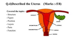
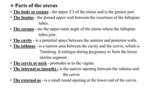
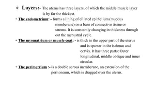
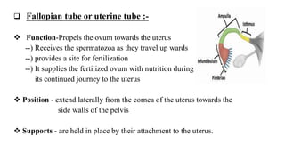
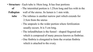
OBSTETRICS AND GYNEACOLOGY
NURSING
Ques no:-4
IMP
QUESTIONS
PRESENTED BY
PAREKH KISHAN
P.B.B.Sc. NURSING
Q.4)Described the Uterus (Marks :-5/8)
Covered the topics :-
- Structure
- Figure
- Position
- Layers
- Parts
- Function
 Introduction
• The uterus (from Latin ”uterus”), or womb is a major female hormone-
responsive secondary sex organ of the reproductive system in humans and
most other mammals.
 Structure:-
----) The uterus is located within the pelvic region immediately
behind and almost overlying the bladder, and in front of
the sigmoid colon.
----) The human uterus is pear-shaped .
----) Size :- 7.6 cm (3.0 in) long,
4.5 cm (1.8 in) broad (side to side), and
3.0 cm (1.2 in) thick.
----) Weight :-A typical adult uterus weights about 60 grams.
 The uterus can be divided anatomically into four regions:
• The Fundus – the uppermost rounded portion of the uterus,
• The Corpus (body),
• The Cervix, and
• The Cervical Canal.
---) The cervix protrudes into the vagina.
---) The uterus is held in position within the pelvis by ligaments,
which are part of the endopelvic fascia.
---) These ligaments include the the pubocervical ligaments,
the cardinal ligaments, and the uterosacral ligaments.
---) It is covered by a sheet-like fold of peritoneum, the broad
ligament.
Position - It leans forward, which is known as anteversion, it
bends forwards on itself, which is known as anteflexion.
• Relation - anteriorly the bladder and posteriorly rectum
• Inferior - Below the uterus is the vagina
• Superior - above the uterus lie the intestine
• Lateral - on both sides of the walls are the broad ligaments, the
fallopian tubes and the ovaries.
• Supports - supported by the pelvic floor and maintained in position by several ligaments.
Ligaments are;
- Peritonial ligament :-
Broad ligament
- Genito inguinal ligament
Round ligament
- Ligaments formed by pelvic fascia
Transverse cervical ligament
Utero sacral ligament
 Parts of the uterus
• The body or corpus - the upper 2/3 of the uterus and is the greater part.
• The fundus - the domed upper wall between the insertions of the fallopian
tubes.
• The cornua - are the upper outer angle of the uterus where the fallopian
tubes join.
• The cavity - is a potential space between the anterior and posterior walls.
• The isthmus - is a narrow area between the cavity and the cervix, which is
7mmlong. It enlarges during pregnancy to form the lower
uterine segment.
• The cervix or neck - protrudes in to the vagina.
• The internal os (mouth) - is the narrow opening between the isthmus and
the cervix
• The external os - is a small round opening at the lower end of the cervix.
 Layers:- The uterus has three layers, of which the middle muscle layer
is by far the thickest.
• The endometrium: - forms a lining of ciliated epithelium (mucous
memberane) on a base of connective tissue or
stroma. It is constantly changing in thickness through
out the menustral cycle.
• The myomatrium or muscle coat: - is thick in the upper part of the uterus
and is sparser in the isthmus and
cervix. It has three parts: Outer
longitudinal, middle oblique and inner
circular.
• The perimetrium :- is a double serous memberane, an extension of the
peritoneum, which is dragged over the uterus.
 Fallopian tube or uterine tube :-
 Function-Propels the ovum towards the uterus
--) Receives the spermatozoa as they travel up wards
--) provides a site for fertilization
--) It supplies the fertilized ovum with nutrition during
its continued journey to the uterus
 Position - extend laterally from the cornea of the uterus towards the
side walls of the pelvis
 Supports - are held in place by their attachment to the uterus.
• Structure - Each tube is 10cm long. It has four portions
of - The interstitial portion is 1.25cm long and lies with in the
Fallopian wall of the uterus. Its lumen is 1 mm wide.
tube - The isthmus is another narrow part which extends for
2.5cm from the uterus
- The ampoule is the wider portion where fertilization
usually occurs. It is 5 cm long.
- The infundibulum is the funnel - shaped fingered end
which is composed of many process known as fimbriae.
- One fimbria is elongated to form the ovarian fimbria
which is attached to the ovary.
 The ovaries
Function: - produce ova and the hormones estrogen and
progesterone
Position: - they are attached to the back of the broad ligament near
the fimbriated end of the fallopian tube.
OVARIE
 Function :-
• The reproductive function of the uterus is to accept a fertilized ovum which
passes through the utero-tubal junction from the fallopian tube.
• The fertilized ovum divides to become a blastocyst, which implants into
the endometrium, and derives nourishment from blood vessels which develop
exclusively for this purpose.
• The fertilized ovum becomes an embryo, attaches to a wall of the uterus,
creates a placenta, and develops into a fetus (gestates) until childbirth. Due to
anatomical barriers such as the pelvis, the uterus is pushed partially into the
abdomen due to its expansion during pregnancy.
• Even during pregnancy the mass of a human uterus amounts to only about a
kilogram (2.2 pounds).
• The uterus also plays a role in sexual response, by directing blood flow to
the pelvis and ovaries, and to the external genitals, including the vagina, labia,
and clitoris.
ANSWER ?

The uterus

  • 1.
    OBSTETRICS AND GYNEACOLOGY NURSING Quesno:-4 IMP QUESTIONS PRESENTED BY PAREKH KISHAN P.B.B.Sc. NURSING
  • 2.
    Q.4)Described the Uterus(Marks :-5/8) Covered the topics :- - Structure - Figure - Position - Layers - Parts - Function
  • 3.
     Introduction • Theuterus (from Latin ”uterus”), or womb is a major female hormone- responsive secondary sex organ of the reproductive system in humans and most other mammals.  Structure:- ----) The uterus is located within the pelvic region immediately behind and almost overlying the bladder, and in front of the sigmoid colon. ----) The human uterus is pear-shaped . ----) Size :- 7.6 cm (3.0 in) long, 4.5 cm (1.8 in) broad (side to side), and 3.0 cm (1.2 in) thick. ----) Weight :-A typical adult uterus weights about 60 grams.
  • 4.
     The uteruscan be divided anatomically into four regions: • The Fundus – the uppermost rounded portion of the uterus, • The Corpus (body), • The Cervix, and • The Cervical Canal. ---) The cervix protrudes into the vagina. ---) The uterus is held in position within the pelvis by ligaments, which are part of the endopelvic fascia. ---) These ligaments include the the pubocervical ligaments, the cardinal ligaments, and the uterosacral ligaments. ---) It is covered by a sheet-like fold of peritoneum, the broad ligament.
  • 5.
    Position - Itleans forward, which is known as anteversion, it bends forwards on itself, which is known as anteflexion. • Relation - anteriorly the bladder and posteriorly rectum • Inferior - Below the uterus is the vagina • Superior - above the uterus lie the intestine • Lateral - on both sides of the walls are the broad ligaments, the fallopian tubes and the ovaries. • Supports - supported by the pelvic floor and maintained in position by several ligaments. Ligaments are; - Peritonial ligament :- Broad ligament - Genito inguinal ligament Round ligament - Ligaments formed by pelvic fascia Transverse cervical ligament Utero sacral ligament
  • 6.
     Parts ofthe uterus • The body or corpus - the upper 2/3 of the uterus and is the greater part. • The fundus - the domed upper wall between the insertions of the fallopian tubes. • The cornua - are the upper outer angle of the uterus where the fallopian tubes join. • The cavity - is a potential space between the anterior and posterior walls. • The isthmus - is a narrow area between the cavity and the cervix, which is 7mmlong. It enlarges during pregnancy to form the lower uterine segment. • The cervix or neck - protrudes in to the vagina. • The internal os (mouth) - is the narrow opening between the isthmus and the cervix • The external os - is a small round opening at the lower end of the cervix.
  • 7.
     Layers:- Theuterus has three layers, of which the middle muscle layer is by far the thickest. • The endometrium: - forms a lining of ciliated epithelium (mucous memberane) on a base of connective tissue or stroma. It is constantly changing in thickness through out the menustral cycle. • The myomatrium or muscle coat: - is thick in the upper part of the uterus and is sparser in the isthmus and cervix. It has three parts: Outer longitudinal, middle oblique and inner circular. • The perimetrium :- is a double serous memberane, an extension of the peritoneum, which is dragged over the uterus.
  • 8.
     Fallopian tubeor uterine tube :-  Function-Propels the ovum towards the uterus --) Receives the spermatozoa as they travel up wards --) provides a site for fertilization --) It supplies the fertilized ovum with nutrition during its continued journey to the uterus  Position - extend laterally from the cornea of the uterus towards the side walls of the pelvis  Supports - are held in place by their attachment to the uterus.
  • 9.
    • Structure -Each tube is 10cm long. It has four portions of - The interstitial portion is 1.25cm long and lies with in the Fallopian wall of the uterus. Its lumen is 1 mm wide. tube - The isthmus is another narrow part which extends for 2.5cm from the uterus - The ampoule is the wider portion where fertilization usually occurs. It is 5 cm long. - The infundibulum is the funnel - shaped fingered end which is composed of many process known as fimbriae. - One fimbria is elongated to form the ovarian fimbria which is attached to the ovary.
  • 10.
     The ovaries Function:- produce ova and the hormones estrogen and progesterone Position: - they are attached to the back of the broad ligament near the fimbriated end of the fallopian tube. OVARIE
  • 11.
     Function :- •The reproductive function of the uterus is to accept a fertilized ovum which passes through the utero-tubal junction from the fallopian tube. • The fertilized ovum divides to become a blastocyst, which implants into the endometrium, and derives nourishment from blood vessels which develop exclusively for this purpose. • The fertilized ovum becomes an embryo, attaches to a wall of the uterus, creates a placenta, and develops into a fetus (gestates) until childbirth. Due to anatomical barriers such as the pelvis, the uterus is pushed partially into the abdomen due to its expansion during pregnancy. • Even during pregnancy the mass of a human uterus amounts to only about a kilogram (2.2 pounds). • The uterus also plays a role in sexual response, by directing blood flow to the pelvis and ovaries, and to the external genitals, including the vagina, labia, and clitoris.
  • 12.