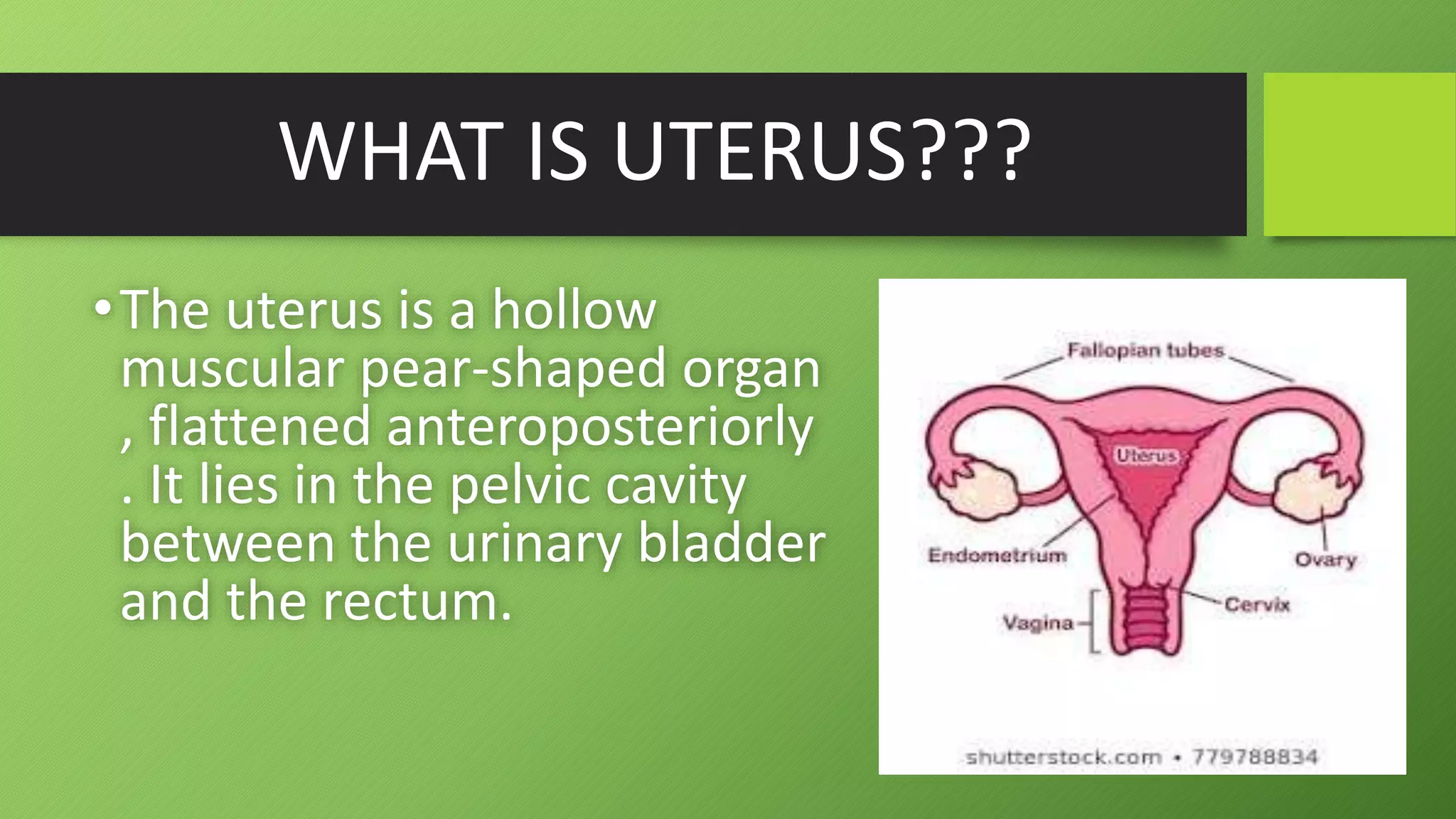
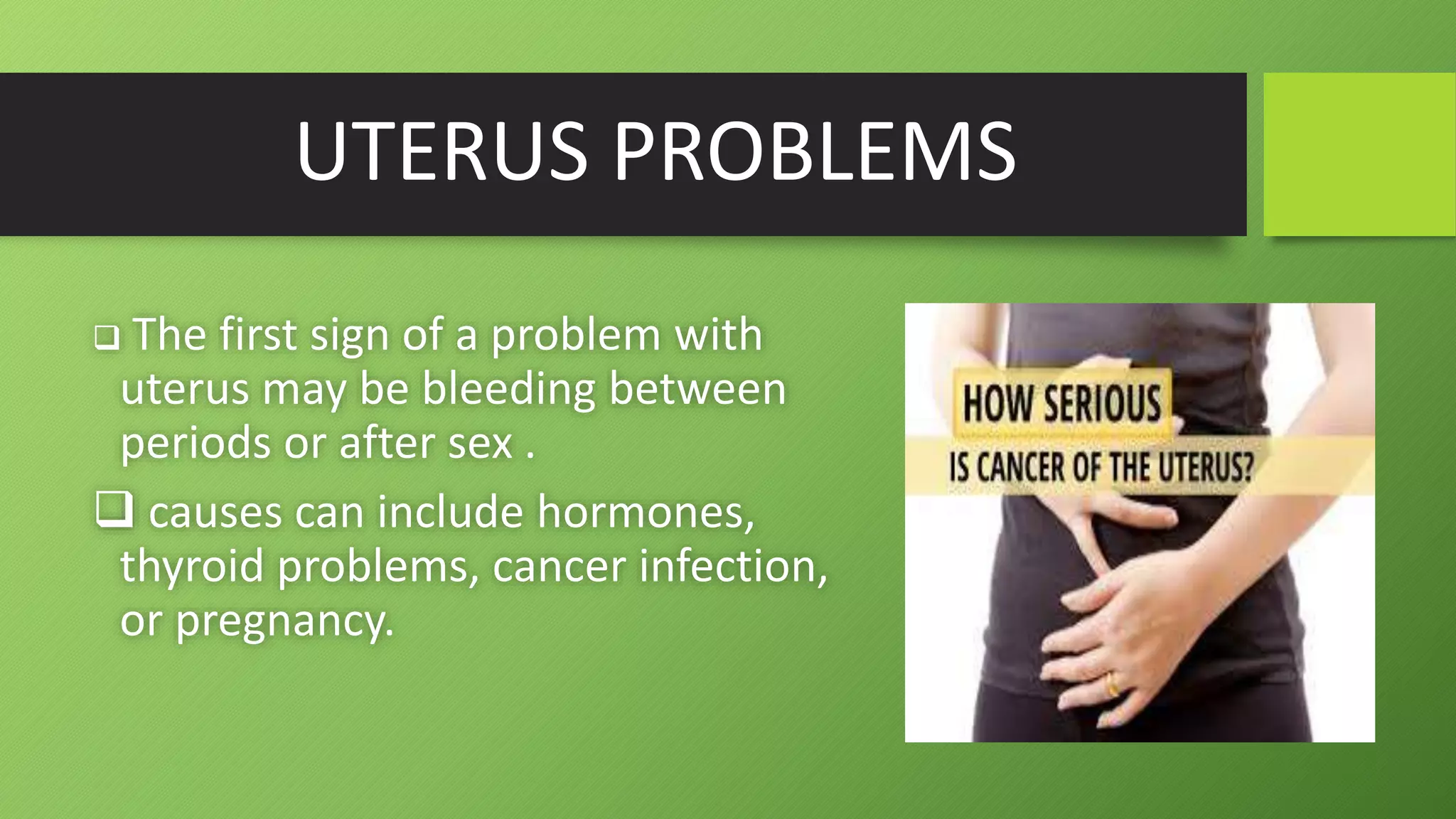
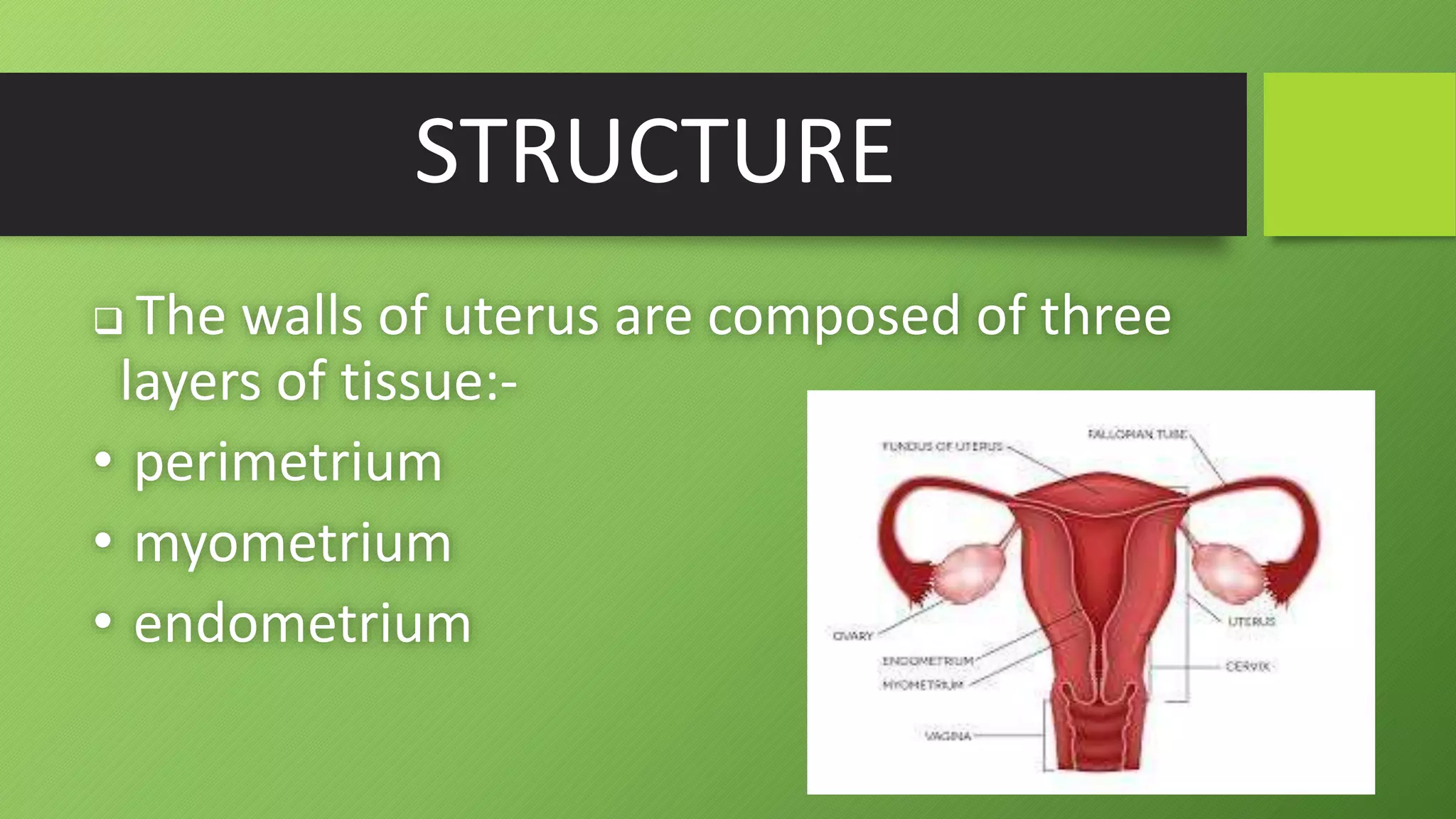
FEMALE UTERUS..
ANATOMY OF UTERUS
BY:- KALPANA
WHAT IS UTERUS???
•The uterus is a hollow
muscular pear-shaped organ
, flattened anteroposteriorly
. It lies in the pelvic cavity
between the urinary bladder
and the rectum.
UTERUS??
The uterus lies in an almost horizontal position.
It is about 7.5cm long, 5cm wide and its walls about
2.5cm thick.
It weights from 30 to 40 grams.
FUNCTION
 uterus also plays a role in sexual response,
by directing blood flow to the pelvis and
ovaries, and to the external genitals,
including the vagina, labia, and clitoris.
 It functions to nourish and house a
fertilized egg until the unborn child, or
offspring, is ready to be delivered.
.
PARTS OF UTERUS :-
•FUNDUS
•BODY
•CERVIX
FUNDUS
 This is the dome-shaped part of the uterus
above the openings of the uterine tubes.
 the fundus of the uterus is the top portion,
opposite form the cervix.
BODY
 This is the main part .
 It is narrowest interiorly at the internal
as where it is continue with the cervix.
CERVIX ( Neck of the uterus)
 This protrudes through the anterior
wall of the vagina, opening into it at the
external as..
UTERUS PROBLEMS
 The first sign of a problem with
uterus may be bleeding between
periods or after sex .
 causes can include hormones,
thyroid problems, cancer infection,
or pregnancy.
STRUCTURE
 The walls of uterus are composed of three
layers of tissue:-
• perimetrium
• myometrium
• endometrium
PERIMETRIUM
• Which is distributed differently on the various surfaces of the
uterus.
• Anteriorly it lies over the fundus and the body where it is folded on
the upper surface of the urinary bladder .
• this fold of peritoneum forms the vesicouterine pouch.
• posteriorly the peritoneum covers the fundus ,the body and the
cervix, the it folds back on to the rectum to form the rectouterine
pouch (of douglas).
MYOMETRIUM
•This is the thickest layer of tissue in the
uterine wall.
• it is a mass of smooth muscle fibres
interlaced with areolar tissue, blood vessel
and nerves.
ENDOMERTIUM
• this consist of columnar epithelium containing a large number of mucus-
secreting tubular glands .
• it is divided functionally into two layers :-
 the functional layer is the upper layer and it thickness and becomes rich
in blood vessels in the first half of the menstrual cycle.
 this layer is shed during menstruation.
 the basal layer lies next to the myometrium ,and is not lost during
menstruation.
 it is the layer from which the fresh functional layer is regenerated during
each cycle .
THANKYOU

Anatomy of Uterus

  • 1.
    FEMALE UTERUS.. ANATOMY OFUTERUS BY:- KALPANA
  • 2.
    WHAT IS UTERUS??? •Theuterus is a hollow muscular pear-shaped organ , flattened anteroposteriorly . It lies in the pelvic cavity between the urinary bladder and the rectum.
  • 3.
    UTERUS?? The uterus liesin an almost horizontal position. It is about 7.5cm long, 5cm wide and its walls about 2.5cm thick. It weights from 30 to 40 grams.
  • 4.
    FUNCTION  uterus alsoplays a role in sexual response, by directing blood flow to the pelvis and ovaries, and to the external genitals, including the vagina, labia, and clitoris.  It functions to nourish and house a fertilized egg until the unborn child, or offspring, is ready to be delivered. .
  • 5.
    PARTS OF UTERUS:- •FUNDUS •BODY •CERVIX
  • 6.
    FUNDUS  This isthe dome-shaped part of the uterus above the openings of the uterine tubes.  the fundus of the uterus is the top portion, opposite form the cervix.
  • 7.
    BODY  This isthe main part .  It is narrowest interiorly at the internal as where it is continue with the cervix.
  • 8.
    CERVIX ( Neckof the uterus)  This protrudes through the anterior wall of the vagina, opening into it at the external as..
  • 9.
    UTERUS PROBLEMS  Thefirst sign of a problem with uterus may be bleeding between periods or after sex .  causes can include hormones, thyroid problems, cancer infection, or pregnancy.
  • 10.
    STRUCTURE  The wallsof uterus are composed of three layers of tissue:- • perimetrium • myometrium • endometrium
  • 11.
    PERIMETRIUM • Which isdistributed differently on the various surfaces of the uterus. • Anteriorly it lies over the fundus and the body where it is folded on the upper surface of the urinary bladder . • this fold of peritoneum forms the vesicouterine pouch. • posteriorly the peritoneum covers the fundus ,the body and the cervix, the it folds back on to the rectum to form the rectouterine pouch (of douglas).
  • 12.
    MYOMETRIUM •This is thethickest layer of tissue in the uterine wall. • it is a mass of smooth muscle fibres interlaced with areolar tissue, blood vessel and nerves.
  • 13.
    ENDOMERTIUM • this consistof columnar epithelium containing a large number of mucus- secreting tubular glands . • it is divided functionally into two layers :-  the functional layer is the upper layer and it thickness and becomes rich in blood vessels in the first half of the menstrual cycle.  this layer is shed during menstruation.  the basal layer lies next to the myometrium ,and is not lost during menstruation.  it is the layer from which the fresh functional layer is regenerated during each cycle .
  • 14.