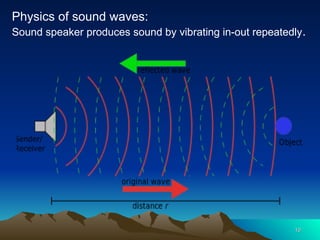
Introduction to Ultrasonography
Dr Gameli Kofi Seadzi
Ultrasound, also called sonography, uses sound
waves to develop images of internal organs of the
body.
A component of the equipment called transducer
emits high-frequency sound, (inaudible to human
ears) and then records the echoes as the sound
waves reflected back.
The reflected wave is used to determine the size,
shape, and consistency of soft tissues and organs.
2
2
• This information is relayed in real time to
produce images on a computer screen.
• Ultrasound technicians, or sonographers,
have special training in how to perform the
test.
• Then a radiologist or doctor will interpret
the ultrasound images.
3
3
Uses of the ultrasound
• Pregnancy.
– Dating: age of pregnancy; due date.
– Type of conception:
• twins or other multiples
• ectopic pregnancies.
– Birth defects and congenital malformations
– Placental characteristics,
– Presentation: breech cephalic or transverse.
– Sex determination
– Size of baby is just before delivery.
4
4
5
5
• Other uses of sonography in medicine
– Diagnostics. Doctors employ ultrasound imaging in
diagnosing a wide variety of conditions affecting the
organs and soft tissues of the body
– Use during medical procedures. Ultrasound
imaging can help doctors during procedures such as
needle biopsies
– Therapeutic applications. Ultrasounds sometimes
are used to detect and treat soft-tissue injuries.
6
6
• Ultrasounds have some diagnostic limitations,
because sound waves do not transmit well
through
–parts of the body that may hold air or gas, such as the
lungs or bowel.
–Fatty tissue in obese patients.
Other types of ultrasound
• Most ultrasounds are done using a transducer
on the surface of the skin.
• Sometimes, a better diagnostic image is
obtained by inserting a special transducer into
one of the body's natural openings:
7
7
– In a transvaginal ultrasound, a transducer wand is
placed in a woman’s vagina to get better images of
her uterus and ovaries.
– A transrectal ultrasound is sometimes used in the
diagnosis of prostate conditions.
– A trans esophageal echocardiogram uses the
transducer probe in the esophagus so that the
sonographer can obtain clearer images of the heart
8
8
9
9
10
10
Physics of sound waves:
Sound speaker produces sound by vibrating in-out repeatedly.
• Sound wave: of peaks and troughs that propagate
through the transmitting medium, eg air.
• Frequency – number of waves passing a given point
per unit time (sec). Measured in cycles per sec = Hz
• Wavelength – the distance between two peaks of the
sound wave.
• Period: the time between two peaks; measured in
seconds
11
11
12
12
13
13
• Frequency X Wavelength = Speed
– The speed of sound is fairly constant in any medium,
and so frequency is inversely proportional to the
wavelength.
– so to increase the frequency reduce the wavelength.
– and to decrease the frequency, increase the
wavelength.
– Sound of lower frequency travel farther from the
source
Amplitude – the height of each wave and depicts the
loudness of the sound.
The Amplitude has no mathematical relationship with
either the frequency or the wavelength.
It is the extent to which particles are displaced in the
transmitting medium eg air.
The amplitude determines the loudness of the sound
14
14
• Sound frequency ranges
– Audible range 20 – 20,000Hz (sonic range)
– Sub-sonic or infra –below 20Hz
– Ultra-sonic – above 20,000Hz
• Ultrasound defined as the sound waves above the
audible limit. i.e. 1 mHz to 20 mHz
• In ultrasonography a transducer produces the
ultrasound by crystals changing shape when electric
current is passed through. (Piezo Electric Effect).
• The transducer can also covert sound waves back to
electricity.
15
15
Behaviour of sound in tissue
Ultrasound transducer
• Converts electrical impulse into (ultra) sound
waves
• US waves sent through tissue and reflected back
to transducer (echo)
• Received wave is converted back into electrical
energy
• Transmitted to the screen monitor in real time
and image formed on screen (the skin line).
16
16
• At standard temperature and pressure (STP)
sound travels at 343m/sec through air.
• Lungs transmit sound at 600m/sec
• Bone transmit at 4,080m/sec
• Soft tissue averages 1,540m/sec
• 13 micro sec - round trip travel time from
transducer to a 1 cm thick tissue and return.
– 26 micro sec = 2 cm of tissue
– 130 micro seconds = ?
– 1300 micro seconds = ?
17
17
• Depth – function of the distance travelled to and fro
transducer
• Brightness – volume or intensity of the returning echo.
louder echo results in brighter image
silent echo produces darker image
• Skin line – processed reflected echo at each linear plain
of reflection.
• Several skin lines used to form a 2D image on the screen.
18
18
• Resolution of image: how detailed the image is; and
ability to differentiate between two images.
• Penetration: how deep the image goes
• Increasing frequency improves resolution.
• Decreasing frequency decreases resolution.
• Converse true for penetration
• Increase frequency reduces penetration..
• Increasing the frequency improves resolution but
reduces penetration.
19
19
Spatial Resolution
Spatial Resolution
• Ability to differentiate between two different points
in space
• Higher frequency better resolution
• Concept of focal zone of transducer – waves
converge then divert. The narrowest portion of
convergence known as the focal zone.
• Best resolution obtained at the focal zone at any
frequency.
20
20
Acoustic Impedance
Acoustic Impedance
• This is the resistance to the travel of sound through a
tissue.
• Sound is reflected or transmitted depending on the
acoustic impedance of the two tissues that are in
proximity.
• The larger the difference two different tissues in close
proximity, the higher the resolution.
21
21
Types of reflection
Types of reflection
• Spectral reflection
– Lots of waves reflected
– Smooth
– Homogenous
– bright
• Diffused reflection
– Wave scattered
– Less amount returned
22
22
Refraction
Refraction
• Bending of sound beam as the speed of travel
changes.
• Slow into fast:
– Produces a wide angle of deflection
• Fast into slow:
– The angle of deflection is narrow
• Refraction is important in the recognition of artefacts.
23
23
Attenuation
Attenuation
• Decrease in the amplitude of sound over a distance.
• For example in audible sounds attenuation results in
not hearing the sound when farther away from the
source.
• Increasing the frequency increases the attenuation of
sound.
24
24
Questions
Questions
25
25

2-Introduction to Ultrasound.pptnotes fred

  • 1.
    Introduction to Ultrasonography Introductionto Ultrasonography Dr Gameli Kofi Seadzi
  • 2.
    Ultrasound, also calledsonography, uses sound waves to develop images of internal organs of the body. A component of the equipment called transducer emits high-frequency sound, (inaudible to human ears) and then records the echoes as the sound waves reflected back. The reflected wave is used to determine the size, shape, and consistency of soft tissues and organs. 2 2
  • 3.
    • This informationis relayed in real time to produce images on a computer screen. • Ultrasound technicians, or sonographers, have special training in how to perform the test. • Then a radiologist or doctor will interpret the ultrasound images. 3 3
  • 4.
    Uses of theultrasound Uses of the ultrasound • Pregnancy. – Dating: age of pregnancy; due date. – Type of conception: • twins or other multiples • ectopic pregnancies. – Birth defects and congenital malformations – Placental characteristics, – Presentation: breech cephalic or transverse. – Sex determination – Size of baby is just before delivery. 4 4
  • 5.
    5 5 • Other usesof sonography in medicine – Diagnostics. Doctors employ ultrasound imaging in diagnosing a wide variety of conditions affecting the organs and soft tissues of the body – Use during medical procedures. Ultrasound imaging can help doctors during procedures such as needle biopsies – Therapeutic applications. Ultrasounds sometimes are used to detect and treat soft-tissue injuries.
  • 6.
    6 6 • Ultrasounds havesome diagnostic limitations, because sound waves do not transmit well through –parts of the body that may hold air or gas, such as the lungs or bowel. –Fatty tissue in obese patients.
  • 7.
    Other types ofultrasound Other types of ultrasound • Most ultrasounds are done using a transducer on the surface of the skin. • Sometimes, a better diagnostic image is obtained by inserting a special transducer into one of the body's natural openings: 7 7
  • 8.
    – In atransvaginal ultrasound, a transducer wand is placed in a woman’s vagina to get better images of her uterus and ovaries. – A transrectal ultrasound is sometimes used in the diagnosis of prostate conditions. – A trans esophageal echocardiogram uses the transducer probe in the esophagus so that the sonographer can obtain clearer images of the heart 8 8
  • 9.
  • 10.
    10 10 Physics of soundwaves: Sound speaker produces sound by vibrating in-out repeatedly.
  • 11.
    • Sound wave:of peaks and troughs that propagate through the transmitting medium, eg air. • Frequency – number of waves passing a given point per unit time (sec). Measured in cycles per sec = Hz • Wavelength – the distance between two peaks of the sound wave. • Period: the time between two peaks; measured in seconds 11 11
  • 12.
  • 13.
    13 13 • Frequency XWavelength = Speed – The speed of sound is fairly constant in any medium, and so frequency is inversely proportional to the wavelength. – so to increase the frequency reduce the wavelength. – and to decrease the frequency, increase the wavelength. – Sound of lower frequency travel farther from the source
  • 14.
    Amplitude – theheight of each wave and depicts the Amplitude – the height of each wave and depicts the loudness of the sound. loudness of the sound. The Amplitude has no mathematical relationship with The Amplitude has no mathematical relationship with either the frequency or the wavelength. either the frequency or the wavelength. It is the extent to which particles are displaced in the It is the extent to which particles are displaced in the transmitting medium eg air. transmitting medium eg air. The amplitude determines the loudness of the sound The amplitude determines the loudness of the sound 14 14
  • 15.
    • Sound frequencyranges – Audible range 20 – 20,000Hz (sonic range) – Sub-sonic or infra –below 20Hz – Ultra-sonic – above 20,000Hz • Ultrasound defined as the sound waves above the audible limit. i.e. 1 mHz to 20 mHz • In ultrasonography a transducer produces the ultrasound by crystals changing shape when electric current is passed through. (Piezo Electric Effect). • The transducer can also covert sound waves back to electricity. 15 15
  • 16.
    Behaviour of soundin tissue Behaviour of sound in tissue Ultrasound transducer • Converts electrical impulse into (ultra) sound waves • US waves sent through tissue and reflected back to transducer (echo) • Received wave is converted back into electrical energy • Transmitted to the screen monitor in real time and image formed on screen (the skin line). 16 16
  • 17.
    • At standardtemperature and pressure (STP) sound travels at 343m/sec through air. • Lungs transmit sound at 600m/sec • Bone transmit at 4,080m/sec • Soft tissue averages 1,540m/sec • 13 micro sec - round trip travel time from transducer to a 1 cm thick tissue and return. – 26 micro sec = 2 cm of tissue – 130 micro seconds = ? – 1300 micro seconds = ? 17 17
  • 18.
    • Depth –function of the distance travelled to and fro transducer • Brightness – volume or intensity of the returning echo. louder echo results in brighter image silent echo produces darker image • Skin line – processed reflected echo at each linear plain of reflection. • Several skin lines used to form a 2D image on the screen. 18 18
  • 19.
    • Resolution ofimage: how detailed the image is; and ability to differentiate between two images. • Penetration: how deep the image goes • Increasing frequency improves resolution. • Decreasing frequency decreases resolution. • Converse true for penetration • Increase frequency reduces penetration.. • Increasing the frequency improves resolution but reduces penetration. 19 19
  • 20.
    Spatial Resolution Spatial Resolution •Ability to differentiate between two different points in space • Higher frequency better resolution • Concept of focal zone of transducer – waves converge then divert. The narrowest portion of convergence known as the focal zone. • Best resolution obtained at the focal zone at any frequency. 20 20
  • 21.
    Acoustic Impedance Acoustic Impedance •This is the resistance to the travel of sound through a tissue. • Sound is reflected or transmitted depending on the acoustic impedance of the two tissues that are in proximity. • The larger the difference two different tissues in close proximity, the higher the resolution. 21 21
  • 22.
    Types of reflection Typesof reflection • Spectral reflection – Lots of waves reflected – Smooth – Homogenous – bright • Diffused reflection – Wave scattered – Less amount returned 22 22
  • 23.
    Refraction Refraction • Bending ofsound beam as the speed of travel changes. • Slow into fast: – Produces a wide angle of deflection • Fast into slow: – The angle of deflection is narrow • Refraction is important in the recognition of artefacts. 23 23
  • 24.
    Attenuation Attenuation • Decrease inthe amplitude of sound over a distance. • For example in audible sounds attenuation results in not hearing the sound when farther away from the source. • Increasing the frequency increases the attenuation of sound. 24 24
  • 25.