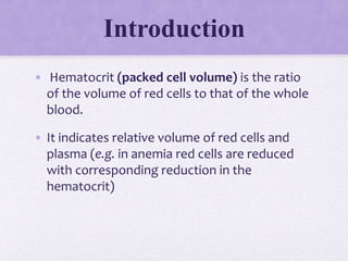
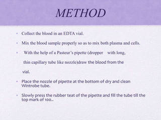
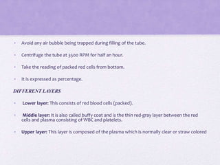
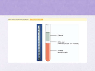
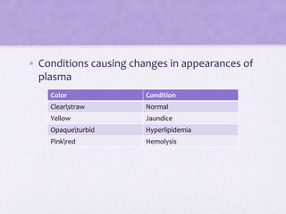
HEMATOCRIT
Introduction
• Hematocrit (packed cell volume) is the ratio
of the volume of red cells to that of the whole
blood.
• It indicates relative volume of red cells and
plasma (e.g. in anemia red cells are reduced
with corresponding reduction in the
hematocrit)
Methods of Estimation of
PCV
• Macromethod using Wintrobe tube
• Micromethod using capillary tube
• Automated analyser
WINTROBE TUBE
It is a special thick walled
• glass tube measuring 11 cm in length
• an internal diameter of 2.5 mm
• capacity of 1 ml.
• This is calibrated at 1 mm intervals from top as 0, 10, 20, 30,...100
for ESR and 100, 90, 80,...10, 0 for PCV.
• The markings are in descending order from the top for PCV
estimation.
Anticoagulants used: EDTA, dry heparin or double oxalate.
PRINCIPLE
• Anticoagulated whole blood is centrifuged at a
standard speed (3500 RPM). RBCs which are
heavier than white cells, platelets and plasma,
sediment at the bottom and the volume of red
cell mass denotes the hematocrit.
METHOD
• Collect the blood in an EDTA vial.
• Mix the blood sample properly so as to mix both plasma and cells.
• With the help of a Pasteur’s pipette (dropper with long,
thin capillary tube like nozzle)draw the blood from the
vial.
• Place the nozzle of pipette at the bottom of dry and clean
Wintrobe tube.
• Slowly press the rubber teat of the pipette and fill the tube till the
top mark of 100..
• Avoid any air bubble being trapped during filling of the tube.
• Centrifuge the tube at 3500 RPM for half an hour.
• Take the reading of packed red cells from bottom.
• It is expressed as percentage.
DIFFERENT LAYERS
• Lower layer: This consists of red blood cells (packed).
• Middle layer: It is also called buffy coat and is the thin red-gray layer between the red
cells and plasma consisting of WBC and platelets.
• Upper layer: This layer is composed of the plasma which is normally clear or straw colored
Color Condition
Clearstraw Normal
Yellow Jaundice
Opaqueturbid Hyperlipidemia
Pinkred Hemolysis
• Conditions causing changes in appearances of
plasma
• Buffy coat gives an approximate indication of the number
of WBCs (normally 0.1 mm of this layer = 1000 WBC/cu mm).
• Thickness of the buffy coat increases with marked
leukocytosis, as in CML, CLL and acute leukemias.
• Absent or minimal buffy coat implies leukopenia.
• WBCs and identification of abnormal cells is easier.
• LE cell test
• Note: Buffy coat is not useful for platelet estimation.
• Other use of Wintrobe tube is for ESR estimation.
Microhematocrit Method
• A heparinized capillary tube is used and the blood is filled by capillary action.
• One end is sealed and it is centrifuged in a special centrifuge.
• As the tubes are not graduated, the PCV is measured using a special chart.
Advantages
• Easy
• Small quantity of blood is needed (useful in pediatric patients).
• Less time
• Amount of plasma trapped in the red cell column is less, thereby minimizing the error.
Disadvantage
• It requires a special (expensive) centrifuge.
Automated Analyser
• Nowadays, automated analysers are available for estimation of hematocrit.
Normal range for PCV
• Hematocrit is expressed as a percentage (e.g. 45%) or as a decimal
fraction (e.g. 0.45).
• It is useful for evaluating absolute values like MCV and MCHC.
• Males 40-50%
• Females 36-46%
• Infants 45-70% (cord blood)
• Causes of Increased PCV
 polycythemia
 Dehydration
 Kidney & heart problems
 Diarrhea
 Burns
 smoking
 Decreased PCV is seen in
 Anemia
 Pregnancy
 Hemolysis

PCV.pptx

  • 1.
  • 2.
    Introduction • Hematocrit (packedcell volume) is the ratio of the volume of red cells to that of the whole blood. • It indicates relative volume of red cells and plasma (e.g. in anemia red cells are reduced with corresponding reduction in the hematocrit)
  • 3.
    Methods of Estimationof PCV • Macromethod using Wintrobe tube • Micromethod using capillary tube • Automated analyser
  • 4.
    WINTROBE TUBE It isa special thick walled • glass tube measuring 11 cm in length • an internal diameter of 2.5 mm • capacity of 1 ml. • This is calibrated at 1 mm intervals from top as 0, 10, 20, 30,...100 for ESR and 100, 90, 80,...10, 0 for PCV. • The markings are in descending order from the top for PCV estimation. Anticoagulants used: EDTA, dry heparin or double oxalate.
  • 5.
    PRINCIPLE • Anticoagulated wholeblood is centrifuged at a standard speed (3500 RPM). RBCs which are heavier than white cells, platelets and plasma, sediment at the bottom and the volume of red cell mass denotes the hematocrit.
  • 6.
    METHOD • Collect theblood in an EDTA vial. • Mix the blood sample properly so as to mix both plasma and cells. • With the help of a Pasteur’s pipette (dropper with long, thin capillary tube like nozzle)draw the blood from the vial. • Place the nozzle of pipette at the bottom of dry and clean Wintrobe tube. • Slowly press the rubber teat of the pipette and fill the tube till the top mark of 100..
  • 7.
    • Avoid anyair bubble being trapped during filling of the tube. • Centrifuge the tube at 3500 RPM for half an hour. • Take the reading of packed red cells from bottom. • It is expressed as percentage. DIFFERENT LAYERS • Lower layer: This consists of red blood cells (packed). • Middle layer: It is also called buffy coat and is the thin red-gray layer between the red cells and plasma consisting of WBC and platelets. • Upper layer: This layer is composed of the plasma which is normally clear or straw colored
  • 9.
    Color Condition Clearstraw Normal YellowJaundice Opaqueturbid Hyperlipidemia Pinkred Hemolysis • Conditions causing changes in appearances of plasma
  • 10.
    • Buffy coatgives an approximate indication of the number of WBCs (normally 0.1 mm of this layer = 1000 WBC/cu mm). • Thickness of the buffy coat increases with marked leukocytosis, as in CML, CLL and acute leukemias. • Absent or minimal buffy coat implies leukopenia. • WBCs and identification of abnormal cells is easier. • LE cell test • Note: Buffy coat is not useful for platelet estimation. • Other use of Wintrobe tube is for ESR estimation.
  • 11.
    Microhematocrit Method • Aheparinized capillary tube is used and the blood is filled by capillary action. • One end is sealed and it is centrifuged in a special centrifuge. • As the tubes are not graduated, the PCV is measured using a special chart. Advantages • Easy • Small quantity of blood is needed (useful in pediatric patients). • Less time • Amount of plasma trapped in the red cell column is less, thereby minimizing the error. Disadvantage • It requires a special (expensive) centrifuge.
  • 12.
    Automated Analyser • Nowadays,automated analysers are available for estimation of hematocrit. Normal range for PCV • Hematocrit is expressed as a percentage (e.g. 45%) or as a decimal fraction (e.g. 0.45). • It is useful for evaluating absolute values like MCV and MCHC. • Males 40-50% • Females 36-46% • Infants 45-70% (cord blood)
  • 13.
    • Causes ofIncreased PCV  polycythemia  Dehydration  Kidney & heart problems  Diarrhea  Burns  smoking
  • 14.
     Decreased PCVis seen in  Anemia  Pregnancy  Hemolysis