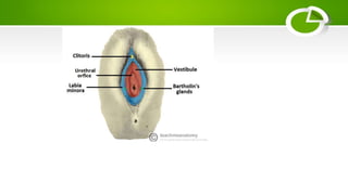
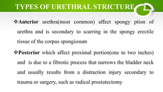
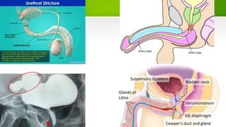
URETHRAL STRICTURE
BY Athanase YAMFASHIJE
INTRODUCTION
The urethra is the tubular structure lined by stratified columnar
epithelium that convey urine from the bladder to an external opening in
the perineum as its primary role in both male and female
It also play an important role in ejaculation for men
 It is lined by mucous secreting glands that act as protection from
corrosive urine contents
Urethral strictures is the narrowing of urethral lumen that is mostly due
to the scaring
SCOPE
 Normal anatomy and physiology
 Epidemiology
 Etiology/Causes of urethral stricture
 Types of urethral stricture
 Symptoms and Signs
 Clinical diagnosis
 Management
 Prevention
NORMAL ANATOMY AND PHYSIOLOGY
MALE URETHRA
The male urethra is approximately 15-20cm long. In addition to
urine, the male urethra transports semen
Anatomically, male urethra is made of three parts(from
proximal to distal)
 Prostatic urethra
 Membranous urethra
 Bulbous/Penile urethra
Prostatic,Membranous, Penile urethra are supplied by the
inferior vesical artery (branch of the internal iliac artery),
bulbourethral artery (branch of the internal pudendal artery) and
direct branches of the internal pudendal artery respectively
The nerve supply to the male urethra is derived from the
prostatic plexus, which contains a mixture of sympathetic,
parasympathetic and visceral afferent fibres
The prostatic and membranous portions drain to the obturator
and internal iliac nodes, while the penile urethra drains to the
deep and superficial inguinal nodes
FEMALE URETHRA
 Short urethra (approximately 4cm)
It begins at the neck of the bladder, and passes inferiorly
through the perineal membrane and muscular pelvic floor
The urethra opens directly onto the perineum, in an area
between the labia minora, known as the vestibule.
 The urethral orifice is located anteriorly to the vaginal opening,
and 2-3cm posteriorly to the clitoris
The distal end of the urethra is marked by the presence
Skene’s glands(correspondant of prostate in male) on either
side of the urethra
The blood supply is via the internal pudendal ertery/vein,
vaginal arteries/veins and inferior vesical branches of the
vaginal arteries/veins
The nerve supply is from the vesical plexus and the pudendal
nerve.Visceral afferents from the urethra run in the pelvic
splanchnic nerves
The proximal and distal female urethra drains into the internal
iliac nodes and superficial inguinal lymph nodes respectively
EPIDEMIOLOGY
 0.6% of the population are at risk
 Common in the elder of over 55 years old
 Males are affected than females
ETIOLOGY/CAUSES
The narrowing of urethra is secondary to the scaring as resul of:
Infections(urethritis and STDs)
inflammatory Processes(like enlarged Prostate)
Post-traumat(endoscopy, intermittent or long-term use of
catheter, injury to bladder or pelvis)
 Post prostate cancer treatment or surgical operation like
circumcision
 Lichen sclerosis
Anterior urethra(most common) affect spongy ption of
urethra and is secondary to scarring in the spongy erectile
tissue of the corpus spongiosum
Posterior which affect proximal portion(one to two inches)
and is due to a fibrotic process that narrows the bladder neck
and usually results from a distraction injury secondary to
trauma or surgery, such as radical prostatectomy
TYPES OF URETHRAL STRICTURE
SYMPTOMS AND SIGNS
 bloody or dark urine/ blood in semen
 Abdominal pain
Decreased urine stream
Incomplete bladder empting
Spraying of the urine stream
Difficulty, staining, or pain when urinating
Increased urge to urinate or more frequent urination
 swelling of the penis, urethral leaking and UTIs in men
loss of bladder control
CLINICAL DIAGNOSIS
Urinalysis
Urinary flow test
Urethral ultrasound to evaluate the length of the stricture
Pelvic ultrasound to look PVR
Pelvic magnetic resonance imaging (MRI) to assess whether
your pelvic bone is affecting or is affected by your condition
Retrograde urethrogram: uses X-ray images to check for a structural
problem or injury of the urethra as well as the length and location of
the stricture along the urethra
Cystoscopy examines your urethra and bladder using a thin, tubelike
device fitted with a lens (cystoscope) to view these organs
Perform biopsy for suspected lichen sclerosus (LS), or if urethral
cancer is suspected
MANAGEMENT
Catheterization
 Dilation
Urethroplasty
Endoscopic urethrotomy
Implanted stent or permanent catheter
Treat the underlying cause especially the infection
REFERENCE
Netter’s atlas of Human Anatomy, 6th ed
Urology-handbook for medical students, 1E 2001
Urology reference guide
https://emedicine.medscape.com/article/450903-overview

Urethral stricture

  • 1.
  • 2.
    INTRODUCTION The urethra isthe tubular structure lined by stratified columnar epithelium that convey urine from the bladder to an external opening in the perineum as its primary role in both male and female It also play an important role in ejaculation for men  It is lined by mucous secreting glands that act as protection from corrosive urine contents Urethral strictures is the narrowing of urethral lumen that is mostly due to the scaring
  • 3.
    SCOPE  Normal anatomyand physiology  Epidemiology  Etiology/Causes of urethral stricture  Types of urethral stricture  Symptoms and Signs  Clinical diagnosis  Management  Prevention
  • 4.
  • 5.
    MALE URETHRA The maleurethra is approximately 15-20cm long. In addition to urine, the male urethra transports semen Anatomically, male urethra is made of three parts(from proximal to distal)  Prostatic urethra  Membranous urethra  Bulbous/Penile urethra
  • 6.
    Prostatic,Membranous, Penile urethraare supplied by the inferior vesical artery (branch of the internal iliac artery), bulbourethral artery (branch of the internal pudendal artery) and direct branches of the internal pudendal artery respectively The nerve supply to the male urethra is derived from the prostatic plexus, which contains a mixture of sympathetic, parasympathetic and visceral afferent fibres The prostatic and membranous portions drain to the obturator and internal iliac nodes, while the penile urethra drains to the deep and superficial inguinal nodes
  • 8.
    FEMALE URETHRA  Shorturethra (approximately 4cm) It begins at the neck of the bladder, and passes inferiorly through the perineal membrane and muscular pelvic floor The urethra opens directly onto the perineum, in an area between the labia minora, known as the vestibule.  The urethral orifice is located anteriorly to the vaginal opening, and 2-3cm posteriorly to the clitoris
  • 9.
    The distal endof the urethra is marked by the presence Skene’s glands(correspondant of prostate in male) on either side of the urethra The blood supply is via the internal pudendal ertery/vein, vaginal arteries/veins and inferior vesical branches of the vaginal arteries/veins The nerve supply is from the vesical plexus and the pudendal nerve.Visceral afferents from the urethra run in the pelvic splanchnic nerves The proximal and distal female urethra drains into the internal iliac nodes and superficial inguinal lymph nodes respectively
  • 11.
    EPIDEMIOLOGY  0.6% ofthe population are at risk  Common in the elder of over 55 years old  Males are affected than females
  • 12.
    ETIOLOGY/CAUSES The narrowing ofurethra is secondary to the scaring as resul of: Infections(urethritis and STDs) inflammatory Processes(like enlarged Prostate) Post-traumat(endoscopy, intermittent or long-term use of catheter, injury to bladder or pelvis)  Post prostate cancer treatment or surgical operation like circumcision  Lichen sclerosis
  • 13.
    Anterior urethra(most common)affect spongy ption of urethra and is secondary to scarring in the spongy erectile tissue of the corpus spongiosum Posterior which affect proximal portion(one to two inches) and is due to a fibrotic process that narrows the bladder neck and usually results from a distraction injury secondary to trauma or surgery, such as radical prostatectomy TYPES OF URETHRAL STRICTURE
  • 15.
    SYMPTOMS AND SIGNS bloody or dark urine/ blood in semen  Abdominal pain Decreased urine stream Incomplete bladder empting Spraying of the urine stream Difficulty, staining, or pain when urinating Increased urge to urinate or more frequent urination  swelling of the penis, urethral leaking and UTIs in men loss of bladder control
  • 16.
    CLINICAL DIAGNOSIS Urinalysis Urinary flowtest Urethral ultrasound to evaluate the length of the stricture Pelvic ultrasound to look PVR Pelvic magnetic resonance imaging (MRI) to assess whether your pelvic bone is affecting or is affected by your condition
  • 17.
    Retrograde urethrogram: usesX-ray images to check for a structural problem or injury of the urethra as well as the length and location of the stricture along the urethra Cystoscopy examines your urethra and bladder using a thin, tubelike device fitted with a lens (cystoscope) to view these organs Perform biopsy for suspected lichen sclerosus (LS), or if urethral cancer is suspected
  • 18.
    MANAGEMENT Catheterization  Dilation Urethroplasty Endoscopic urethrotomy Implantedstent or permanent catheter Treat the underlying cause especially the infection
  • 20.
    REFERENCE Netter’s atlas ofHuman Anatomy, 6th ed Urology-handbook for medical students, 1E 2001 Urology reference guide https://emedicine.medscape.com/article/450903-overview https://emedicine.medscape.com/article/450903-overview