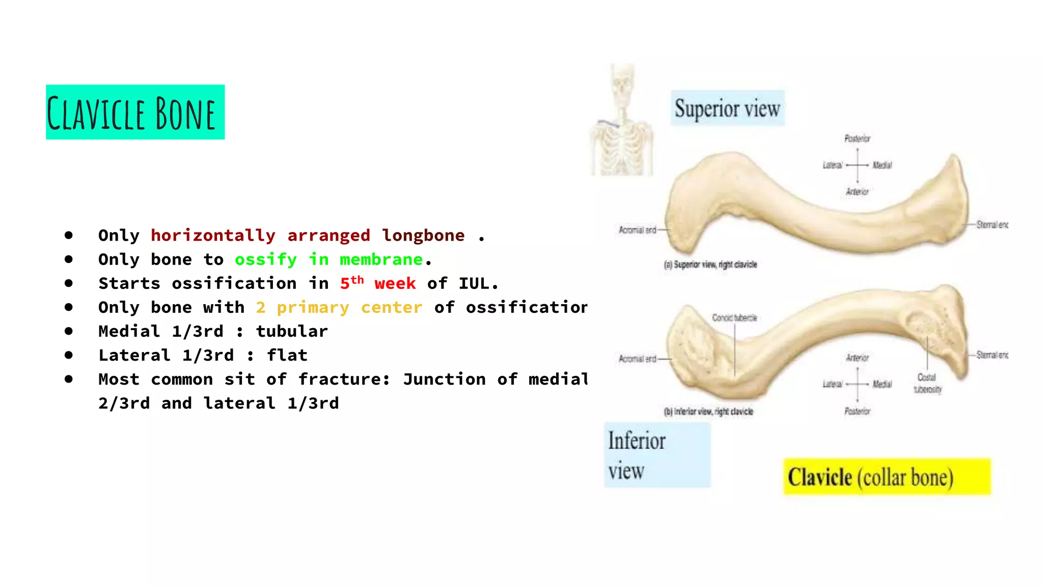
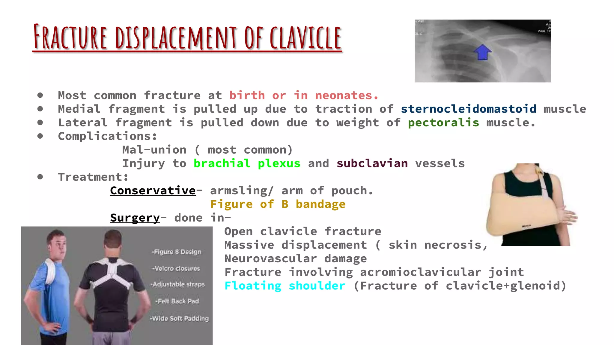
Upper Limb Fractures
(Clavicle & Shoulder Joint)
------ Soushrita Purkait
3rd professional MBBS, part 1
N. R. S Medical College and Hospital
Clavicle Trauma
Clavicle Bone
● Only horizontally arranged longbone .
● Only bone to ossify in membrane.
● Starts ossification in 5th week of IUL.
● Only bone with 2 primary center of ossification.
● Medial 1/3rd : tubular
● Lateral 1/3rd : flat
● Most common sit of fracture: Junction of medial
2/3rd and lateral 1/3rd
Fracture displacement of clavicle
● Most common fracture at birth or in neonates.
● Medial fragment is pulled up due to traction of sternocleidomastoid muscle
● Lateral fragment is pulled down due to weight of pectoralis muscle.
● Complications:
Mal-union ( most common)
Injury to brachial plexus and subclavian vessels
● Treatment:
Conservative- armsling/ arm of pouch.
Figure of B bandage
Surgery- done in-
Open clavicle fracture
Massive displacement ( skin necrosis)
Neurovascular damage
Fracture involving acromioclavicular joint
Floating shoulder (Fracture of clavicle+glenoid)
Shoulder Joint
Trauma
Shoulder joint
● It is a ball ( head of humerus )and socket
(glenoid) type of joint.
● Most common joint to dislocate.
● The stabilisers of shoulder joint are
STATIC
DYNAMIC
Capsule Rotator cuff muscle
Labrum (Fibro- supraspinatus
cartilagenous infraspinatus
ring around teres minor
glenoid) subscapularis
Glenohumoral
Liguments Deltoid and long head of
Superior biceps
Middle
Inferior
Anterior Dislocation Of Shoulder
● Emergency
● SUBTYPES:
Pre-glenoid
Subcoracoid (most common)
Subclavicular
Intrathoracic.
● CINICAL PRESENTATIONS:
Attitude of limb-
-Arm by the side of the body, abducted
and externally rotated
-Shoulder conture is lost
Contd.
● TESTS:
1. Hamiltons ruler test : Place a ruler on the
lateral aspect of arm and check is it touching the
acromian process and lateral epicondyle of arm.
2. Dugas test : check if patient can touch the
opposite shoulder.
3. Callaways test: Compare the girth of affected and
un affected side. If girth is more in affected side then
it indicates dislocation.
4.Bryants test: Vertical axillary circumference
increases.
● Tests for neurovascular injury: Axillary nerve may get
damaged.
Axillary function test : Motor – supplies deltoid ,
can’t be tested.
Sensory- REGIMENTAL BADGE
SIGN – Parasthesia / burning sensation over teres minor
Contd.
● RADIOLOGY:
X-Ray AP view : head is dislocated.
X-Ray Lateral view :confirmatory
● MANAGEMENT: Reduce the dislocation.
MODIFIED KOCHERS TECHNIQUE: (most common)
1. Traction
2. External rotation
3. Adduction
4. Medial roration
STIMSON MANEUVER : Traction of dislocated
limb
HIPPOCRATIC METHOD : Obsolete
Contd.
● COMPLICATIONS:
- Most common complication: Recurrent shoulder
dislocation.
- Most common late complication: Recurrent shoulder
dislocation.
- Most common early complication: Axillary nerve
injury.
● LESIONS:
1. Bankert’s lesion: (most common) avulsion of labrum
form antero-inferior glenoid rim.
2. Bony Bankert’s lesion : If some part of bone also get
avulsed.
3. Hill Sach’s lesion : Because of recurrent dislocation
head of humerus hits the glenoid repeatedly leading to
indentation on the postero-lateral head of humerus.
Posterior dislocation Of Shoulder
● MECHANISM OF INJURY:
-High voltage electric shock
- Seizure Hand abducted
- Fall on outstretched hand & internally
rotated
Very difficult to
diagnose clinically
● RADIOLOGY: X-Ray shows light bulb sign.
● LESIONS: - Reverse Bankert (Antero-inferior)
- Reverse Hill Sach’s (Antero-medial)
Inferior Dislocation Of Shoulder
● Aka Luxatio erecta.
● Extremely rare.
● MECHANISM OF INJURY: Hyperabduction injury
Patient present with erected
hand by the side of the head.
● TESTS FOR SHOULDER INSTABILITY:
1. Anterior:
Fulcrum test
Crank test
Apprehension test (Abduct and externally rotate the shoulder)
2. Posterior: Jerk test
3. Inferior : Sulcus test
● SURGERIES FOR SHOULDER IMSTABILITY:
Putti-Plat procedure: Double breasting the subscapsularis muscle using capsule.
Bristow-Latarjet procedure: Osteotomization of coracoid process and
repositioning on the anterior surface of glenoid to prevent dislocation.
Upper limb trauma ( clavicle & shoulder fracture)

Upper limb trauma ( clavicle & shoulder fracture)

  • 1.
    Upper Limb Fractures (Clavicle& Shoulder Joint) ------ Soushrita Purkait 3rd professional MBBS, part 1 N. R. S Medical College and Hospital
  • 2.
  • 3.
    Clavicle Bone ● Onlyhorizontally arranged longbone . ● Only bone to ossify in membrane. ● Starts ossification in 5th week of IUL. ● Only bone with 2 primary center of ossification. ● Medial 1/3rd : tubular ● Lateral 1/3rd : flat ● Most common sit of fracture: Junction of medial 2/3rd and lateral 1/3rd
  • 4.
    Fracture displacement ofclavicle ● Most common fracture at birth or in neonates. ● Medial fragment is pulled up due to traction of sternocleidomastoid muscle ● Lateral fragment is pulled down due to weight of pectoralis muscle. ● Complications: Mal-union ( most common) Injury to brachial plexus and subclavian vessels ● Treatment: Conservative- armsling/ arm of pouch. Figure of B bandage Surgery- done in- Open clavicle fracture Massive displacement ( skin necrosis) Neurovascular damage Fracture involving acromioclavicular joint Floating shoulder (Fracture of clavicle+glenoid)
  • 5.
  • 6.
    Shoulder joint ● Itis a ball ( head of humerus )and socket (glenoid) type of joint. ● Most common joint to dislocate. ● The stabilisers of shoulder joint are STATIC DYNAMIC Capsule Rotator cuff muscle Labrum (Fibro- supraspinatus cartilagenous infraspinatus ring around teres minor glenoid) subscapularis Glenohumoral Liguments Deltoid and long head of Superior biceps Middle Inferior
  • 7.
    Anterior Dislocation OfShoulder ● Emergency ● SUBTYPES: Pre-glenoid Subcoracoid (most common) Subclavicular Intrathoracic. ● CINICAL PRESENTATIONS: Attitude of limb- -Arm by the side of the body, abducted and externally rotated -Shoulder conture is lost
  • 8.
    Contd. ● TESTS: 1. Hamiltonsruler test : Place a ruler on the lateral aspect of arm and check is it touching the acromian process and lateral epicondyle of arm. 2. Dugas test : check if patient can touch the opposite shoulder. 3. Callaways test: Compare the girth of affected and un affected side. If girth is more in affected side then it indicates dislocation. 4.Bryants test: Vertical axillary circumference increases. ● Tests for neurovascular injury: Axillary nerve may get damaged. Axillary function test : Motor – supplies deltoid , can’t be tested. Sensory- REGIMENTAL BADGE SIGN – Parasthesia / burning sensation over teres minor
  • 9.
    Contd. ● RADIOLOGY: X-Ray APview : head is dislocated. X-Ray Lateral view :confirmatory ● MANAGEMENT: Reduce the dislocation. MODIFIED KOCHERS TECHNIQUE: (most common) 1. Traction 2. External rotation 3. Adduction 4. Medial roration STIMSON MANEUVER : Traction of dislocated limb HIPPOCRATIC METHOD : Obsolete
  • 10.
    Contd. ● COMPLICATIONS: - Mostcommon complication: Recurrent shoulder dislocation. - Most common late complication: Recurrent shoulder dislocation. - Most common early complication: Axillary nerve injury. ● LESIONS: 1. Bankert’s lesion: (most common) avulsion of labrum form antero-inferior glenoid rim. 2. Bony Bankert’s lesion : If some part of bone also get avulsed. 3. Hill Sach’s lesion : Because of recurrent dislocation head of humerus hits the glenoid repeatedly leading to indentation on the postero-lateral head of humerus.
  • 11.
    Posterior dislocation OfShoulder ● MECHANISM OF INJURY: -High voltage electric shock - Seizure Hand abducted - Fall on outstretched hand & internally rotated Very difficult to diagnose clinically ● RADIOLOGY: X-Ray shows light bulb sign. ● LESIONS: - Reverse Bankert (Antero-inferior) - Reverse Hill Sach’s (Antero-medial)
  • 12.
    Inferior Dislocation OfShoulder ● Aka Luxatio erecta. ● Extremely rare. ● MECHANISM OF INJURY: Hyperabduction injury Patient present with erected hand by the side of the head. ● TESTS FOR SHOULDER INSTABILITY: 1. Anterior: Fulcrum test Crank test Apprehension test (Abduct and externally rotate the shoulder) 2. Posterior: Jerk test 3. Inferior : Sulcus test ● SURGERIES FOR SHOULDER IMSTABILITY: Putti-Plat procedure: Double breasting the subscapsularis muscle using capsule. Bristow-Latarjet procedure: Osteotomization of coracoid process and repositioning on the anterior surface of glenoid to prevent dislocation.