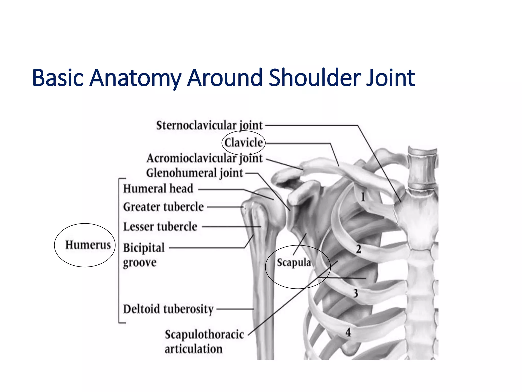
The document discusses injuries around the shoulder joint, including:
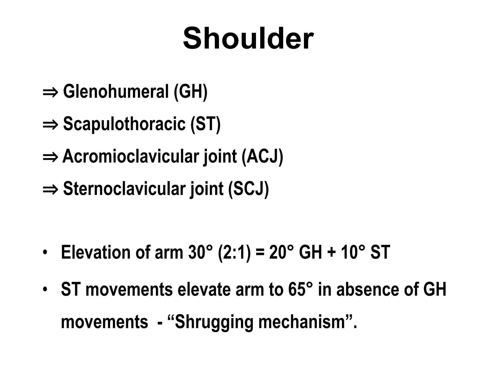
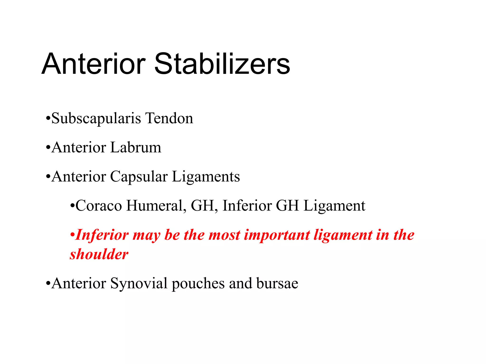
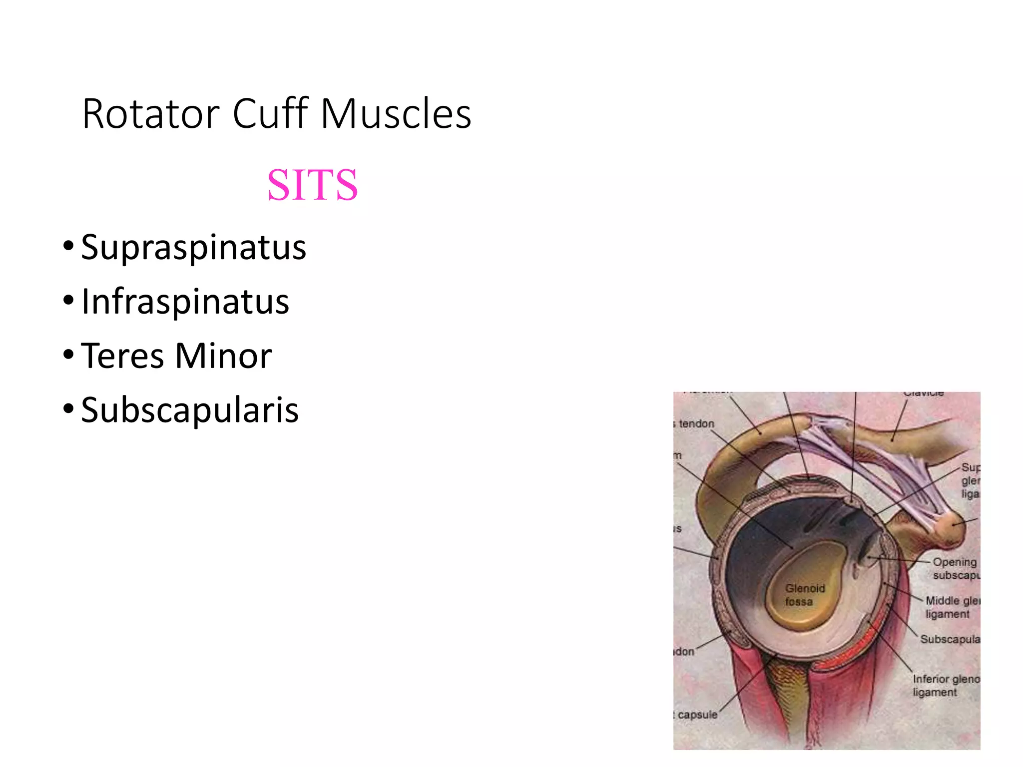
- Basic anatomy of the shoulder joint and its stabilizing muscles and ligaments.
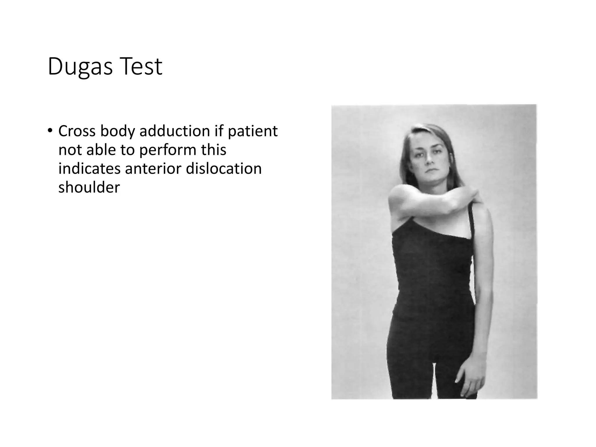
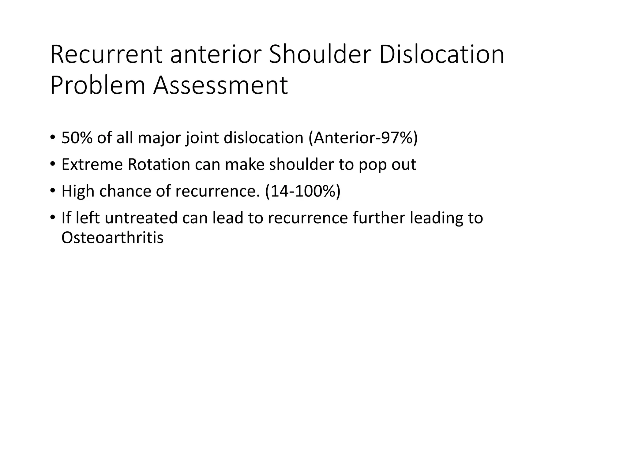
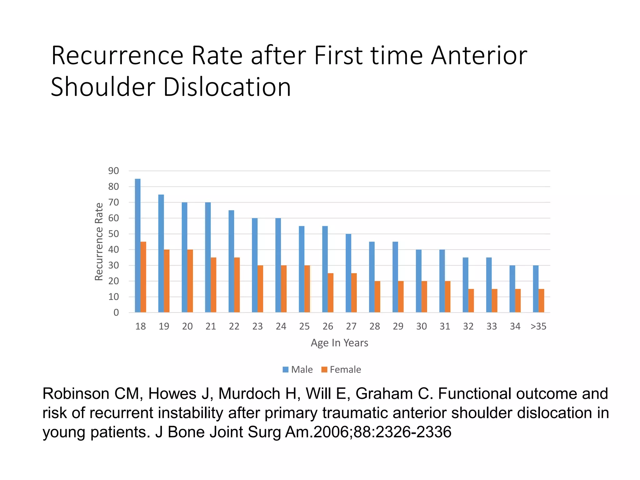
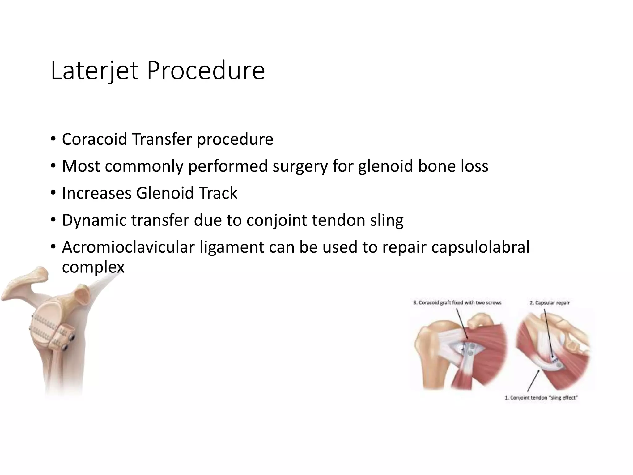
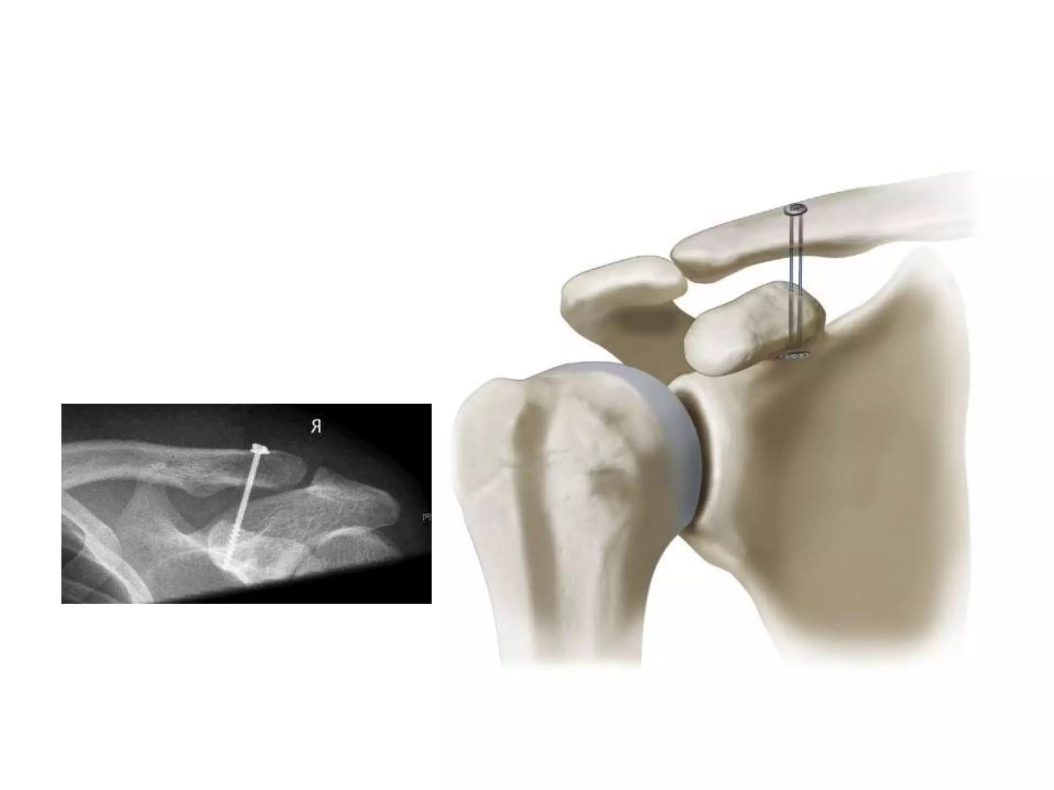
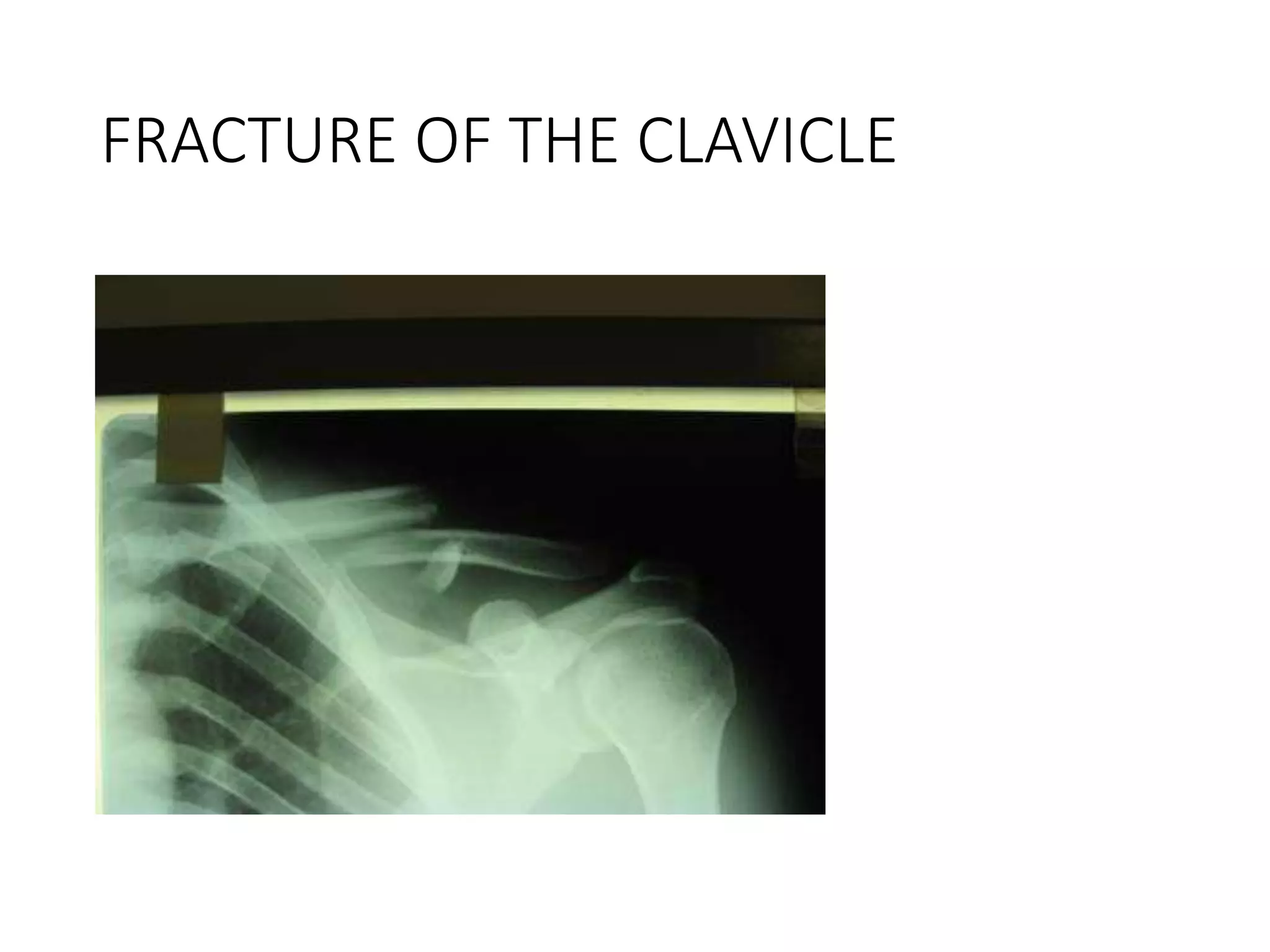
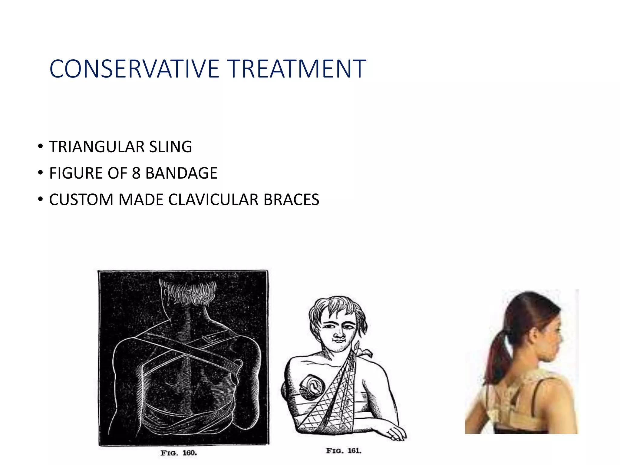
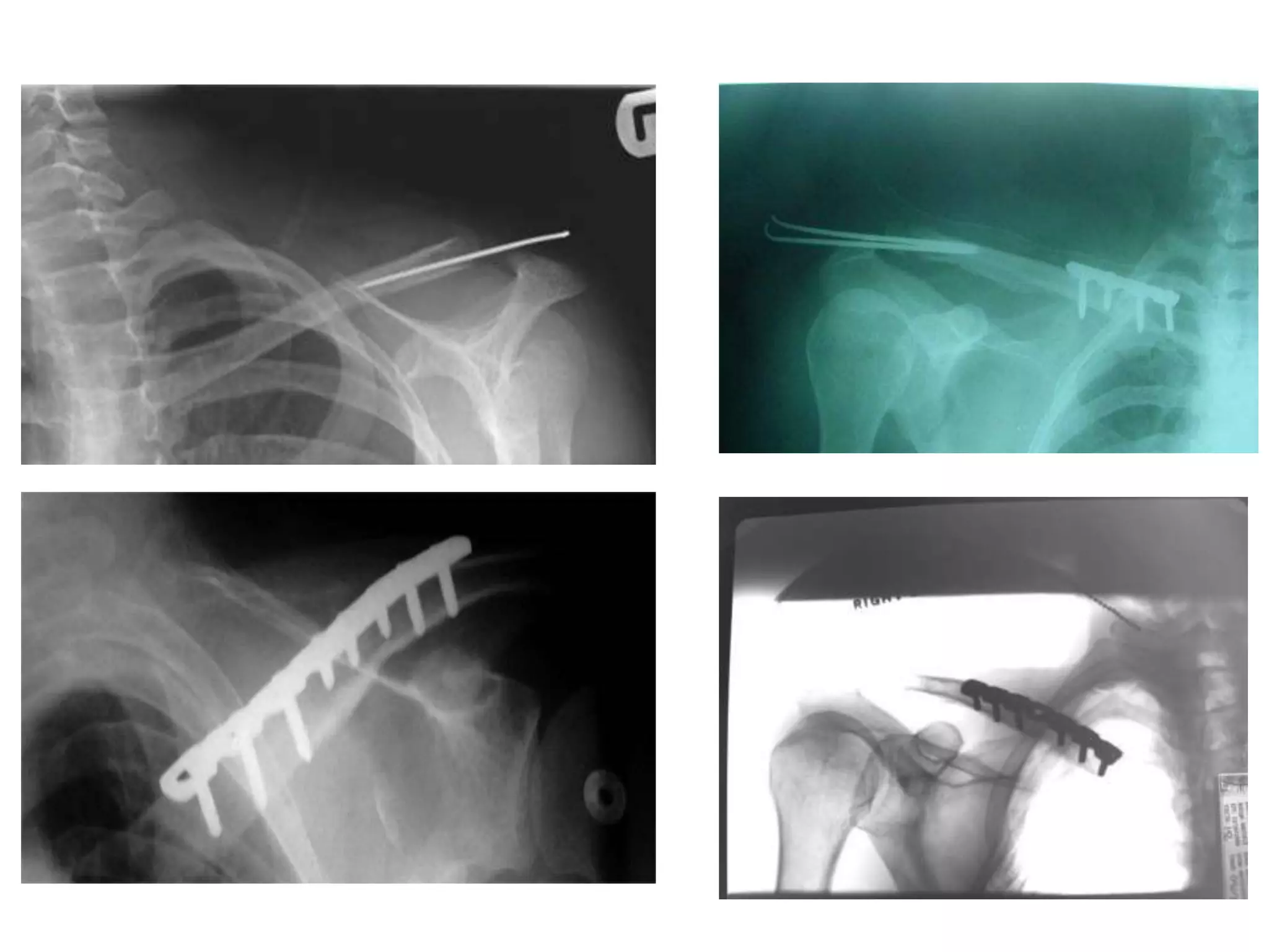
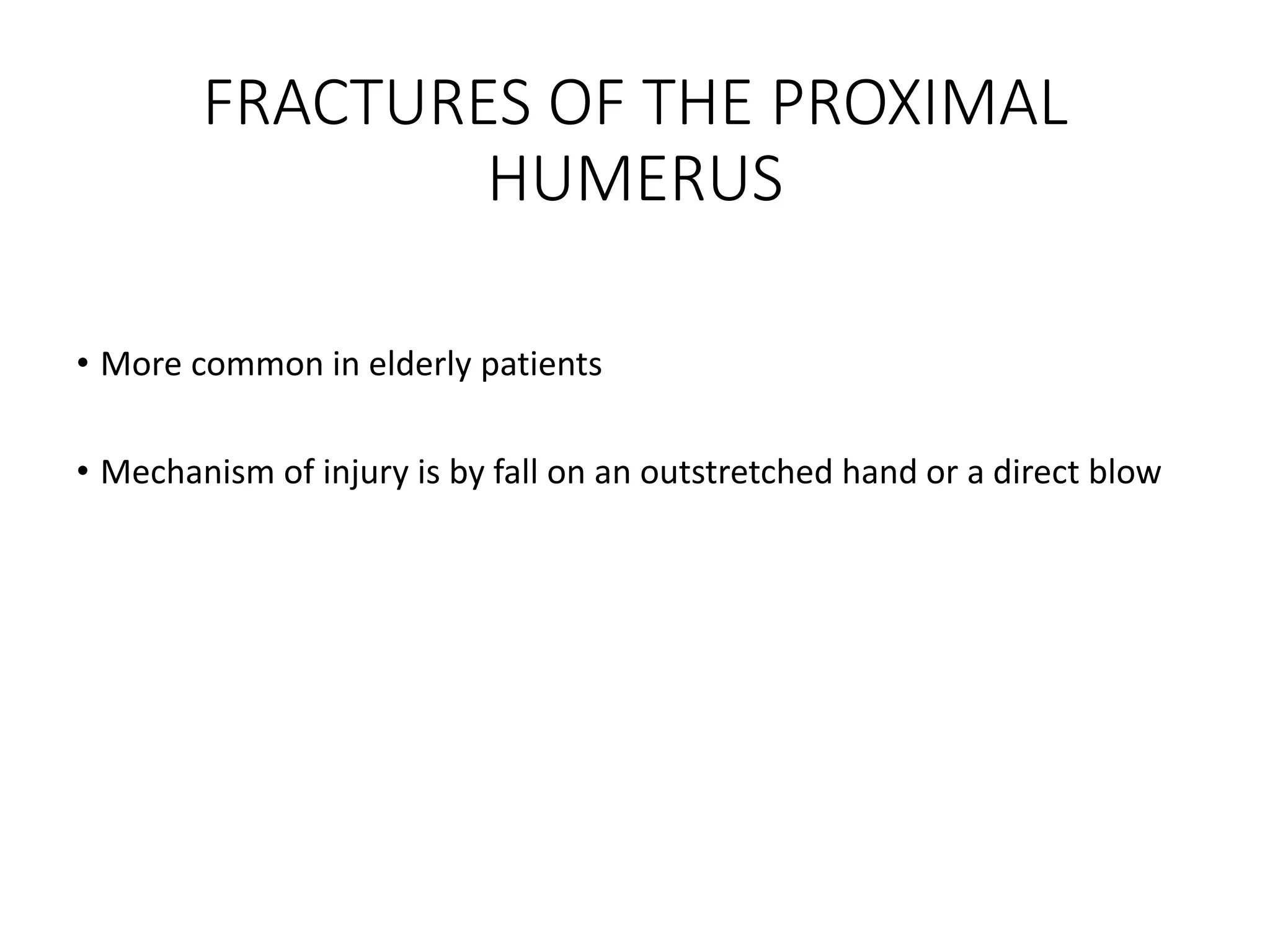
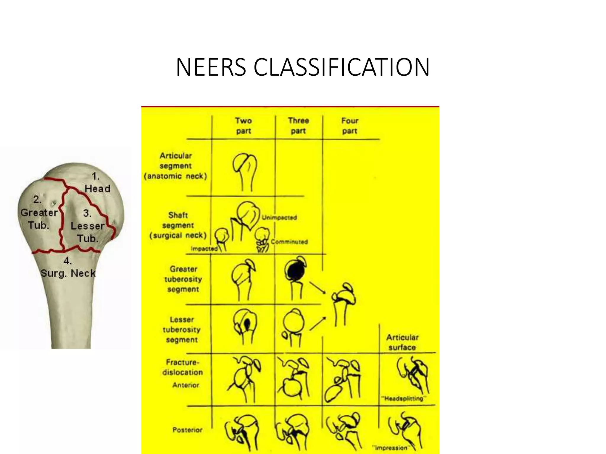
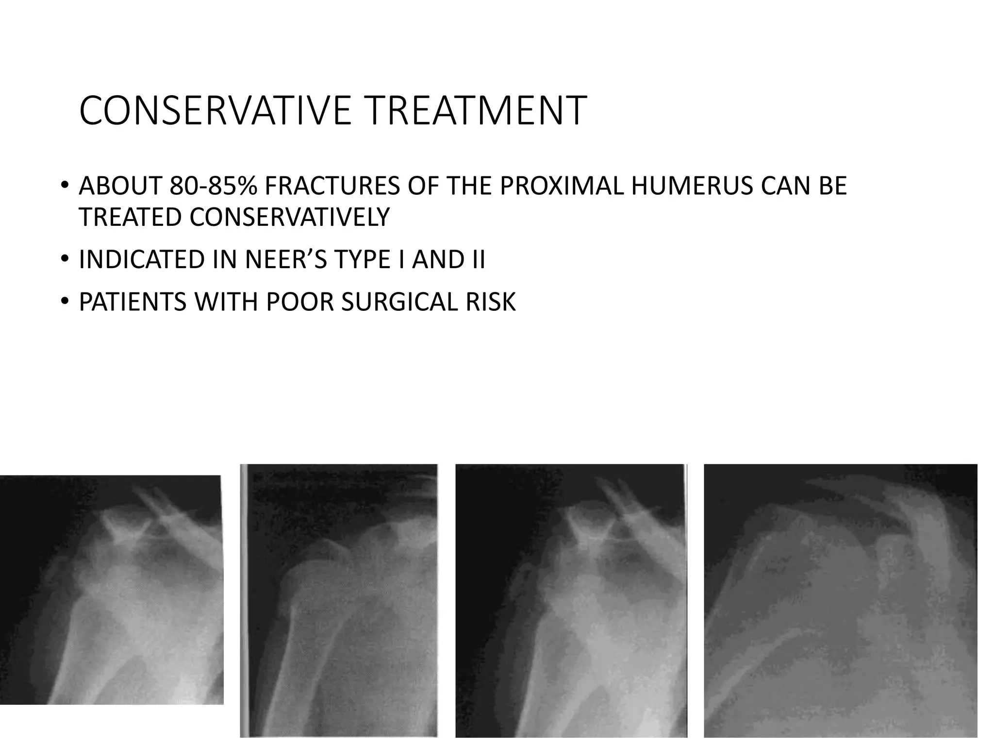
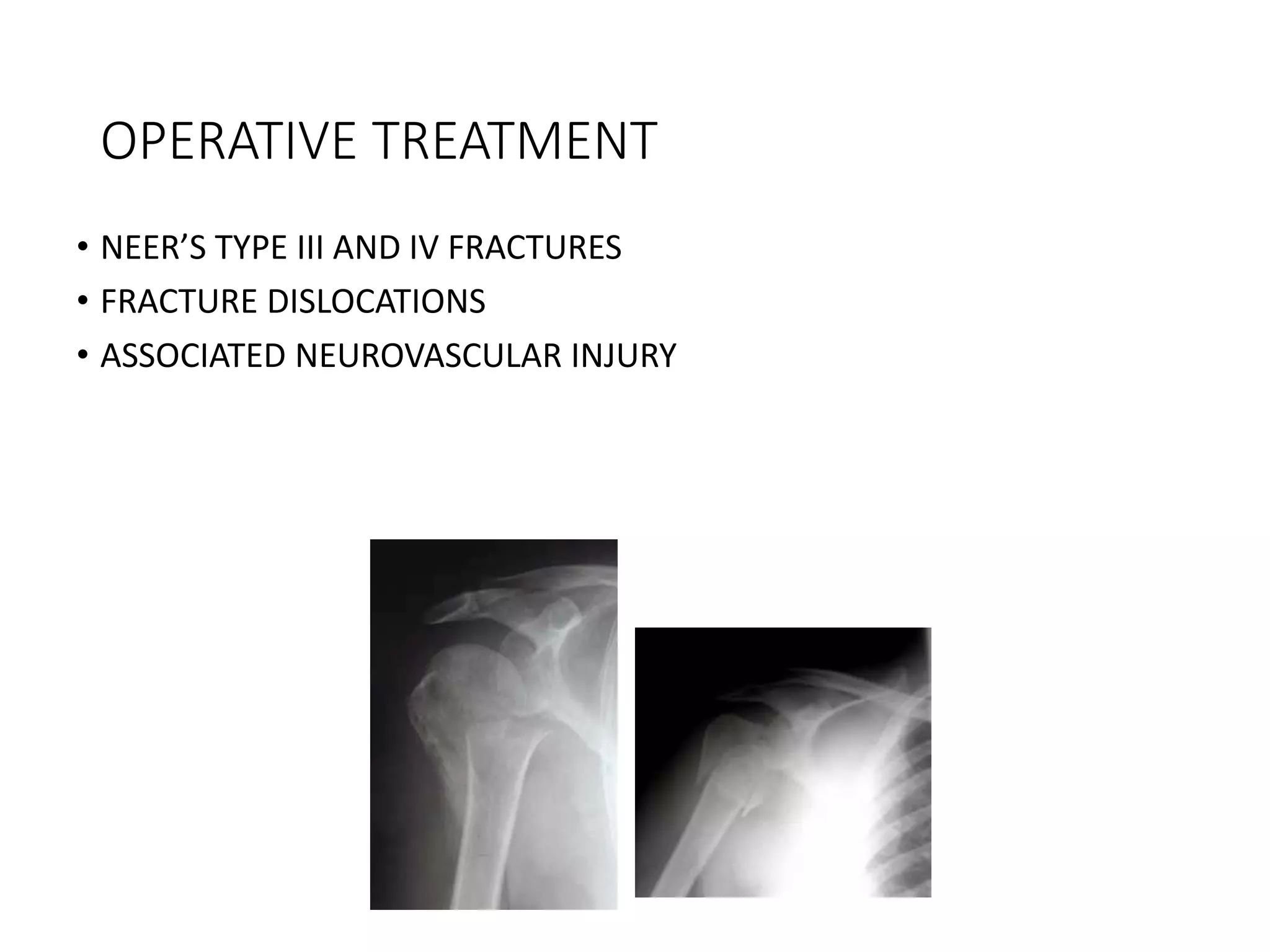
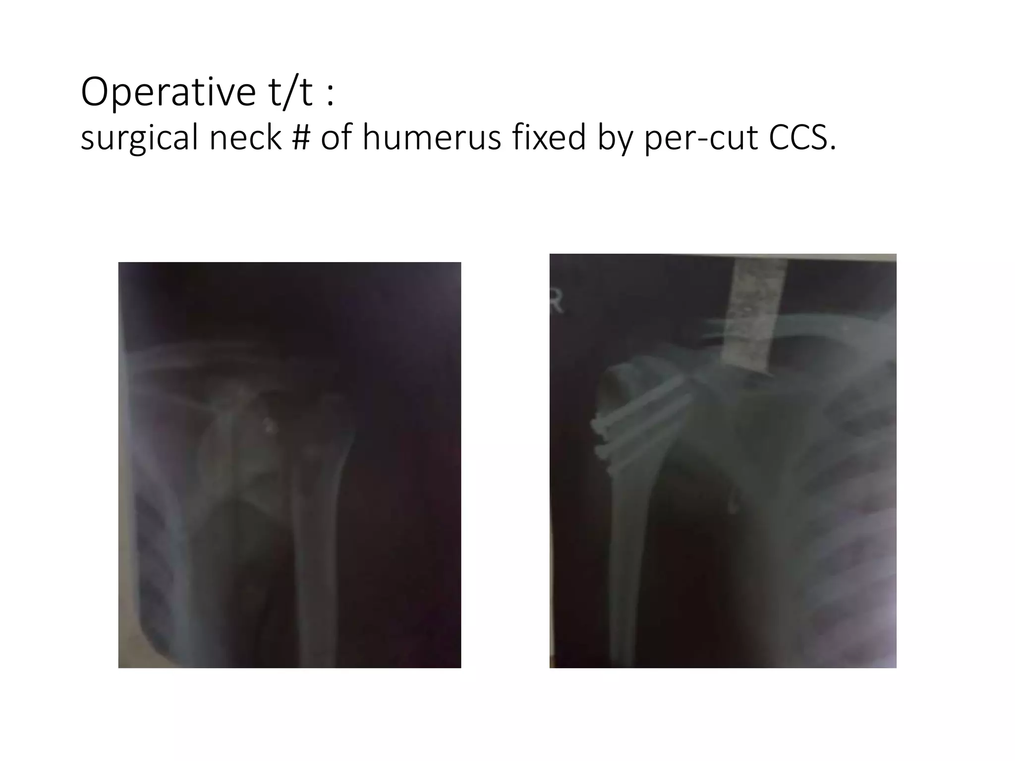
- Common injuries like anterior dislocation, fractures of the clavicle and proximal humerus.
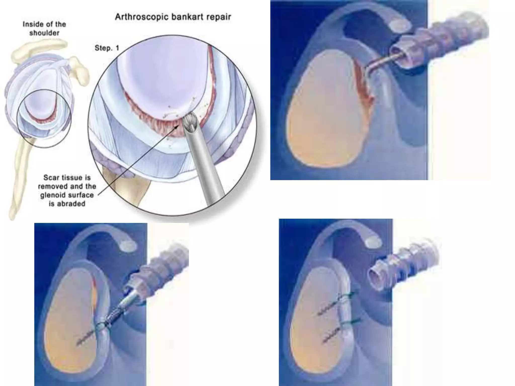
- Treatment approaches for these injuries including closed/open reduction, immobilization, and surgical repair or reconstruction depending on the injury type and chronicity.
Arthroscopic ACL Reconstruction By Dr Shekhar ShrivastavDelhiArthroscopy
Arthroscopic Acl Reconstruction By Dr Shekhar Shrivastav.
HOW NORMAL KNEE WORKS ?
The knee is the largest joint in the body, and one of the most easily injured. It is made up of the lower end of the thigh bone(femur), the upper end of the shin bone (tibia), and the knee cap (patella), which slides in a groove on the end of the femur. Four bands of tissue, the anterior and posterior cruciate ligaments, and the medial and lateral collateral ligaments connect the femur and the tibia and provide joint stability. The surfaces where the femur, tibia and patella touch are covered with articular cartilage, a smooth substance that cushions the bones and enables them to glide freely. Semicircular rings of tough fibrous-cartilage tissue called the lateral and medial menisci act as shock absorbers and stabilizers.
WHAT IS THE ROLE OF ACL ?
ACL along with other ligaments of the knee joint and meniscus provides stability to the knee joint.
WHAT IS LIGAMENT RECONSTRUCTION ( ACL ) ?
Ligament reconstruction involves replacing the torn ligament with a tendon (graft) from your knee and fixing the graft in place with screws. This procedure is performed with the use of the arthroscope. The anterior cruciate ligament (ACL) is the most common ligament requiring reconstruction procedures. The torn ligament is excised arthroscopically and new ligament is prepared by ligament grafts taken from your own body. Bony tunnels are prepared in femur and tibia using specialized instruments through which the new ligament is passed and fixed with special screws. This procedure requires relative rest or leave from your work or studies for about 2-3 weeks after which you will be allowed normal day to day activities.
WHEN CAN THE PATIENT BE AMBULATED AFTER SURGERY ?
The patient can walk from the same evening of the surgery. Initially the patient is advised to walk with a brace and a walking cane. Strengthening and range of motion exercises for the knee are started from the next day. The patient is discharged from the hospital 2nd or 3rd day after surgery. The patient can walk without support by 10-14 days depending on muscle strengthening. Slow Jogging and other strenuous activities are permitted after 3 months and the patient can return to active sports only 8-9 months after surgery.
Torn ACL Reconstructed ACL
For Further Queries contact your Orthopedic Surgeon at
+ 91 9971192233
Total Hip replacement for Ankylosing Spondylitis: Planning & Execution Vaibhav Bagaria
Performing Total Hip replacement in Ankylosing Spondylitis requires a well thought of strategy. Preoperative planning, Inventory ordering, positioning, cup and stem orientation all play a role.
Management of Shoulder dislocations and shoulder instability in sports BhaskarBorgohain4
acute shoulder dislocation is one of the most common sports injuries especially in contact sports. recurrent dislocations are quite common after anterior dislocation of shoulder especially in young athletes who are engaged in sports with lots of overhead activities during their games. Bankarts lesion, Hill sachs lesion are common predisposing factors for recurrence. Simple acute first time dislocations may be reduced on the field by a trained person but further referral is must for detail evaluation. recurrent dislocation can be reduced on field too by less trained. complicated dislocations, neurovascular deficits, fracture dislocation are to be referred to hospital immediately. Practical scientific algorithms are presented for their appropriate management here.
28,000 ankle sprains occur daily in the US (Kaminski 2013)
Ankle is the 2nd most commonly injured body site. (Ferran 2006)
Ankle sprains are the most common type of ankle injury. (Ferran 2006)
A sprained ankle can happen to athletes and non-athletes,
children and adults.
Inversion injury most common mechanism (Ferran 2006)
Only risk factor is previous ankle sprain (Ferran 2006)
Sex , generalized joint laxity or anatomical foot types are
not risk factors. (Beynnon et al. 2002 )
Basics of patellofemoral instability for postgraduates. Gives brief introduction about patellofemoral joint anatomy, causes, examintaion and treatment for patellofemoral instability
give complete information of shoulder dislocation ,types,risks fracture, different management methods and recent advanced .also include management merthod of posterior and inferior stability
Elbow joint is a complex multiarticular joint. Its stability is provided by multiple factors , however unstable elbow is not uncommon .
"Types of elbow instability, how to suspect , diagnose and how to treat" .
All these will be discussed at the lecture which will be presented by Dr. Ahmed Saleh (assistant Lecturer at Mansoura University Hospitals.
Rasamanikya is a excellent preparation in the field of Rasashastra, it is used in various Kushtha Roga, Shwasa, Vicharchika, Bhagandara, Vatarakta, and Phiranga Roga. In this article Preparation& Comparative analytical profile for both Formulationon i.e Rasamanikya prepared by Kushmanda swarasa & Churnodhaka Shodita Haratala. The study aims to provide insights into the comparative efficacy and analytical aspects of these formulations for enhanced therapeutic outcomes.
Integrating Ayurveda into Parkinson’s Management: A Holistic ApproachAyurveda ForAll
Explore the benefits of combining Ayurveda with conventional Parkinson's treatments. Learn how a holistic approach can manage symptoms, enhance well-being, and balance body energies. Discover the steps to safely integrate Ayurvedic practices into your Parkinson’s care plan, including expert guidance on diet, herbal remedies, and lifestyle modifications.
Adv. biopharm. APPLICATION OF PHARMACOKINETICS : TARGETED DRUG DELIVERY SYSTEMSAkankshaAshtankar
MIP 201T & MPH 202T
ADVANCED BIOPHARMACEUTICS & PHARMACOKINETICS : UNIT 5
APPLICATION OF PHARMACOKINETICS : TARGETED DRUG DELIVERY SYSTEMS By - AKANKSHA ASHTANKAR
Knee anatomy and clinical tests 2024.pdfvimalpl1234
This includes all relevant anatomy and clinical tests compiled from standard textbooks, Campbell,netter etc..It is comprehensive and best suited for orthopaedicians and orthopaedic residents.
Here is the updated list of Top Best Ayurvedic medicine for Gas and Indigestion and those are Gas-O-Go Syp for Dyspepsia | Lavizyme Syrup for Acidity | Yumzyme Hepatoprotective Capsules etc
- Video recording of this lecture in English language: https://youtu.be/kqbnxVAZs-0
- Video recording of this lecture in Arabic language: https://youtu.be/SINlygW1Mpc
- Link to download the book free: https://nephrotube.blogspot.com/p/nephrotube-nephrology-books.html
- Link to NephroTube website: www.NephroTube.com
- Link to NephroTube social media accounts: https://nephrotube.blogspot.com/p/join-nephrotube-on-social-media.html
Muktapishti is a traditional Ayurvedic preparation made from Shoditha Mukta (Purified Pearl), is believed to help regulate thyroid function and reduce symptoms of hyperthyroidism due to its cooling and balancing properties. Clinical evidence on its efficacy remains limited, necessitating further research to validate its therapeutic benefits.
ABDOMINAL TRAUMA in pediatrics part one.drhasanrajab
Abdominal trauma in pediatrics refers to injuries or damage to the abdominal organs in children. It can occur due to various causes such as falls, motor vehicle accidents, sports-related injuries, and physical abuse. Children are more vulnerable to abdominal trauma due to their unique anatomical and physiological characteristics. Signs and symptoms include abdominal pain, tenderness, distension, vomiting, and signs of shock. Diagnosis involves physical examination, imaging studies, and laboratory tests. Management depends on the severity and may involve conservative treatment or surgical intervention. Prevention is crucial in reducing the incidence of abdominal trauma in children.
Title: Sense of Smell
Presenter: Dr. Faiza, Assistant Professor of Physiology
Qualifications:
MBBS (Best Graduate, AIMC Lahore)
FCPS Physiology
ICMT, CHPE, DHPE (STMU)
MPH (GC University, Faisalabad)
MBA (Virtual University of Pakistan)
Learning Objectives:
Describe the primary categories of smells and the concept of odor blindness.
Explain the structure and location of the olfactory membrane and mucosa, including the types and roles of cells involved in olfaction.
Describe the pathway and mechanisms of olfactory signal transmission from the olfactory receptors to the brain.
Illustrate the biochemical cascade triggered by odorant binding to olfactory receptors, including the role of G-proteins and second messengers in generating an action potential.
Identify different types of olfactory disorders such as anosmia, hyposmia, hyperosmia, and dysosmia, including their potential causes.
Key Topics:
Olfactory Genes:
3% of the human genome accounts for olfactory genes.
400 genes for odorant receptors.
Olfactory Membrane:
Located in the superior part of the nasal cavity.
Medially: Folds downward along the superior septum.
Laterally: Folds over the superior turbinate and upper surface of the middle turbinate.
Total surface area: 5-10 square centimeters.
Olfactory Mucosa:
Olfactory Cells: Bipolar nerve cells derived from the CNS (100 million), with 4-25 olfactory cilia per cell.
Sustentacular Cells: Produce mucus and maintain ionic and molecular environment.
Basal Cells: Replace worn-out olfactory cells with an average lifespan of 1-2 months.
Bowman’s Gland: Secretes mucus.
Stimulation of Olfactory Cells:
Odorant dissolves in mucus and attaches to receptors on olfactory cilia.
Involves a cascade effect through G-proteins and second messengers, leading to depolarization and action potential generation in the olfactory nerve.
Quality of a Good Odorant:
Small (3-20 Carbon atoms), volatile, water-soluble, and lipid-soluble.
Facilitated by odorant-binding proteins in mucus.
Membrane Potential and Action Potential:
Resting membrane potential: -55mV.
Action potential frequency in the olfactory nerve increases with odorant strength.
Adaptation Towards the Sense of Smell:
Rapid adaptation within the first second, with further slow adaptation.
Psychological adaptation greater than receptor adaptation, involving feedback inhibition from the central nervous system.
Primary Sensations of Smell:
Camphoraceous, Musky, Floral, Pepperminty, Ethereal, Pungent, Putrid.
Odor Detection Threshold:
Examples: Hydrogen sulfide (0.0005 ppm), Methyl-mercaptan (0.002 ppm).
Some toxic substances are odorless at lethal concentrations.
Characteristics of Smell:
Odor blindness for single substances due to lack of appropriate receptor protein.
Behavioral and emotional influences of smell.
Transmission of Olfactory Signals:
From olfactory cells to glomeruli in the olfactory bulb, involving lateral inhibition.
Primitive, less old, and new olfactory systems with different path
Local Advanced Lung Cancer: Artificial Intelligence, Synergetics, Complex Sys...Oleg Kshivets
Overall life span (LS) was 1671.7±1721.6 days and cumulative 5YS reached 62.4%, 10 years – 50.4%, 20 years – 44.6%. 94 LCP lived more than 5 years without cancer (LS=2958.6±1723.6 days), 22 – more than 10 years (LS=5571±1841.8 days). 67 LCP died because of LC (LS=471.9±344 days). AT significantly improved 5YS (68% vs. 53.7%) (P=0.028 by log-rank test). Cox modeling displayed that 5YS of LCP significantly depended on: N0-N12, T3-4, blood cell circuit, cell ratio factors (ratio between cancer cells-CC and blood cells subpopulations), LC cell dynamics, recalcification time, heparin tolerance, prothrombin index, protein, AT, procedure type (P=0.000-0.031). Neural networks, genetic algorithm selection and bootstrap simulation revealed relationships between 5YS and N0-12 (rank=1), thrombocytes/CC (rank=2), segmented neutrophils/CC (3), eosinophils/CC (4), erythrocytes/CC (5), healthy cells/CC (6), lymphocytes/CC (7), stick neutrophils/CC (8), leucocytes/CC (9), monocytes/CC (10). Correct prediction of 5YS was 100% by neural networks computing (error=0.000; area under ROC curve=1.0).
4. Shoulder
⇒ Glenohumeral (GH)
⇒ Scapulothoracic (ST)
⇒ Acromioclavicular joint (ACJ)
⇒ Sternoclavicular joint (SCJ)
• Elevation of arm 30° (2:1) = 20° GH + 10° ST
• ST movements elevate arm to 65° in absence of GH
movements - “Shrugging mechanism”.
5. Shoulder Anatomy
• Most mobile joint of body
• Glenoid to humeral head ratio is 4:1
• Make shoulder most mobile but relatively unstable joint
• Stability is almost totally dependent upon the synergism of the
musculotendinous units
6. • The muscles and a lack of
restrictive bony or
ligamentous structure give
the shoulder tremendous
range of motion.
• It also makes the
shoulder very
vulnerable to outside
forces
7. Anterior Stabilizers
•Subscapularis Tendon
•Anterior Labrum
•Anterior Capsular Ligaments
•Coraco Humeral, GH, Inferior GH Ligament
•Inferior may be the most important ligament in the
shoulder
•Anterior Synovial pouches and bursae
16. STATIC STABILIZERS
Normal anatomical structures which are constraints to humeral head translation
like ligaments.
DYANAMIC STABILIZERS
Structures whose normal physiological action creates a stabilizing effect like
muscles
17. DISLOCATION OF THE SHOULDER
JOINT
• ANTERIOR
• POSTERIOR
• INFERIOR (LUXATIO ERECTAE)
18. MECHANISM OF INJURY AND CLINICAL
FEATURES OF ANTERIOR
GLENOHUMERAL DISLOCATIONS
• Caused by fall on an outstretched hand
Clinically characterized by
• Shoulder in abduction and external rotation
• Positive Dugas test
• Positive Hamilton ruler test
• Positive Callaway’s test
28. NON-OPERATIVE TREATMENT PROTOCOL
• All patients< 30 yrs shoulder immobilized for 3 wks
• Patients 30-40yrs shoulder immobilized for 1-2 wks
• Patients >40 yrs the shoulder immobilized for 1 wks
29. Recurrent anterior Shoulder Dislocation
Problem Assessment
• 50% of all major joint dislocation (Anterior-97%)
• Extreme Rotation can make shoulder to pop out
• High chance of recurrence. (14-100%)
• If left untreated can lead to recurrence further leading to
Osteoarthritis
30. Recurrence Rate after First time Anterior
Shoulder Dislocation
0
10
20
30
40
50
60
70
80
90
18 19 20 21 22 23 24 25 26 27 28 29 30 31 32 33 34 >35
Recurrence
Rate
Age In Years
Male Female
Robinson CM, Howes J, Murdoch H, Will E, Graham C. Functional outcome and
risk of recurrent instability after primary traumatic anterior shoulder dislocation in
young patients. J Bone Joint Surg Am.2006;88:2326-2336
31. COMPLICATIONS
• Neurovascular damage
• Recurrence
Age is the single most important factor for developing recurrent
instability.
• Patient <20 yrs.recurrence is > 85%
• Patient > 40 yrs. recurrence is < 6%
35. FOR RECURRENT ANTERIOR INSTABILITY
• Medially displaced labrum mobilized and reattached to glenoid rim with suture
anchors- ARTHROSCOPIC BANKART PROCEDURE
36.
37. Laterjet Procedure
• Coracoid Transfer procedure
• Most commonly performed surgery for glenoid bone loss
• Increases Glenoid Track
• Dynamic transfer due to conjoint tendon sling
• Acromioclavicular ligament can be used to repair capsulolabral
complex
41. MECHANISM OF INJURY AND SITE OF
FRACTURE
• DIRECT IMPACT
• INDIRECT IMPACT : Fall on an utstretched hand
SITE OF FRACTURE
• 85% occur at junction of middle and outer thirds
• Lateral and medial end fractures uncommon
47. CONSERVATIVE TREATMENT
• ABOUT 80-85% FRACTURES OF THE PROXIMAL HUMERUS CAN BE
TREATED CONSERVATIVELY
• INDICATED IN NEER’S TYPE I AND II
• PATIENTS WITH POOR SURGICAL RISK
48. OPERATIVE TREATMENT
• NEER’S TYPE III AND IV FRACTURES
• FRACTURE DISLOCATIONS
• ASSOCIATED NEUROVASCULAR INJURY
Inherently unstable Joint- Shallow glenoid that Articulated with a part of humeral head
Trauma, Contact Sports, Motor Vehicle Accidents being common causes
Age at the time of initial dislocation –Younger patient more likely to have recurrence
Limited evidence supports surgery in first time dislocators esp young, male and involved in contact sport or high demand activities