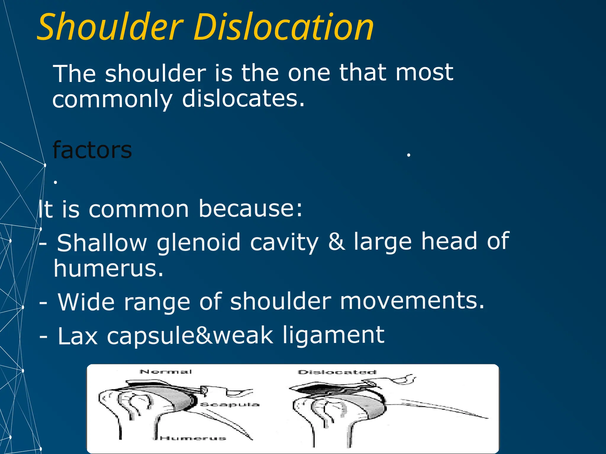
Shoulder joint
dislocation
Done by:
mohammed alqadi
Supervision by
Dr /Ibtissam Al ariqi
Shoulder joint dislocation
• Breef anatomy about shoulder region
• Incidence
• Defination
• Classification
• Etiology
• Clinical features
• Investigation
• Management
• Complications
Shoulder joint anatomy
• The shoulder region is mainly composed of :
Three bony structures:
1- The Clavicle
2- The Humerus
3- The Scapula
Muscles
The rotator cuff
is a set of four
muscles that
motor the
shoulder
joint. These
muscles are: -
Subscapularis,
Supraspinatus,
Infraspinatus,
and Teres
Minor.
Bones and Ligaments
Glenohumeral ligament
Coracohumeral
ligament
Coracoacromial ligament
Coracoclavicular ligament
Ligamen
ts
Shoulder joint stabilization
• Connective tissue
• glenoid labrum: cartilaginous ring, surrounds
glenoid fossa
• increases contact area between head of
humerus and glenoid fossa.
• increases joint stability
Shoulder Dislocation
The shoulder is the one that most
commonly dislocates.
factors .
.
lt is common because:
- Shallow glenoid cavity & large head of
humerus.
- Wide range of shoulder movements.
- Lax capsule&weak ligament
Cont:..
Dislocation : is displacement of a bone from a
joint.
Shoulder dislocation: is displacement of the
head of the humerus from the glenoid cavity of
the scapula at the gleno humeral joint .
1. Shoulder dislocation classification:
Anterior dislocation.
Posterior dislocation
Inferior dislocation
• 1-traumatic
• 2-non-traumatic:
• a-congenital malformation of the
articular surface.
• b- hyperflexibility of the joint due to
laxity of the connective tissue.
• C_Neuromuscular disorder
Causes
1- Anterior dislocation
Dislocation forword
Mechanism of injury:
caused by a fall on the hand.
The head of the humerus is driven forward, tearing
the capsule and producing avulsion of the glenoid
labrum (Bankart lesion). then the arm drops,bringing
the head of humerus to its subcoracoid position.
Anterior dislocation subdivisions:
Sub coracoid Sub glenoid Sub clavicular
Clinical features
Symptoms
. History of trauma.
(see above)
the humerus is "
. Pain.
. Swelling.
. lnability to move the
affected limb (all
shoulder movements
are limited and painful).
Signs
• .
1 lnspection:
• .
- Swelling
• - The arm appears to take origin from a point just
• under junction of middle and outer thirds of the
• .
clavicle
• - Deformity:
• o The patient holds the injured limb at the etbow by the
• .
other hand in position of slight abduction
• .
o Loss of shoulder contour (flattening)
• o Loss of axillary concavity
• 2.Palpation:
• - Head is palpable anteriorly (ln the
subcoracoid or subclavicular region)
• - Empty space under the acromion
(empty glenoid cavity).
• 3. Movemen!. complete limitation of
shoulder movements.
• - Axillary nerve ) Deltoid muscle
wasting & sensory loss over its lateral
aspect of arm.
• - Axillary artery )
X-Ray
AP-Xray will show the
humeral head is
displaced anteriorly and
medially .
lateral view
Will show the humeral
head out of line with the
socket.
Anterior dislocation treatment
Is an emergency we must examine the pulse and
nerve
It should be reduced in less than 24 hours .Strong
analgesics are needed to relive the pain of a
dislocation and the anxiety associated with it.
●Some reduction methods need anasthesia
apply a sling arm for3-4day .
●A fracture dislocation will probably require
surgery.
Without a fracture … closed reduction is usually
adequate
active exercises are start as soon as possible to
prevent stiffness of joint.
Treatement.(cont)….
Various of methods of reduction:
1)Stimon's technique.
2)Hippocratic method.
3)Kocher's method.
Stimon's technique.
The patient is left prone with the arm
hanging over the side of the bed. After
15 or 20 minutes the shoulder may
reduce.
Hippocratic methods
• Traction is applied to the arm with
the shoulder is slight abduction while
an assistant applied firm counter-
tracthion to the body.
The heel of the foot is placed against the
humeral head in the axilla.
And longitudinal traction is applied to the arm.
Kocher's method
The elbow is bent to 90 and hold close to
the body. No traction should be applied .
The arm is slowly rotated 75 laterally.The
point of the elbow is lefted forward and
the arm is rotated medially.
Treatement cont..
When the patient is fully awake, active
abduction is gently tested to exclude an
axillary nerve injury and rotator cuff tear. The
median, radial, ulnar and musculocutaneous
nerves are also teste and the pulse is felt.
The arm is rested in a sling for about three
weeks in those under 30 years of age (who
are most prone to recurrence) and for only a
week in those over 30 (who are most prone
to stiffness). Then movements are begun, but
combined abduction and lateral rotation must
be avoided for at least 3 weeks. Throughout
this period, elbow and finger movements are
practised every day.
Complication
Early……Rotator cuff tear, nerve injury,
vascular injury, fracture dislocation.
Late……..Shoulder stiffness, unreduced
dislocation, recurrent dislocation.
Posterior dislocation of the
shoulder:
less than 2 per cent of all dislocations
around the shoulder.
Mechanism of injury
Indirect force
producing marked internal rotation and
adduction(during a fit or convulsion or
electric shock)
Direct….A fall on to the fixed adductor
arm
A fall on the outstretched
hand.
Clinical picture:
Usually missed . several well-marked clinical
features. arm is held in internal rotation
and is locked in that the position. The front
of the shoulder looks flat with a prominent
coracoid, but swelling may obscure this
deformity; seen from above, however, the
posterior displacement is usually
apparent .
X-ray of Posterior shoulder dislocation
AP_x-ray
shows a head-on projection giving the
classic ‘electric light-bulb’ appearance.
The head of the humerus looks
abnormal in shape and away from
glenoid fossa.
Treatment of posterior dislocation
(
The acute dislocation is reduced usually
)
under general anaesthesia
By pulling on the arm with the shoulder in
.
adduction a few minutes are allowed for the
head of the humerus to disengage and the
arm is rotated laterally while the humeral
.
head is pushed forward if reduction feels
,
stable the arm is immobilized in sling
,
otherwise the shoulder is held widely
abducted and laterally roated in airplane
-
type splint for 3 6week to allow capsule to
heal in shortest postion .
:
Complication
Unreduced dislocation
Recurrent dislocation subluxation.
Inferior shoulder dislocation
(luxatio erecta)
Mechanism of injury :
Caused by sever hyper abduction force .the humeral head
is lifted across inferior rim of glenoid cavity with humeral
shaft pointing upward .
Soft tissue avulsion of capsule , tendon injury &Muscle
tearing
Fracture of the glenoid or (proximal of humerus damage
to brachial plxus & axillary artery .
clinical picture:
Pt arm locked in full abduction . On
examination the humeral head may feel
on or below axilla . We must examine
pulse and nerve
X-ray
The humeral shaft is
shown in the abducted
position with the head
sitting below the glenoid.
Treatement
Reduction by pulling head upward in the
line of abducted arm with counter –
traction downward over the top of the
shoulder.
If the humeral head is stuck in soft tissue
with soft tissue injury we must do open
reduction .
We must examin a vascular and nerve
befor and after reduction
Acromioclavicular joint injuries
Occur either:
1-Subluxation (luxation = dislocation) here the
coracoclaviculer ligament is intact.
2- Dislocation :coracoclaviculer ligament is torn
Mechanism of injury:
A fall on the shoulder with the arm
adducted may strain or tear the
acromioclavicular ligment and upwards
subluxation of the clavicle
Clinicaly
Brusing Tenderness Pain Movement is
limited.
Treatement
subluxation do not require any special
treatement .The arm is rested in a sling 1week
Dislocation:
A well-tried technique is to repair the
coracoclavicular lig.and hold the reducd with
temporary coracoclavicular screw.
Shoulder is rested for 2 weeks and excersie
The screw is removed after 8 weeks
Complication
Rotator cuff syndrome
Unreduced dislocation.
Ossification of the ligaments
Osteoarthritis
THANK YOU

shoulder dislocation by Mohammed alqadi_١٠٤٢١٩.pptx

  • 1.
    Shoulder joint dislocation Done by: mohammedalqadi Supervision by Dr /Ibtissam Al ariqi
  • 2.
    Shoulder joint dislocation •Breef anatomy about shoulder region • Incidence • Defination • Classification • Etiology • Clinical features • Investigation • Management • Complications
  • 3.
    Shoulder joint anatomy •The shoulder region is mainly composed of : Three bony structures: 1- The Clavicle 2- The Humerus 3- The Scapula
  • 4.
    Muscles The rotator cuff isa set of four muscles that motor the shoulder joint. These muscles are: - Subscapularis, Supraspinatus, Infraspinatus, and Teres Minor.
  • 5.
    Bones and Ligaments Glenohumeralligament Coracohumeral ligament Coracoacromial ligament Coracoclavicular ligament Ligamen ts
  • 6.
    Shoulder joint stabilization •Connective tissue • glenoid labrum: cartilaginous ring, surrounds glenoid fossa • increases contact area between head of humerus and glenoid fossa. • increases joint stability
  • 7.
    Shoulder Dislocation The shoulderis the one that most commonly dislocates. factors . . lt is common because: - Shallow glenoid cavity & large head of humerus. - Wide range of shoulder movements. - Lax capsule&weak ligament
  • 8.
    Cont:.. Dislocation : isdisplacement of a bone from a joint. Shoulder dislocation: is displacement of the head of the humerus from the glenoid cavity of the scapula at the gleno humeral joint . 1. Shoulder dislocation classification: Anterior dislocation. Posterior dislocation Inferior dislocation
  • 9.
    • 1-traumatic • 2-non-traumatic: •a-congenital malformation of the articular surface. • b- hyperflexibility of the joint due to laxity of the connective tissue. • C_Neuromuscular disorder Causes
  • 10.
    1- Anterior dislocation Dislocationforword Mechanism of injury: caused by a fall on the hand. The head of the humerus is driven forward, tearing the capsule and producing avulsion of the glenoid labrum (Bankart lesion). then the arm drops,bringing the head of humerus to its subcoracoid position. Anterior dislocation subdivisions: Sub coracoid Sub glenoid Sub clavicular
  • 11.
    Clinical features Symptoms . Historyof trauma. (see above) the humerus is " . Pain. . Swelling. . lnability to move the affected limb (all shoulder movements are limited and painful).
  • 12.
    Signs • . 1 lnspection: •. - Swelling • - The arm appears to take origin from a point just • under junction of middle and outer thirds of the • . clavicle • - Deformity: • o The patient holds the injured limb at the etbow by the • . other hand in position of slight abduction • . o Loss of shoulder contour (flattening) • o Loss of axillary concavity
  • 13.
    • 2.Palpation: • -Head is palpable anteriorly (ln the subcoracoid or subclavicular region) • - Empty space under the acromion (empty glenoid cavity). • 3. Movemen!. complete limitation of shoulder movements. • - Axillary nerve ) Deltoid muscle wasting & sensory loss over its lateral aspect of arm. • - Axillary artery )
  • 14.
    X-Ray AP-Xray will showthe humeral head is displaced anteriorly and medially . lateral view Will show the humeral head out of line with the socket.
  • 15.
    Anterior dislocation treatment Isan emergency we must examine the pulse and nerve It should be reduced in less than 24 hours .Strong analgesics are needed to relive the pain of a dislocation and the anxiety associated with it. ●Some reduction methods need anasthesia apply a sling arm for3-4day . ●A fracture dislocation will probably require surgery. Without a fracture … closed reduction is usually adequate active exercises are start as soon as possible to prevent stiffness of joint.
  • 17.
    Treatement.(cont)…. Various of methodsof reduction: 1)Stimon's technique. 2)Hippocratic method. 3)Kocher's method.
  • 18.
    Stimon's technique. The patientis left prone with the arm hanging over the side of the bed. After 15 or 20 minutes the shoulder may reduce.
  • 19.
    Hippocratic methods • Tractionis applied to the arm with the shoulder is slight abduction while an assistant applied firm counter- tracthion to the body.
  • 20.
    The heel ofthe foot is placed against the humeral head in the axilla. And longitudinal traction is applied to the arm.
  • 21.
    Kocher's method The elbowis bent to 90 and hold close to the body. No traction should be applied . The arm is slowly rotated 75 laterally.The point of the elbow is lefted forward and the arm is rotated medially.
  • 24.
    Treatement cont.. When thepatient is fully awake, active abduction is gently tested to exclude an axillary nerve injury and rotator cuff tear. The median, radial, ulnar and musculocutaneous nerves are also teste and the pulse is felt. The arm is rested in a sling for about three weeks in those under 30 years of age (who are most prone to recurrence) and for only a week in those over 30 (who are most prone to stiffness). Then movements are begun, but combined abduction and lateral rotation must be avoided for at least 3 weeks. Throughout this period, elbow and finger movements are practised every day.
  • 25.
    Complication Early……Rotator cuff tear,nerve injury, vascular injury, fracture dislocation. Late……..Shoulder stiffness, unreduced dislocation, recurrent dislocation.
  • 26.
    Posterior dislocation ofthe shoulder: less than 2 per cent of all dislocations around the shoulder. Mechanism of injury Indirect force producing marked internal rotation and adduction(during a fit or convulsion or electric shock) Direct….A fall on to the fixed adductor arm A fall on the outstretched hand.
  • 27.
    Clinical picture: Usually missed. several well-marked clinical features. arm is held in internal rotation and is locked in that the position. The front of the shoulder looks flat with a prominent coracoid, but swelling may obscure this deformity; seen from above, however, the posterior displacement is usually apparent .
  • 28.
    X-ray of Posteriorshoulder dislocation AP_x-ray shows a head-on projection giving the classic ‘electric light-bulb’ appearance. The head of the humerus looks abnormal in shape and away from glenoid fossa.
  • 29.
    Treatment of posteriordislocation ( The acute dislocation is reduced usually ) under general anaesthesia By pulling on the arm with the shoulder in . adduction a few minutes are allowed for the head of the humerus to disengage and the arm is rotated laterally while the humeral . head is pushed forward if reduction feels , stable the arm is immobilized in sling , otherwise the shoulder is held widely abducted and laterally roated in airplane - type splint for 3 6week to allow capsule to heal in shortest postion . : Complication Unreduced dislocation Recurrent dislocation subluxation.
  • 30.
    Inferior shoulder dislocation (luxatioerecta) Mechanism of injury : Caused by sever hyper abduction force .the humeral head is lifted across inferior rim of glenoid cavity with humeral shaft pointing upward . Soft tissue avulsion of capsule , tendon injury &Muscle tearing Fracture of the glenoid or (proximal of humerus damage to brachial plxus & axillary artery . clinical picture: Pt arm locked in full abduction . On examination the humeral head may feel on or below axilla . We must examine pulse and nerve
  • 31.
    X-ray The humeral shaftis shown in the abducted position with the head sitting below the glenoid.
  • 32.
    Treatement Reduction by pullinghead upward in the line of abducted arm with counter – traction downward over the top of the shoulder. If the humeral head is stuck in soft tissue with soft tissue injury we must do open reduction . We must examin a vascular and nerve befor and after reduction
  • 33.
    Acromioclavicular joint injuries Occureither: 1-Subluxation (luxation = dislocation) here the coracoclaviculer ligament is intact. 2- Dislocation :coracoclaviculer ligament is torn Mechanism of injury: A fall on the shoulder with the arm adducted may strain or tear the acromioclavicular ligment and upwards subluxation of the clavicle Clinicaly Brusing Tenderness Pain Movement is limited.
  • 34.
    Treatement subluxation do notrequire any special treatement .The arm is rested in a sling 1week Dislocation: A well-tried technique is to repair the coracoclavicular lig.and hold the reducd with temporary coracoclavicular screw. Shoulder is rested for 2 weeks and excersie The screw is removed after 8 weeks Complication Rotator cuff syndrome Unreduced dislocation. Ossification of the ligaments Osteoarthritis
  • 35.