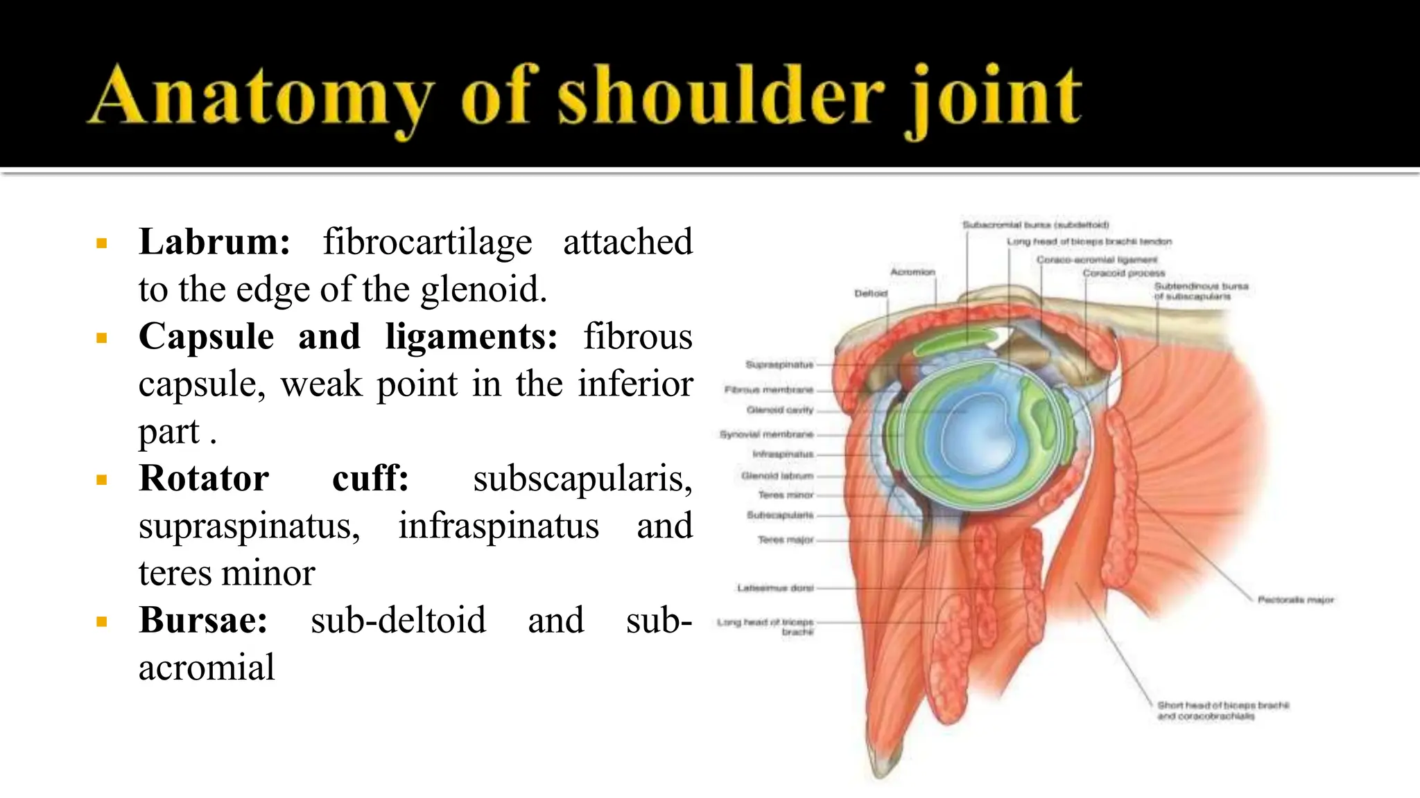
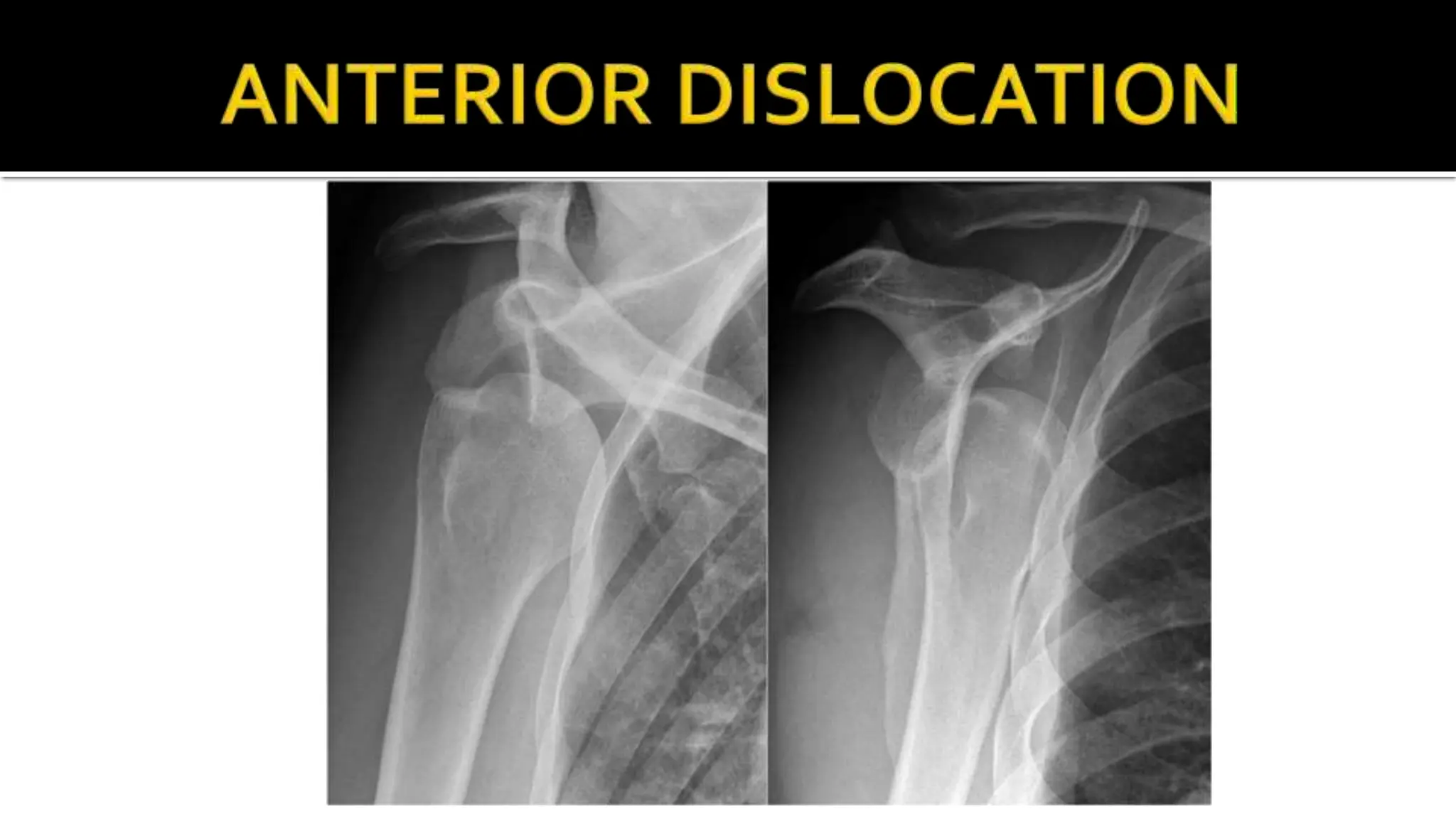
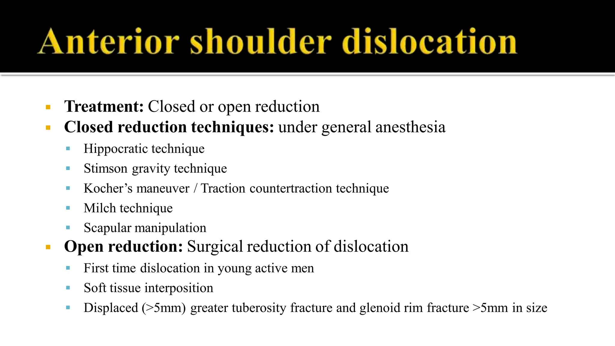
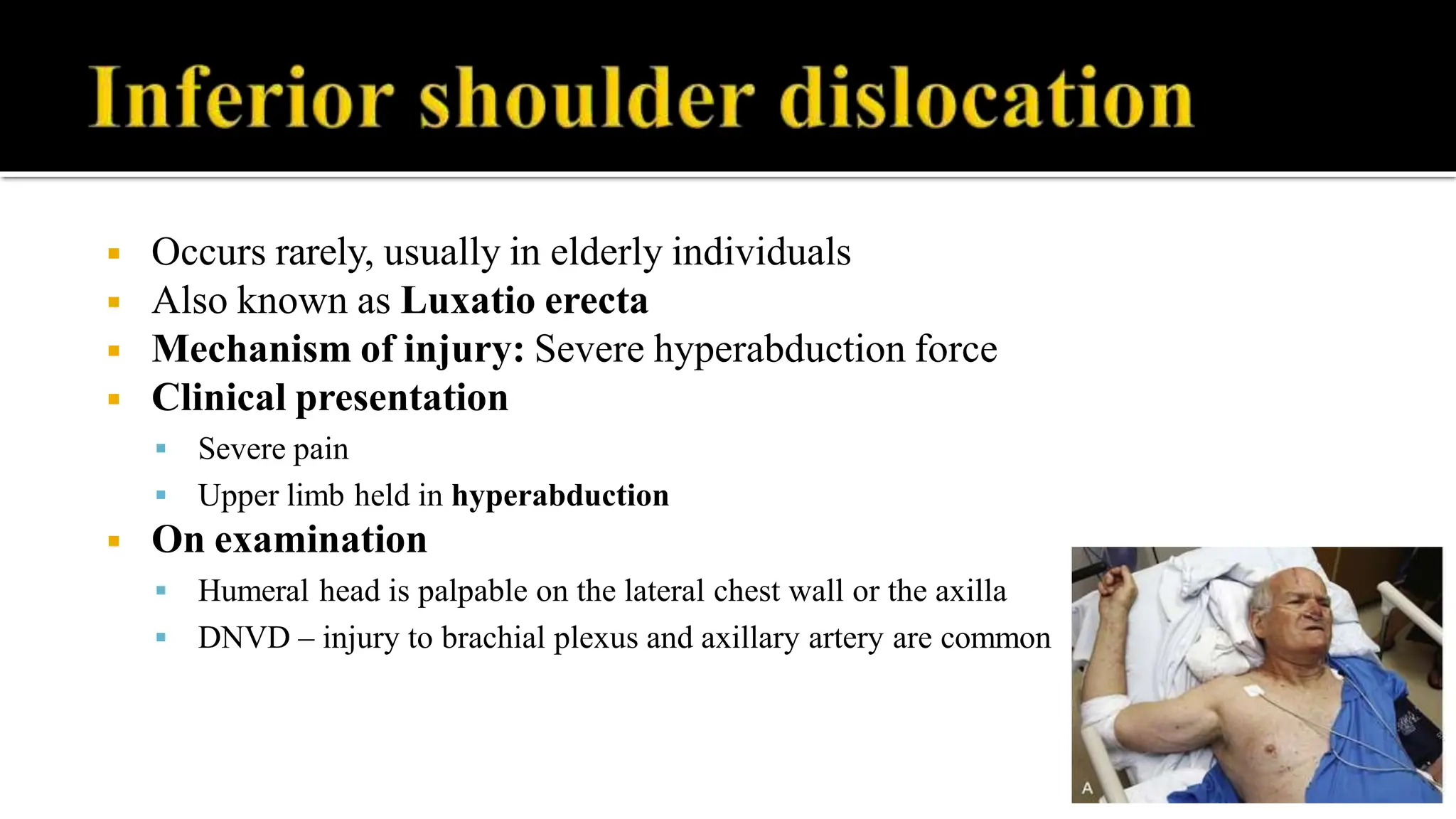
Dr. M.Praveen M.S.ORTHO.,FEIORA.,
Assistant Professor,
Dept. of Orthopaedics,
Srinivasan Medical College and Hospital.
◾ Glenohumeral joint – ball and socket type of synovial joint
◾ Most mobile joint in the body, but compromised in its stability
◾ Glenoid cavity: shallow; anteverted and inferiorly angulated from
the long axis of the scapula
◾ Labrum: fibrocartilage attached
to the edge of the glenoid.
◾ Capsule and ligaments: fibrous
capsule, weak point in the inferior
part .
◾ Rotator cuff: subscapularis,
supraspinatus, infraspinatus and
teres minor
◾ Bursae: sub-deltoid and sub-
acromial
◾ Capsulolabral complex
◾ Rotator cuff
◾ Negative intra-articular pressure
◾ Synovial fluid adhesion-cohesion
◾ Increased range of movements of shoulder
joint
◾ Factors responsible for shoulder dislocation:
 The shallowness of the glenoid socket
 Extraordinary range of movement
 Underlying conditions like ligamentous laxity, glenoid dysplasia
 Vulnerability of the joint during stressful activities of the upper limb
Shoulder dislocation
Anterior dislocation
1. Subcoracoid
2. Subglenoid
3. Infraclavicular
4. Intrathoracic
Posterior dislocation
1. Subacromial
2. Subglenoid
3. Subspinous
Inferior dislocation
Infraglenoid dislocation.
Also known as Luxatio
erecta
◾ Represent
dislocation
96% of shoulder
◾ Mechanism of injury
 Indirect injury: fall on outstretched
hand with shoulder in Abduction, ER
and extension.
 Direct injury: anteriorly directed
force to the posterior shoulder
◾ Clinical presentation
 Severe pain, inability to move the
upper limb
 Limb will be held in abduction and
external rotation
◾ On examination
 Loss of normal contour of shoulder –
flattening / squaring of the shoulder
 Prominent acromion process
 Palpable globular mass anteriorly
 DNVD - Regiment badge sign due to
axillary nerve injury
◾ Special tests:
 Vertical circumference of the axilla – increased
 Hamilton ruler test
 Dugas’test-touch opp shoulder with arm on chest
◾ Pathological lesions around glenohumeral joint
 Bankart’s lesion – avulsion of the labrum off the glenoid rim +/- glenoid rim
fracture (Bony bankart lesion)
 Hill – Sach’s lesion – posterolateral defect caused by glenoid impression on the
humeral head
 Erosion of glenoid rim
 Cause of recurrent dislocation
◾ Investigaions:
 X-ray Anteroposterior and axillary
views - to confirm the diagnosis
 CT scan – to diagnose bony lesions
 MRI – to diagnose ligamentous laxity
and soft tissue injuries
 Arthrography – to evaluate rotator
cuff tears
◾ APView
◾ Treatment: Closed or open reduction
◾ Closed reduction techniques: under general anesthesia
 Hippocratic technique
 Stimson gravity technique
 Kocher’s maneuver / Traction countertraction technique
 Milch technique
 Scapular manipulation
◾ Open reduction: Surgical reduction of dislocation
 First time dislocation in young active men
 Soft tissue interposition
 Displaced (>5mm) greater tuberosity fracture and glenoid rim fracture >5mm in size
Stimson method
Milchmethod
Hippocraticmethod
Kocher method
◾ Post-reduction protocol:
 Repeat X-ray to confirm the reduction
 Immobilize the shoulder using Universal shoulder immobilizer for 3weeks
 Physiotherapy after 3 weeks
◾ Complications:
◾ Early:
 Fractures around the joint – Humeral head, GT, glenoid rim, acromion and coracoid
 Soft tissue injuries – Rotator cuff tears, capsular tears
 Neurovascular injuries – axillary artery and nerve, musculocutaneous nerve; usually occurs in
adults
◾ Late:
 Recurrent dislocation, unreduced dislocation
 Shoulder stiffness due to post-traumatic arthitis
◾ Represent 2% - 4% of shoulder dislocation
◾ Mechanism of injury:
 Direct or Indirect trauma
 Electric shock or convulsive mechanisms
◾ Clinical presentation:
 Pain, restricted movements, arm held in internal rotation and adduction
◾ On examination:
 Palpable mass posterior to the shoulder
 Prominent coracoid process
 Flat shoulder contour, empty glenoid
 DNVD –Axillary nerve
◾ Investigations:
 X-ray shoulder anteroposterior and axillary view
 CT scan – to evaluate any associated fractures
◾ Treatment: Closed or open reduction
◾ Complications:
◾ Early:
 Fractures around the shoulder joint
 Neurovascular injury
◾ Late:
 Unreduced or recurrent dislocation
 Anterior dislocation – usually results due to
overtightening the posterior structures
Light bulb appearance
◾ Complications:
◾ Early:
 Fractures around the shoulder joint
 Neurovascular injury
◾ Late:
 Unreduced or recurrent dislocation
 Anterior dislocation – usually results due to overtightening the posterior structures
◾ Occurs rarely, usually in elderly individuals
◾ Also known as Luxatio erecta
◾ Mechanism of injury: Severe hyperabduction force
◾ Clinical presentation
 Severe pain
 Upper limb held in hyperabduction
◾ On examination
 Humeral head is palpable on the lateral chest wall or the axilla
 DNVD – injury to brachial plexus and axillary artery are common
◾ Investigations:
 X-ray AP view – diagnostic; look for associated fractures
around the shoulder joint
 MRI – soft tissue injuries
◾ Treatment: Closed or open reduction
◾ Complications:
 Rotator cuff avulsions and tear, pectoralis injury
 Proximal humeral fractures
 Neurovascular injury – axillary artery and brachial plexus;
usually recovers after the reduction
◾ Any doubts can be raised now
THANK YOU..

SHOULDER DISLOCATION - Types, Clinical Features

  • 1.
    Dr. M.Praveen M.S.ORTHO.,FEIORA., AssistantProfessor, Dept. of Orthopaedics, Srinivasan Medical College and Hospital.
  • 2.
    ◾ Glenohumeral joint– ball and socket type of synovial joint ◾ Most mobile joint in the body, but compromised in its stability ◾ Glenoid cavity: shallow; anteverted and inferiorly angulated from the long axis of the scapula
  • 4.
    ◾ Labrum: fibrocartilageattached to the edge of the glenoid. ◾ Capsule and ligaments: fibrous capsule, weak point in the inferior part . ◾ Rotator cuff: subscapularis, supraspinatus, infraspinatus and teres minor ◾ Bursae: sub-deltoid and sub- acromial
  • 5.
    ◾ Capsulolabral complex ◾Rotator cuff ◾ Negative intra-articular pressure ◾ Synovial fluid adhesion-cohesion
  • 6.
    ◾ Increased rangeof movements of shoulder joint
  • 7.
    ◾ Factors responsiblefor shoulder dislocation:  The shallowness of the glenoid socket  Extraordinary range of movement  Underlying conditions like ligamentous laxity, glenoid dysplasia  Vulnerability of the joint during stressful activities of the upper limb
  • 8.
    Shoulder dislocation Anterior dislocation 1.Subcoracoid 2. Subglenoid 3. Infraclavicular 4. Intrathoracic Posterior dislocation 1. Subacromial 2. Subglenoid 3. Subspinous Inferior dislocation Infraglenoid dislocation. Also known as Luxatio erecta
  • 9.
    ◾ Represent dislocation 96% ofshoulder ◾ Mechanism of injury  Indirect injury: fall on outstretched hand with shoulder in Abduction, ER and extension.  Direct injury: anteriorly directed force to the posterior shoulder
  • 10.
    ◾ Clinical presentation Severe pain, inability to move the upper limb  Limb will be held in abduction and external rotation ◾ On examination  Loss of normal contour of shoulder – flattening / squaring of the shoulder  Prominent acromion process  Palpable globular mass anteriorly  DNVD - Regiment badge sign due to axillary nerve injury
  • 11.
    ◾ Special tests: Vertical circumference of the axilla – increased  Hamilton ruler test  Dugas’test-touch opp shoulder with arm on chest
  • 12.
    ◾ Pathological lesionsaround glenohumeral joint  Bankart’s lesion – avulsion of the labrum off the glenoid rim +/- glenoid rim fracture (Bony bankart lesion)  Hill – Sach’s lesion – posterolateral defect caused by glenoid impression on the humeral head  Erosion of glenoid rim  Cause of recurrent dislocation
  • 14.
    ◾ Investigaions:  X-rayAnteroposterior and axillary views - to confirm the diagnosis  CT scan – to diagnose bony lesions  MRI – to diagnose ligamentous laxity and soft tissue injuries  Arthrography – to evaluate rotator cuff tears
  • 15.
  • 17.
    ◾ Treatment: Closedor open reduction ◾ Closed reduction techniques: under general anesthesia  Hippocratic technique  Stimson gravity technique  Kocher’s maneuver / Traction countertraction technique  Milch technique  Scapular manipulation ◾ Open reduction: Surgical reduction of dislocation  First time dislocation in young active men  Soft tissue interposition  Displaced (>5mm) greater tuberosity fracture and glenoid rim fracture >5mm in size
  • 18.
  • 19.
  • 20.
  • 21.
  • 22.
    ◾ Post-reduction protocol: Repeat X-ray to confirm the reduction  Immobilize the shoulder using Universal shoulder immobilizer for 3weeks  Physiotherapy after 3 weeks ◾ Complications: ◾ Early:  Fractures around the joint – Humeral head, GT, glenoid rim, acromion and coracoid  Soft tissue injuries – Rotator cuff tears, capsular tears  Neurovascular injuries – axillary artery and nerve, musculocutaneous nerve; usually occurs in adults ◾ Late:  Recurrent dislocation, unreduced dislocation  Shoulder stiffness due to post-traumatic arthitis
  • 23.
    ◾ Represent 2%- 4% of shoulder dislocation ◾ Mechanism of injury:  Direct or Indirect trauma  Electric shock or convulsive mechanisms ◾ Clinical presentation:  Pain, restricted movements, arm held in internal rotation and adduction ◾ On examination:  Palpable mass posterior to the shoulder  Prominent coracoid process  Flat shoulder contour, empty glenoid  DNVD –Axillary nerve
  • 24.
    ◾ Investigations:  X-rayshoulder anteroposterior and axillary view  CT scan – to evaluate any associated fractures ◾ Treatment: Closed or open reduction ◾ Complications: ◾ Early:  Fractures around the shoulder joint  Neurovascular injury ◾ Late:  Unreduced or recurrent dislocation  Anterior dislocation – usually results due to overtightening the posterior structures Light bulb appearance
  • 25.
    ◾ Complications: ◾ Early: Fractures around the shoulder joint  Neurovascular injury ◾ Late:  Unreduced or recurrent dislocation  Anterior dislocation – usually results due to overtightening the posterior structures
  • 26.
    ◾ Occurs rarely,usually in elderly individuals ◾ Also known as Luxatio erecta ◾ Mechanism of injury: Severe hyperabduction force ◾ Clinical presentation  Severe pain  Upper limb held in hyperabduction ◾ On examination  Humeral head is palpable on the lateral chest wall or the axilla  DNVD – injury to brachial plexus and axillary artery are common
  • 27.
    ◾ Investigations:  X-rayAP view – diagnostic; look for associated fractures around the shoulder joint  MRI – soft tissue injuries ◾ Treatment: Closed or open reduction ◾ Complications:  Rotator cuff avulsions and tear, pectoralis injury  Proximal humeral fractures  Neurovascular injury – axillary artery and brachial plexus; usually recovers after the reduction
  • 28.
    ◾ Any doubtscan be raised now THANK YOU..