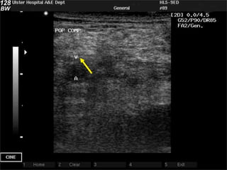
DVT ultrasound
           SAH & RNSH 2011
    Critical Care Ultrasound Course
Thanks to Cindy Lucas… and yet again to
             Daniel Lichtenstein


                                      1
DVT
!   Anatomy
!   Below knee DVT?
!   Compression US:
    !   Technique
    !   Sites
!   What the scan can tell you
!   Pitfalls
Anatomy of the deep veins
     Below the knee           Above the knee
!Anterior tibial       !   Popliteal Vein in popliteal
                           fossa
!Posterior tibial
                       !   From confluence of 3 calf
!Peroneal                  veins
!Gastrocnemial         !   To adductor canal
!Soleal
                       !   (Superficial) Femoral
                       !   Profunda Femoris joins
                           4cm below inguinal
!Variable                  ligament
!Paired                !   Common Femoral
!Tricky!               !   Long/ great saphenous
!Relevance   of DVT?
Here’s the problem
  It would be nice to scan            But it’s hard!
    the below knee veins     !   Variable
!Incr sensitivity            !   Paired
!Incr accuracy               !   Tiny
                             !   Tricky
!Variable                    !   And most of them don’t
                                 ermbolize
!Paired
                             !   (But some do…)
!Tricky!
!Relevance   of DVT?
Previous top tip: just
look for above knee
    Leave the calves to the
        sonographers!


                              6
But Lichtenstein came up with a solution

!   Except for the anterior tibials, the below
    knee veins travel all in a line, a couple cm
    below the interosseous membrane
!   together with their arteries: 2 veins for
    each artery = 6 vessels, all lined up
!   We can see them from the front of the leg!
!   Probe between the tibia & fibula
Now we have 2
        options
1.   Just above knee: leave the
     calves to the sonographers!
2.       Below knee (anterior
              approach)
                                   8
DVT scan:
Technique


            9
Probe & preset?
!   Ideally linear probe / vein preset
!   But curved probe / FAST preset works
    too
!   Don’t need Doppler
Compression US
!   Probe in transverse position
!   Just squash the vein!
!   If it squashes easily & completely, there
    is no DVT
!   If it doesn’t, there’s a DVT
Normal veins
!        Completely compressible
!   Press hard enough to just indent the
                  artery
Features of DVT
!   Gold standard sign: vein not completely
    compressible
!   You might see thrombus
!   Vein might fail to augment on Doppler
Test


       1
       6
DVT?
DVT?
DVT?
DVT?
DVT?
DVT?
DVT?
Compression US
!   Pros                        Cons
!   Easy            !   Misses external iliac
                        DVT
!   Rapid
                    !   Misses small non-
!   B mode only
                        occlusive DVT
!   98% sensitive
Which sites can I compress?


                        2
                        3
Which sites can I
            compress?
!   Internal Jugular V
!   Subclavian V
!   IVC
!   Saphenofemoral confluence (up fem)
!   Lower (superf) femoral near adductor hiatus
!   Long saphenous V
!   Short saphenous V
!   Popliteal vein & trifurcation
!   Beloe knee veins
Which sites should I
              compress?
!   Up to you
!   The more veins you scan, the more sensitive
    you are… eg UL veins add 4% in PE
!   The fewer you scan, the less irritating it is
!   3-point scan is reasonable
    1.   Upper femoral (confluence)
    2.   Lower femoral (near adductor hiatus)
    3.   Popliteal (irritating if supine) …or …below knee
         (weird at first)
1: Groin
!   Probe in transverse position
!   Start just below inguinal ligament
!   ‘Mickey Mouse’ sign
    ! Femoral A
    ! Saphenofemoral confluence

    ! Then compress
2: insertion of LSV / GSV
3: fem V just above knee
!   Adductor hiatus
!   Medial to the bone
!   Hand behind, presses forward
4: popliteal fossa
!   Lie patient on side, or lift leg
!   Popliteal vein
    ! Superficial to popliteal artery
    ! visualise bone beneath

    ! follow it to the trifurcation
5: below the knee
!   Supine patient
!   Probe transverse
!   Between tibia & fibula
Handy Hints as you go
     down the leg
1.   Decrease greyscale (dynamic range)
2.   Decrease frequency
3.   Increase depth as you go
4.   Obese: change to curved probe
5.   Sit with legs over bed / stand up
6.   Valsalva (humming works)
7.   Doppler …
Pitfalls
!   Duplicate venous systems (duplex
    popliteal up to 35%)
!   Non occlusive thrombus
!   LSV, SSV
!   Ant tibial veins
!   However … ‘90% = 100%’
One more time: Handy Hints
 !   You don’t need Doppler
 !   You don’t need linear probe
 !   But you won’t be 100%
 !   Below-knee isn’t that hard
 !   Sitting up / standing
 !   Valsalva (humming works)
DVT US: Summary
!   Compression US
!   Groin
!   Just above knee
!   Below knee

5 scanning for dvt

  • 1.
    DVT ultrasound SAH & RNSH 2011 Critical Care Ultrasound Course Thanks to Cindy Lucas… and yet again to Daniel Lichtenstein 1
  • 2.
    DVT ! Anatomy ! Below knee DVT? ! Compression US: ! Technique ! Sites ! What the scan can tell you ! Pitfalls
  • 3.
    Anatomy of thedeep veins
  • 4.
    Anatomy of thedeep veins Below the knee Above the knee !Anterior tibial ! Popliteal Vein in popliteal fossa !Posterior tibial ! From confluence of 3 calf !Peroneal veins !Gastrocnemial ! To adductor canal !Soleal ! (Superficial) Femoral ! Profunda Femoris joins 4cm below inguinal !Variable ligament !Paired ! Common Femoral !Tricky! ! Long/ great saphenous !Relevance of DVT?
  • 5.
    Here’s the problem It would be nice to scan But it’s hard! the below knee veins ! Variable !Incr sensitivity ! Paired !Incr accuracy ! Tiny ! Tricky !Variable ! And most of them don’t ermbolize !Paired ! (But some do…) !Tricky! !Relevance of DVT?
  • 6.
    Previous top tip:just look for above knee Leave the calves to the sonographers! 6
  • 7.
    But Lichtenstein cameup with a solution ! Except for the anterior tibials, the below knee veins travel all in a line, a couple cm below the interosseous membrane ! together with their arteries: 2 veins for each artery = 6 vessels, all lined up ! We can see them from the front of the leg! ! Probe between the tibia & fibula
  • 8.
    Now we have2 options 1. Just above knee: leave the calves to the sonographers! 2. Below knee (anterior approach) 8
  • 9.
  • 10.
    Probe & preset? ! Ideally linear probe / vein preset ! But curved probe / FAST preset works too ! Don’t need Doppler
  • 11.
    Compression US ! Probe in transverse position ! Just squash the vein! ! If it squashes easily & completely, there is no DVT ! If it doesn’t, there’s a DVT
  • 14.
    Normal veins ! Completely compressible ! Press hard enough to just indent the artery
  • 15.
    Features of DVT ! Gold standard sign: vein not completely compressible ! You might see thrombus ! Vein might fail to augment on Doppler
  • 16.
    Test 1 6
  • 17.
  • 18.
  • 19.
  • 20.
  • 21.
  • 22.
  • 23.
  • 24.
    Compression US ! Pros Cons ! Easy ! Misses external iliac DVT ! Rapid ! Misses small non- ! B mode only occlusive DVT ! 98% sensitive
  • 25.
    Which sites canI compress? 2 3
  • 26.
    Which sites canI compress? ! Internal Jugular V ! Subclavian V ! IVC ! Saphenofemoral confluence (up fem) ! Lower (superf) femoral near adductor hiatus ! Long saphenous V ! Short saphenous V ! Popliteal vein & trifurcation ! Beloe knee veins
  • 27.
    Which sites shouldI compress? ! Up to you ! The more veins you scan, the more sensitive you are… eg UL veins add 4% in PE ! The fewer you scan, the less irritating it is ! 3-point scan is reasonable 1. Upper femoral (confluence) 2. Lower femoral (near adductor hiatus) 3. Popliteal (irritating if supine) …or …below knee (weird at first)
  • 28.
    1: Groin ! Probe in transverse position ! Start just below inguinal ligament ! ‘Mickey Mouse’ sign ! Femoral A ! Saphenofemoral confluence ! Then compress
  • 34.
    2: insertion ofLSV / GSV
  • 35.
    2: insertion ofLSV / GSV
  • 36.
    3: fem Vjust above knee ! Adductor hiatus ! Medial to the bone ! Hand behind, presses forward
  • 43.
    4: popliteal fossa ! Lie patient on side, or lift leg ! Popliteal vein ! Superficial to popliteal artery ! visualise bone beneath ! follow it to the trifurcation
  • 49.
    5: below theknee ! Supine patient ! Probe transverse ! Between tibia & fibula
  • 53.
    Handy Hints asyou go down the leg 1. Decrease greyscale (dynamic range) 2. Decrease frequency 3. Increase depth as you go 4. Obese: change to curved probe 5. Sit with legs over bed / stand up 6. Valsalva (humming works) 7. Doppler …
  • 54.
    Pitfalls ! Duplicate venous systems (duplex popliteal up to 35%) ! Non occlusive thrombus ! LSV, SSV ! Ant tibial veins ! However … ‘90% = 100%’
  • 55.
    One more time:Handy Hints ! You don’t need Doppler ! You don’t need linear probe ! But you won’t be 100% ! Below-knee isn’t that hard ! Sitting up / standing ! Valsalva (humming works)
  • 56.
    DVT US: Summary ! Compression US ! Groin ! Just above knee ! Below knee