This document provides an overview of ulnar nerve injury, including:
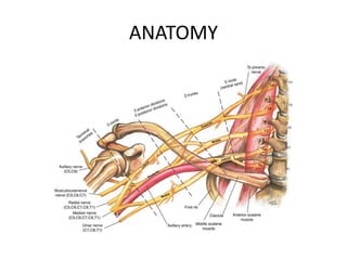
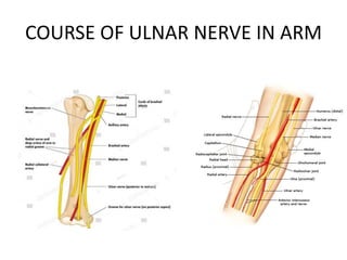
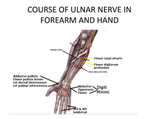
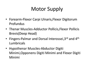
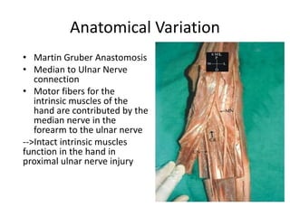
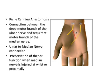
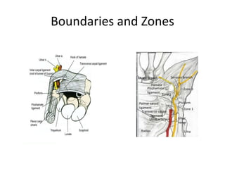
- Anatomy of the ulnar nerve and the muscles it innervates
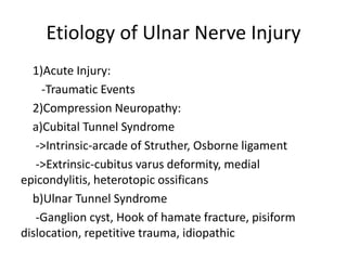
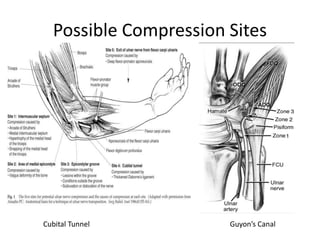
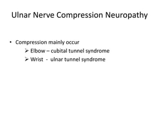
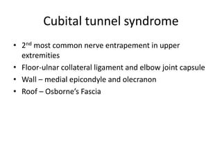
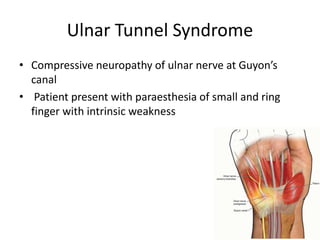
- Common causes of ulnar nerve injury such as trauma, compression neuropathies like cubital tunnel syndrome
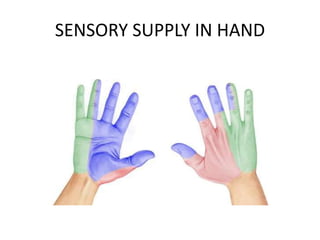
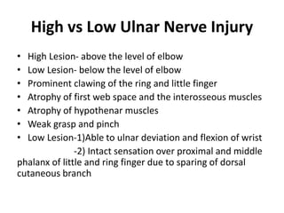
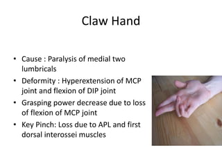
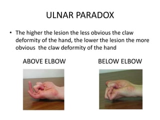
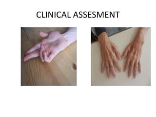
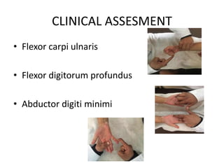
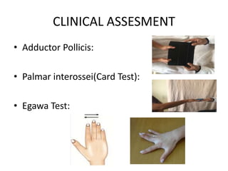
- Clinical signs of ulnar nerve injury including clawing of the ring and little fingers
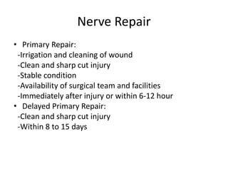
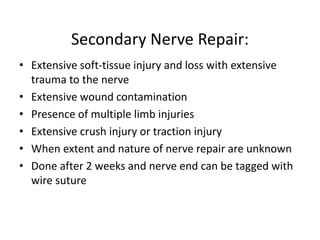
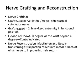
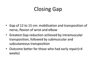
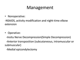
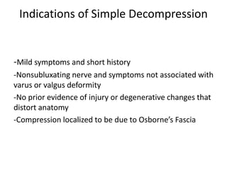
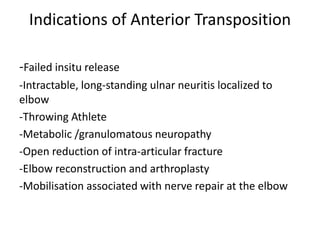
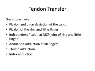
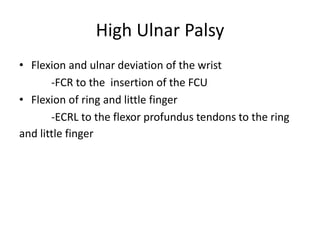
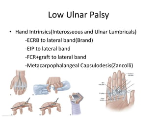
- Surgical management options like nerve repair, grafting, decompression for compression neuropathies, and tendon transfers to restore hand function
- Specific procedures for cubital tunnel syndrome and ulnar tunnel syndrome decompression