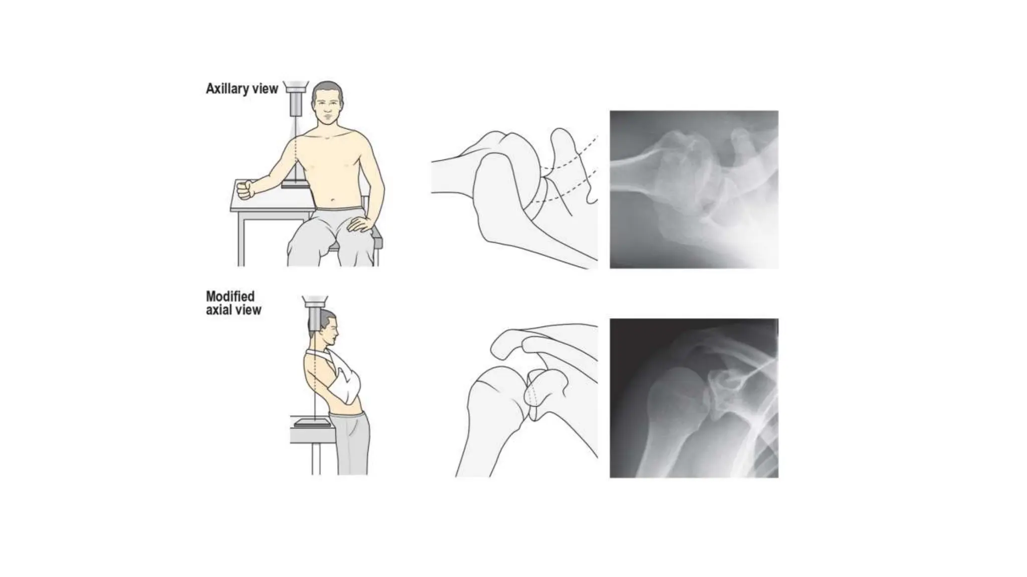
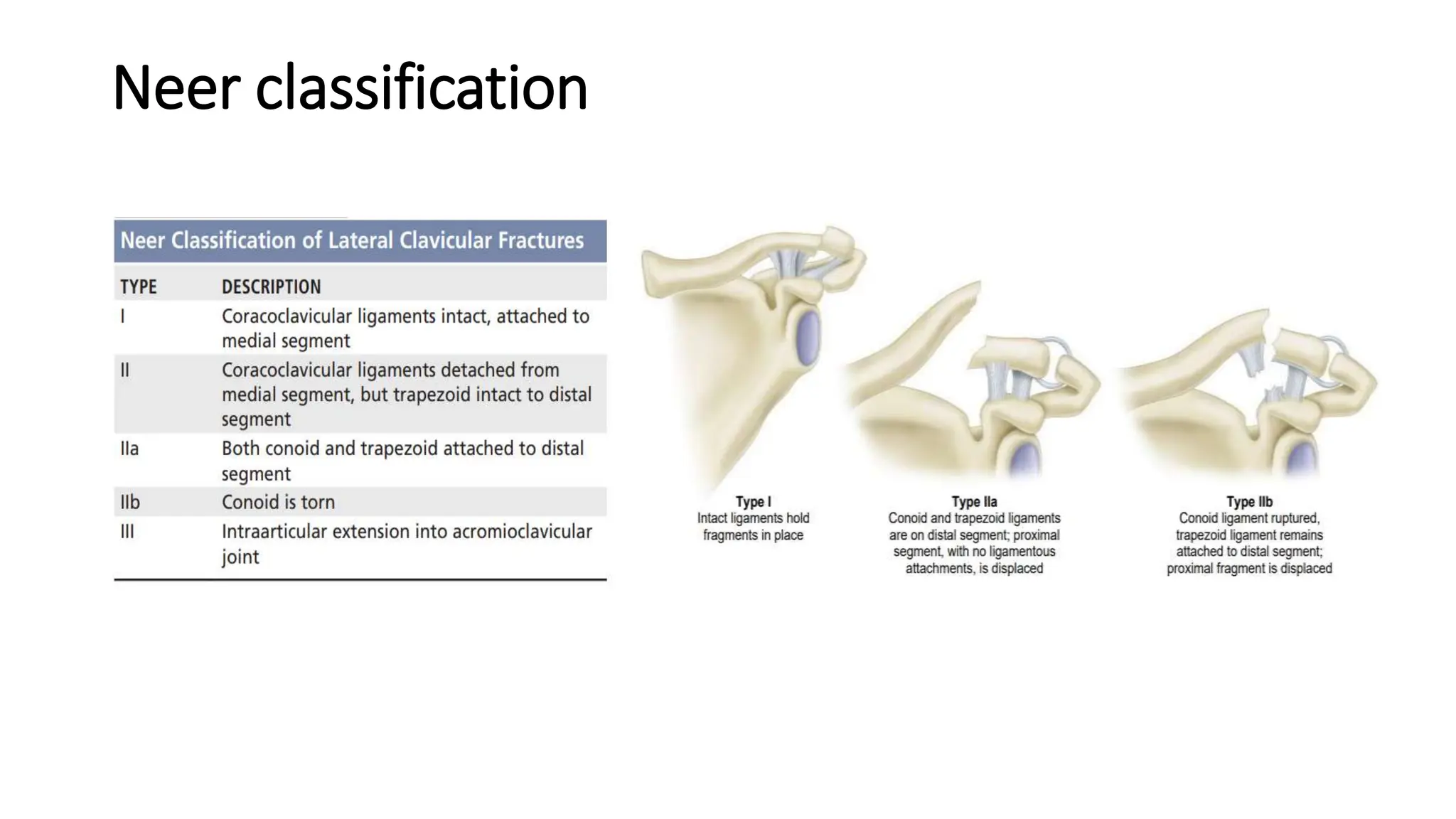
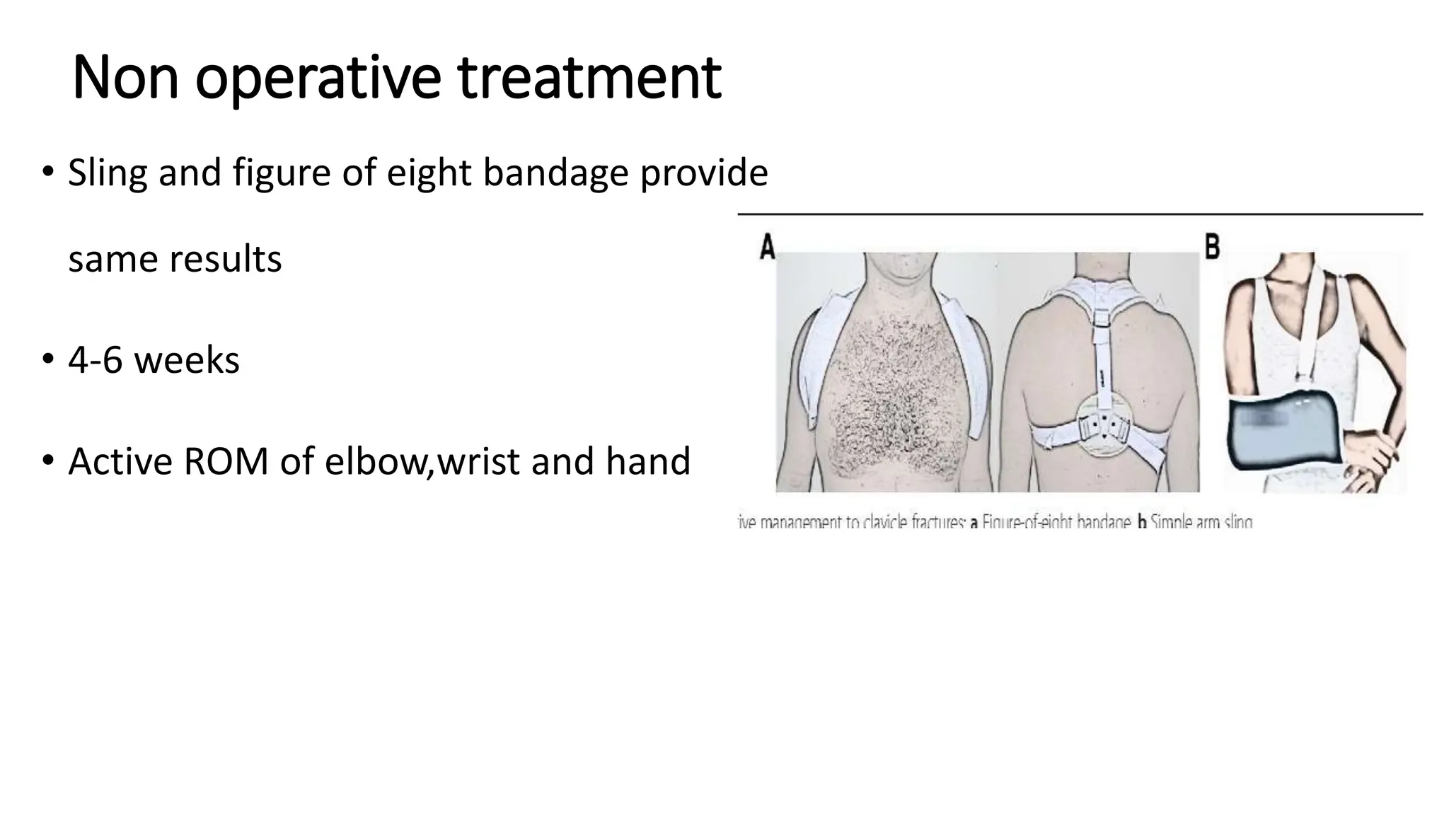
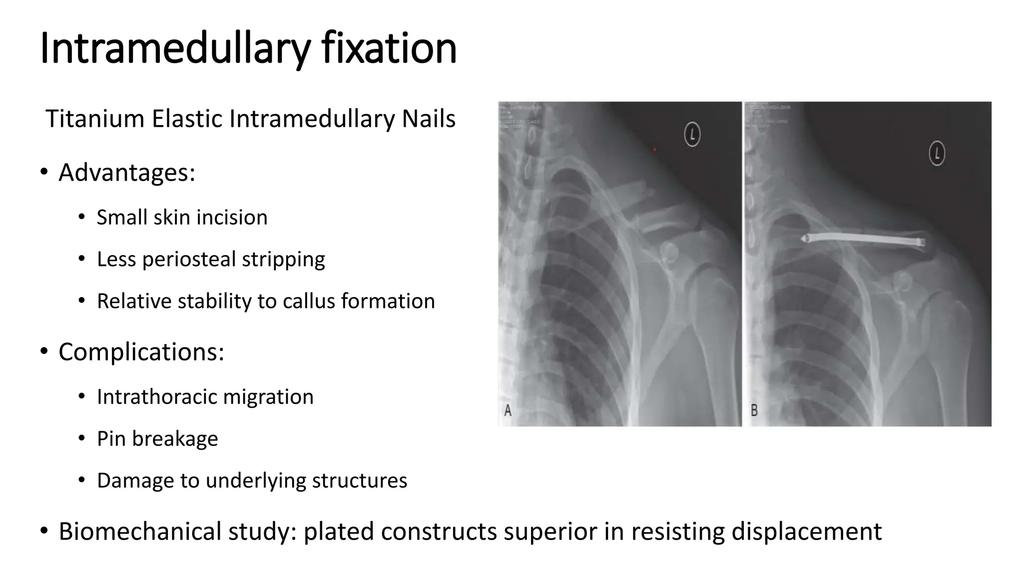
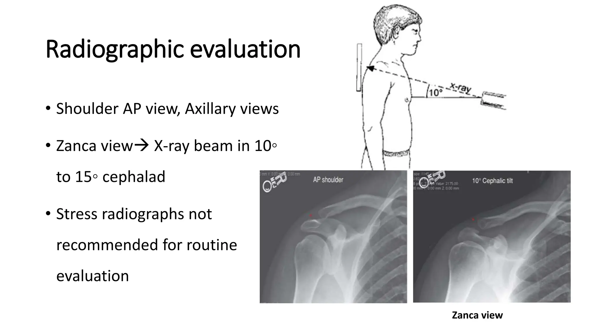
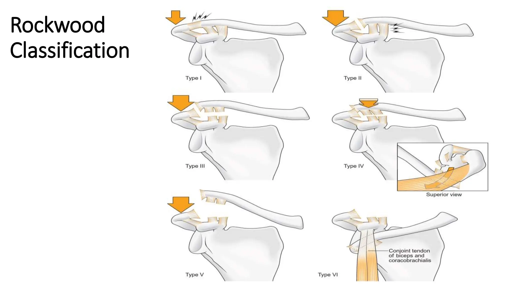
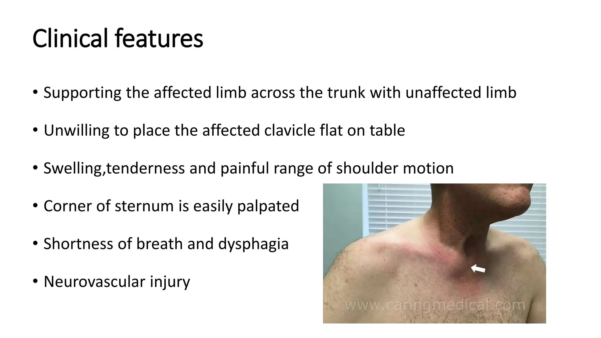
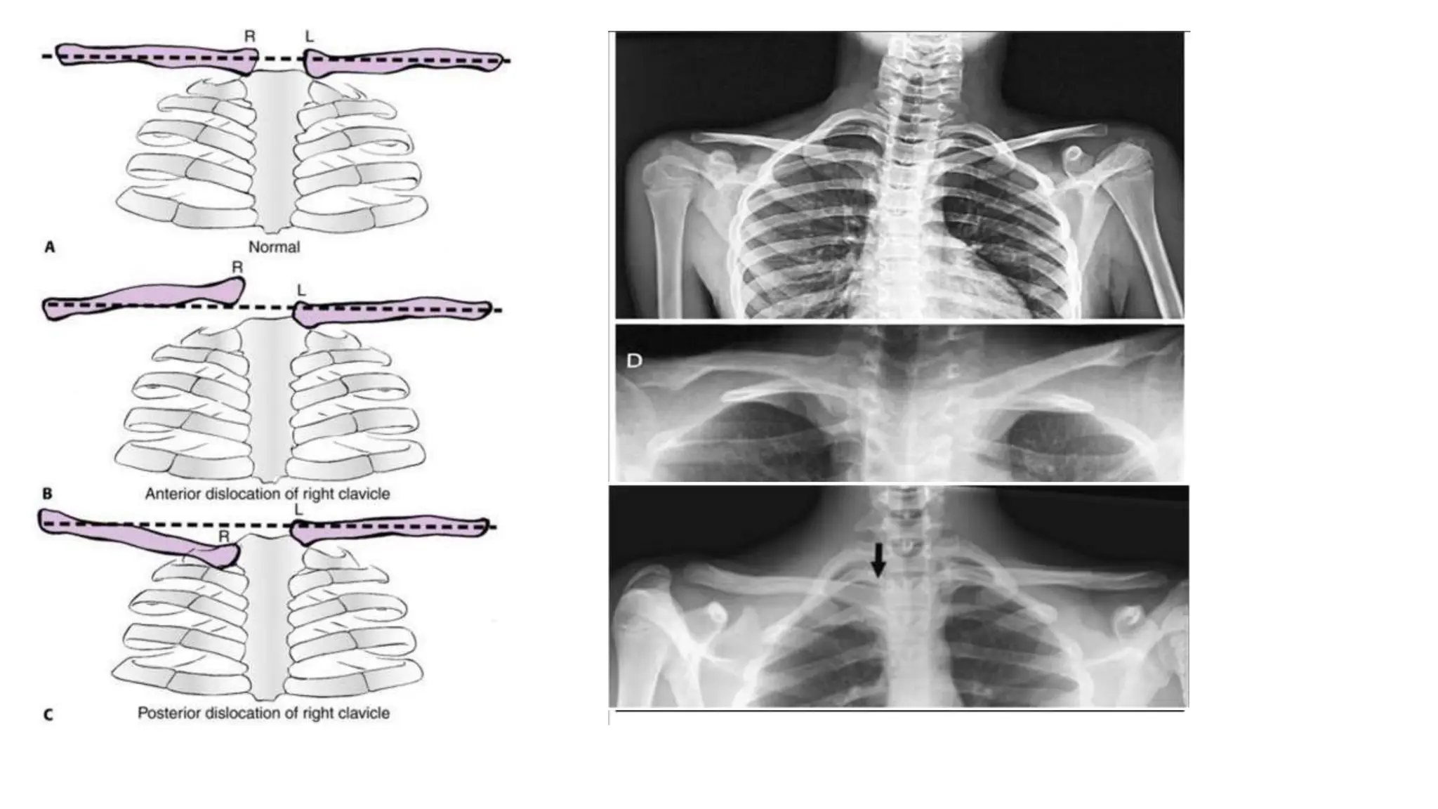
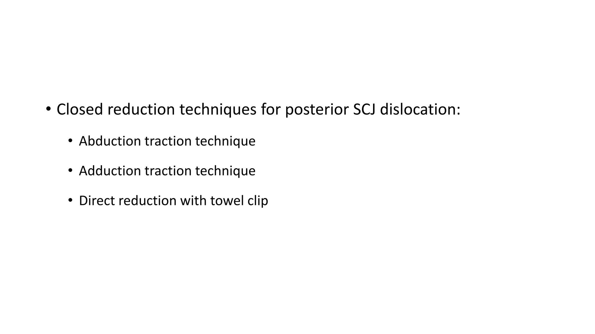
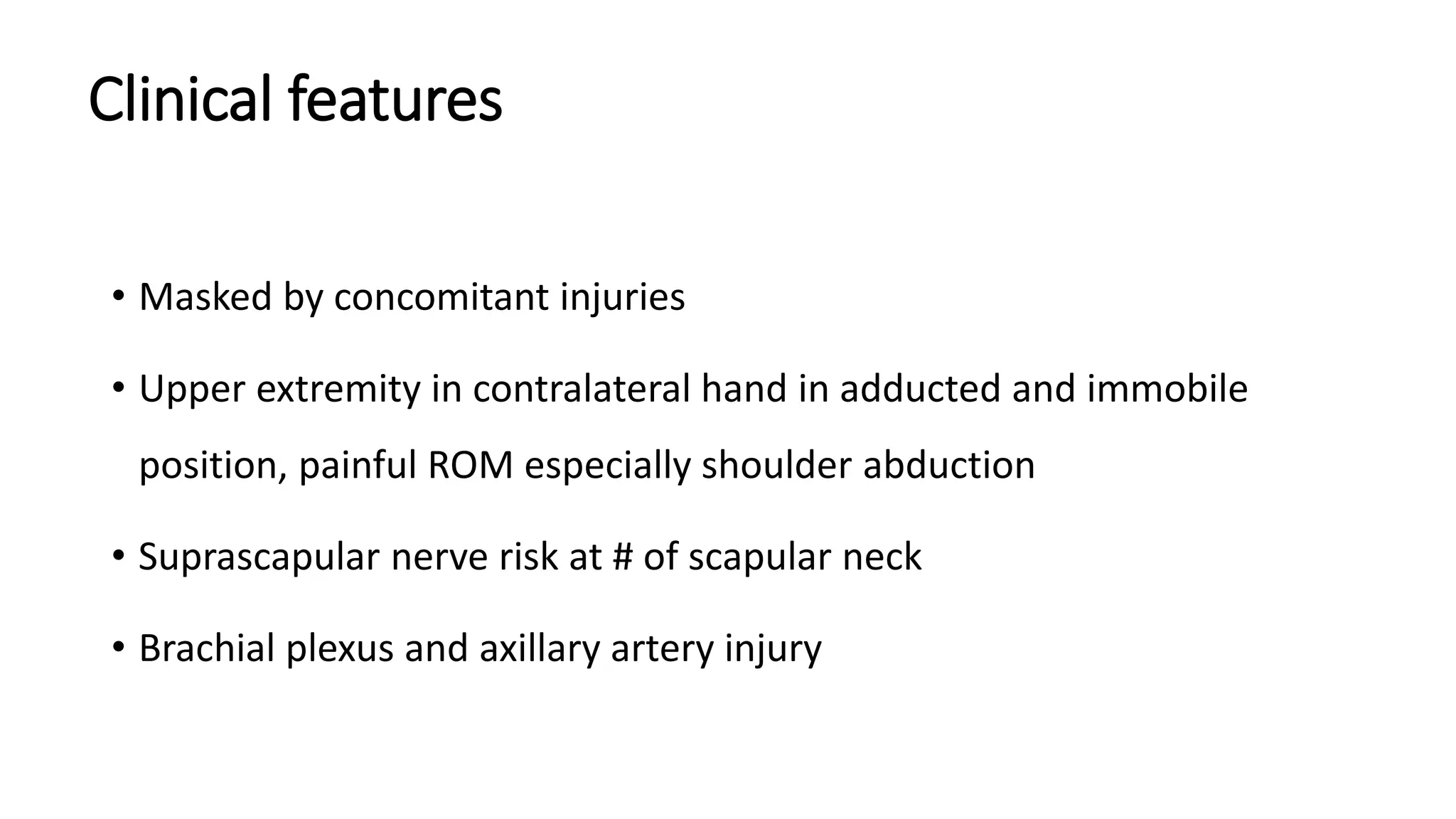
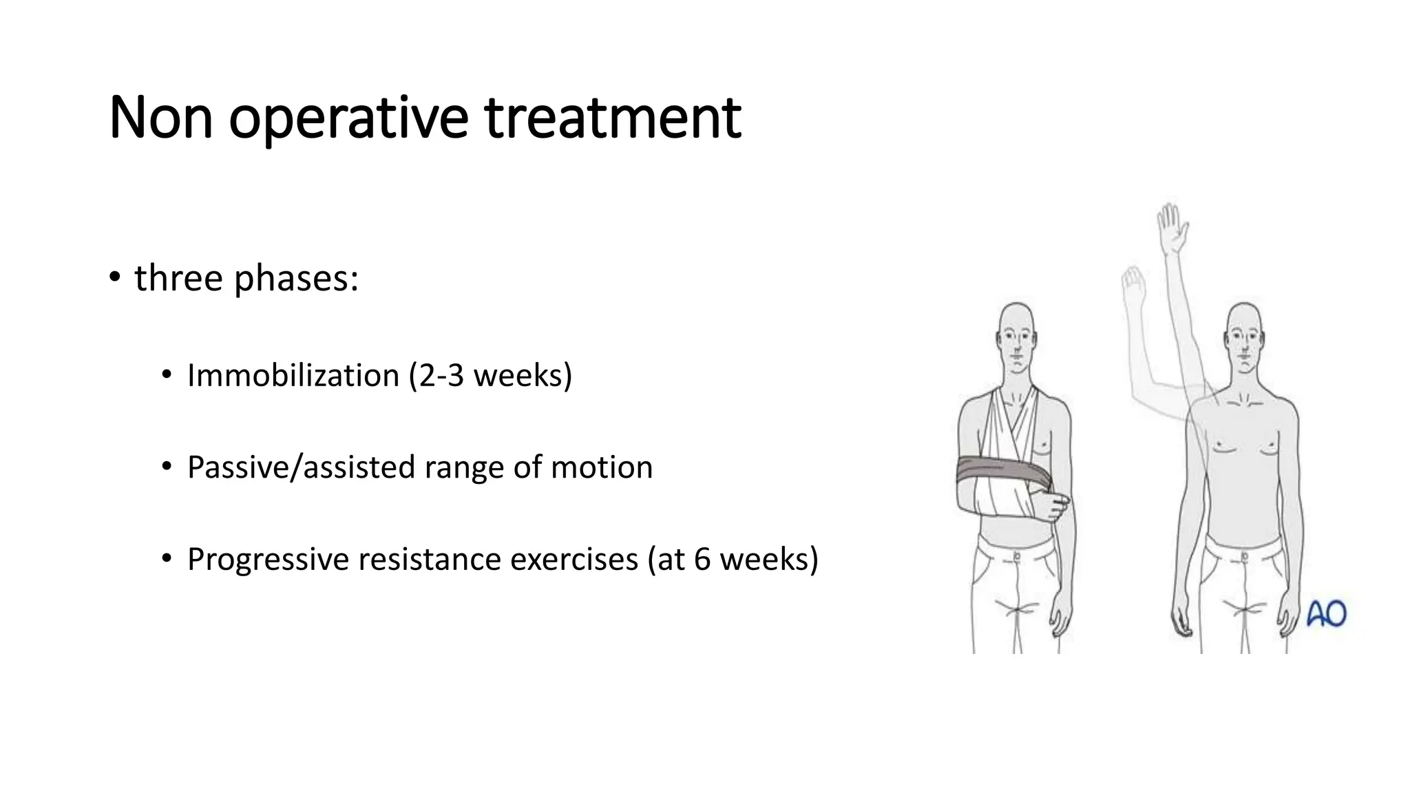
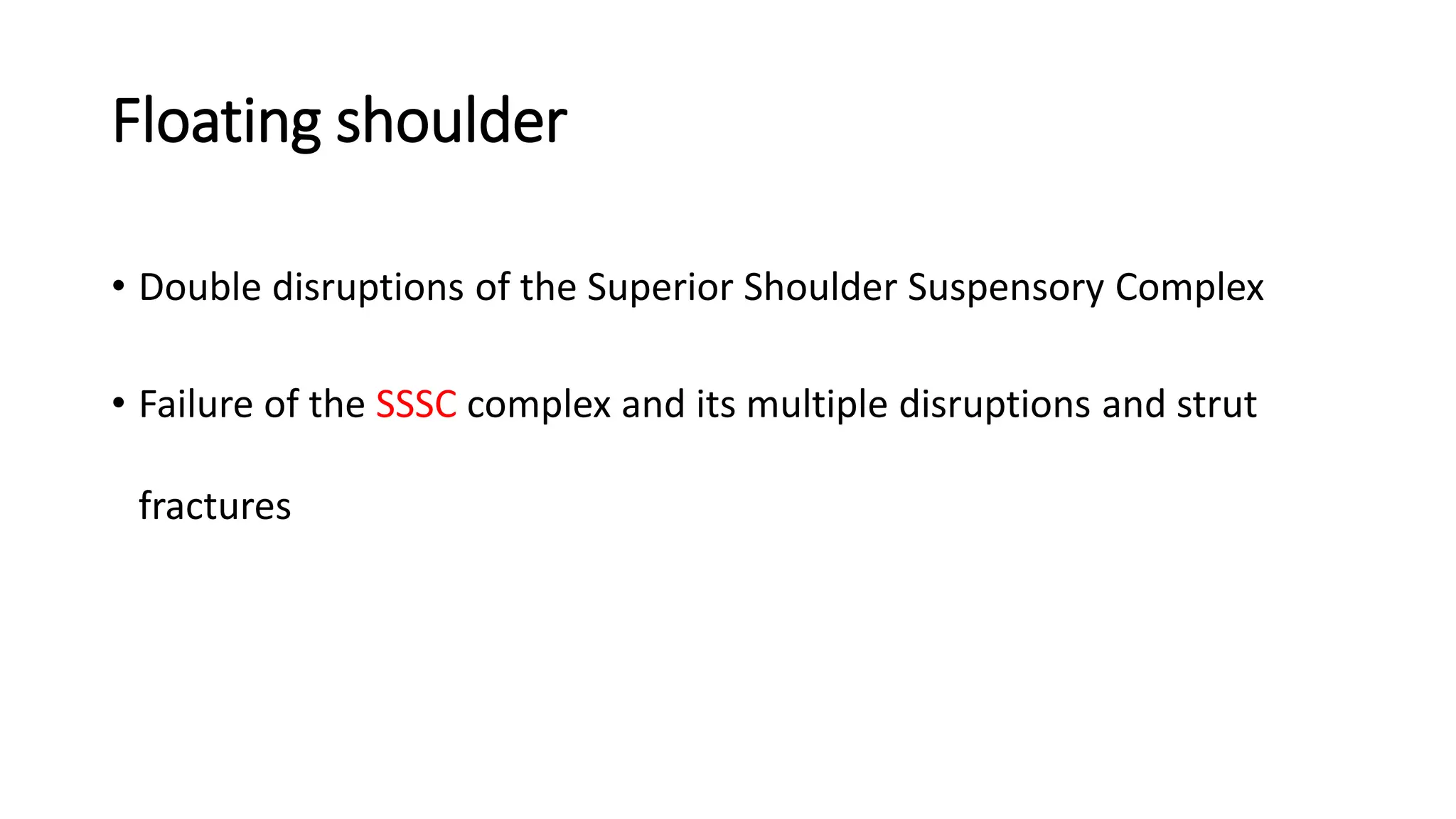
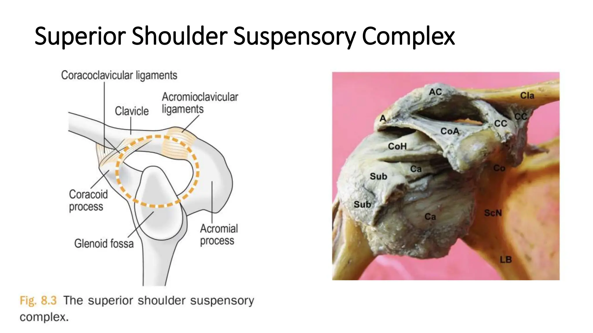
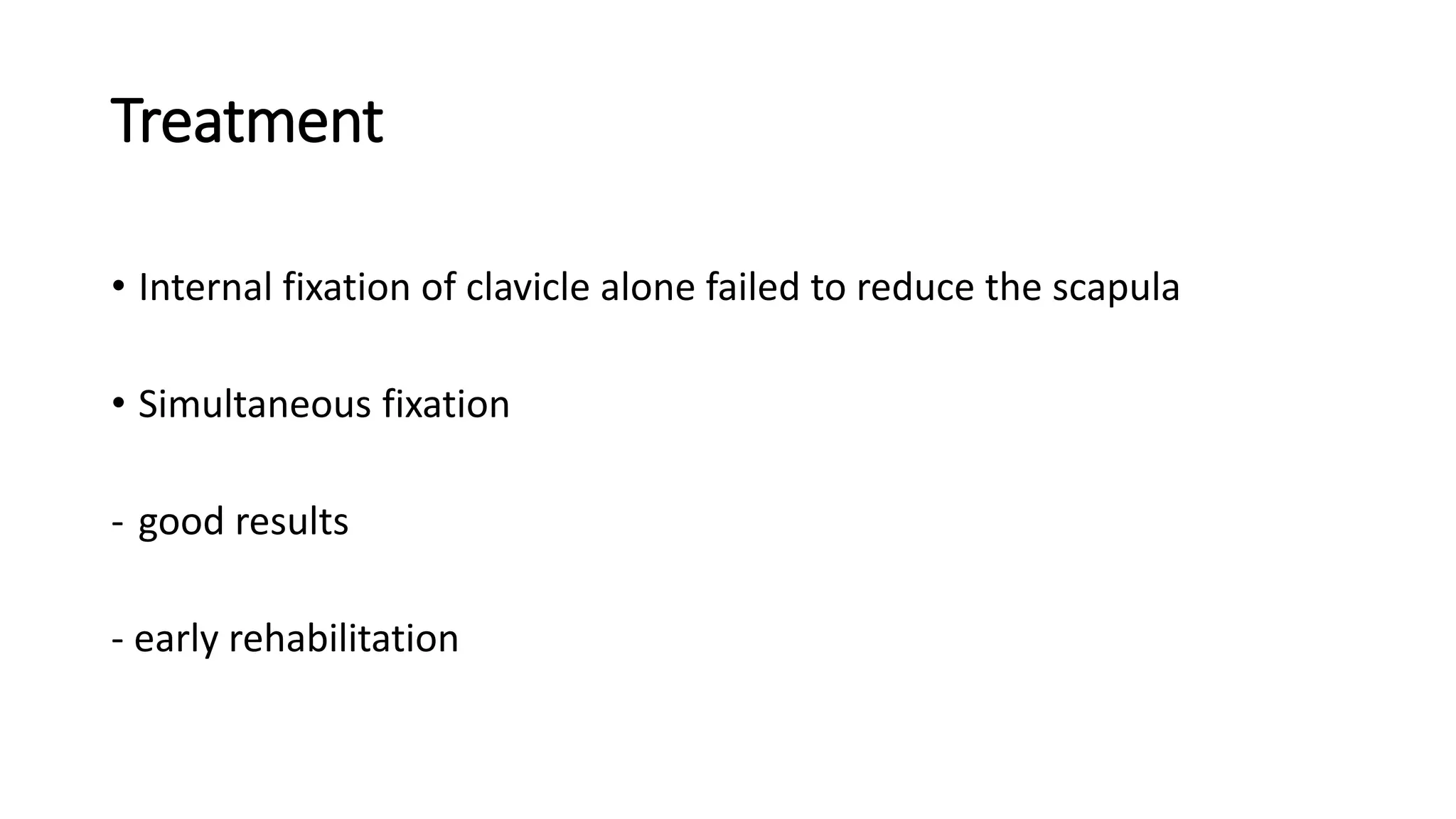
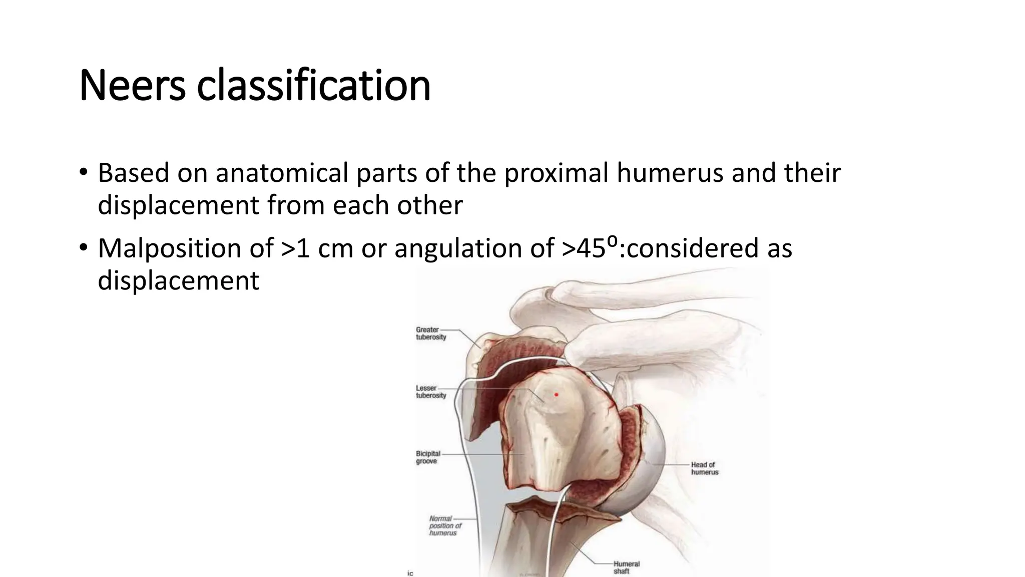
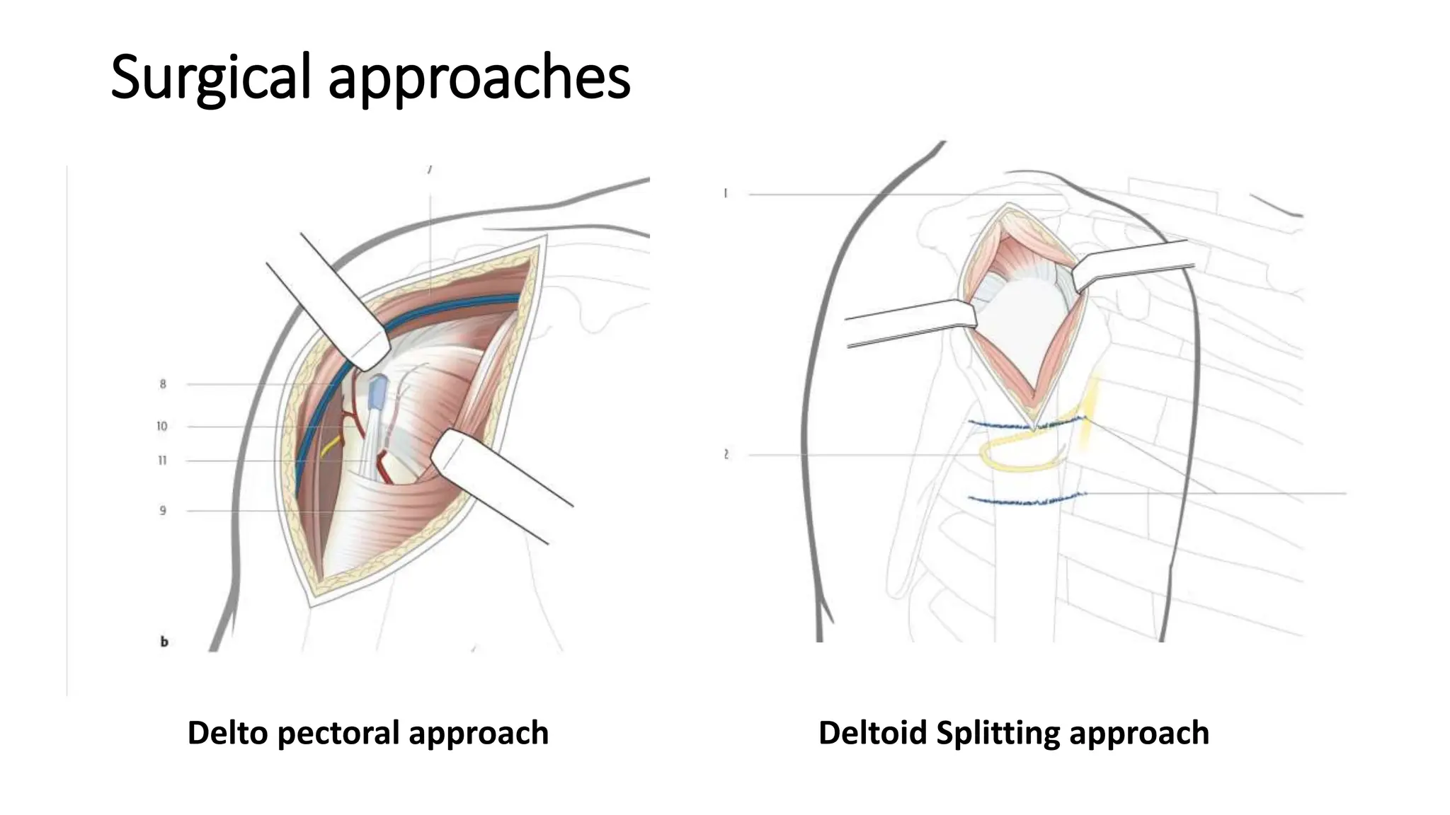
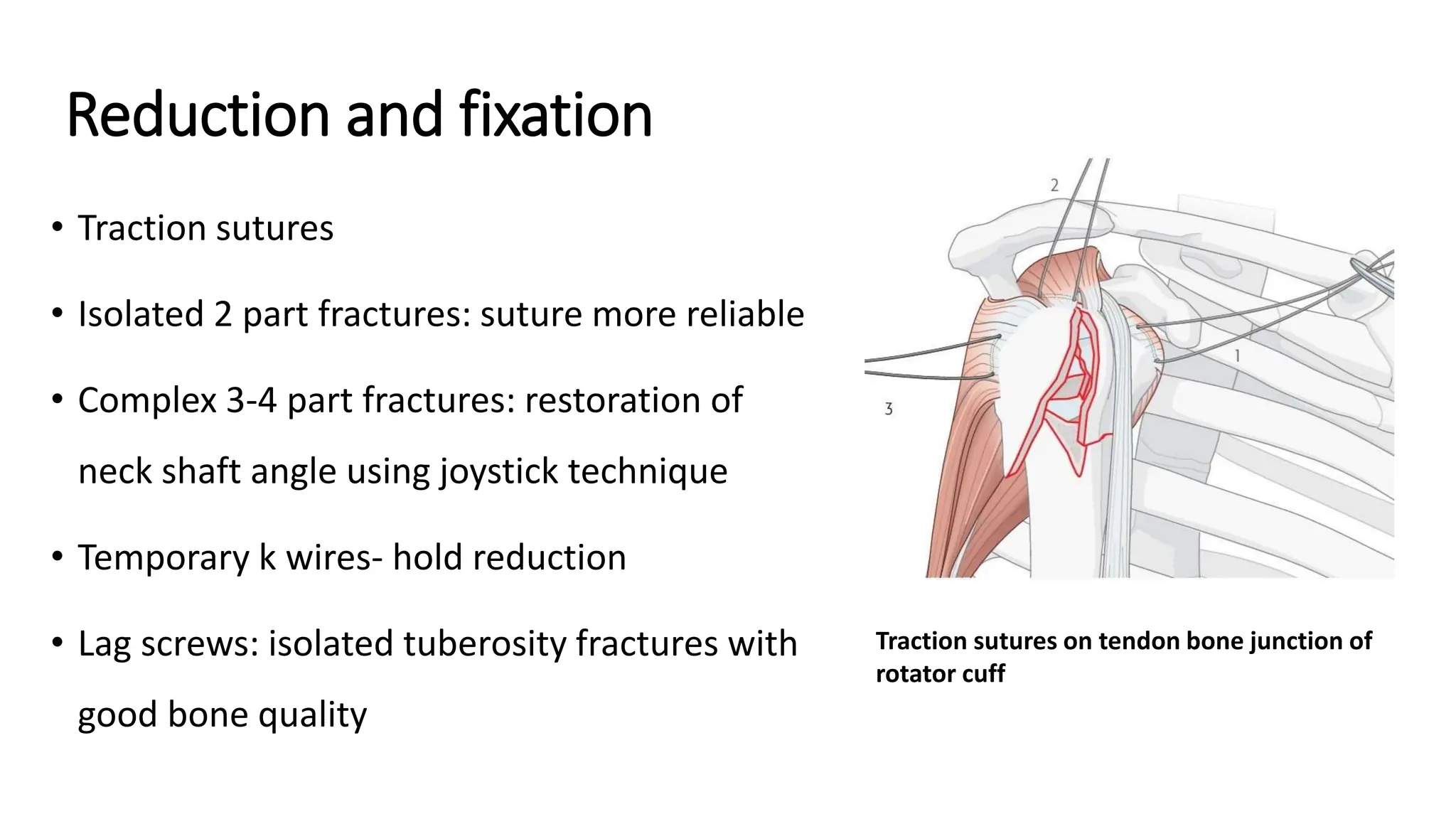
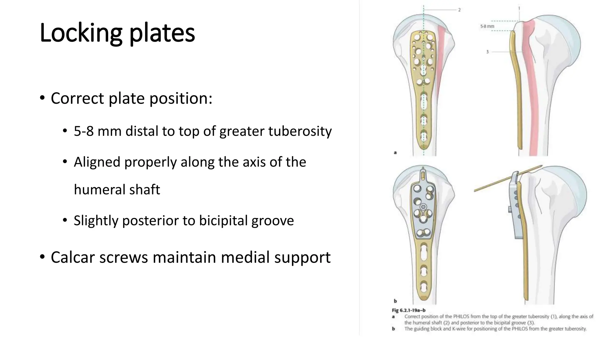
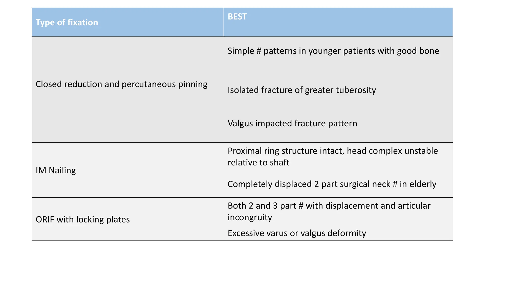
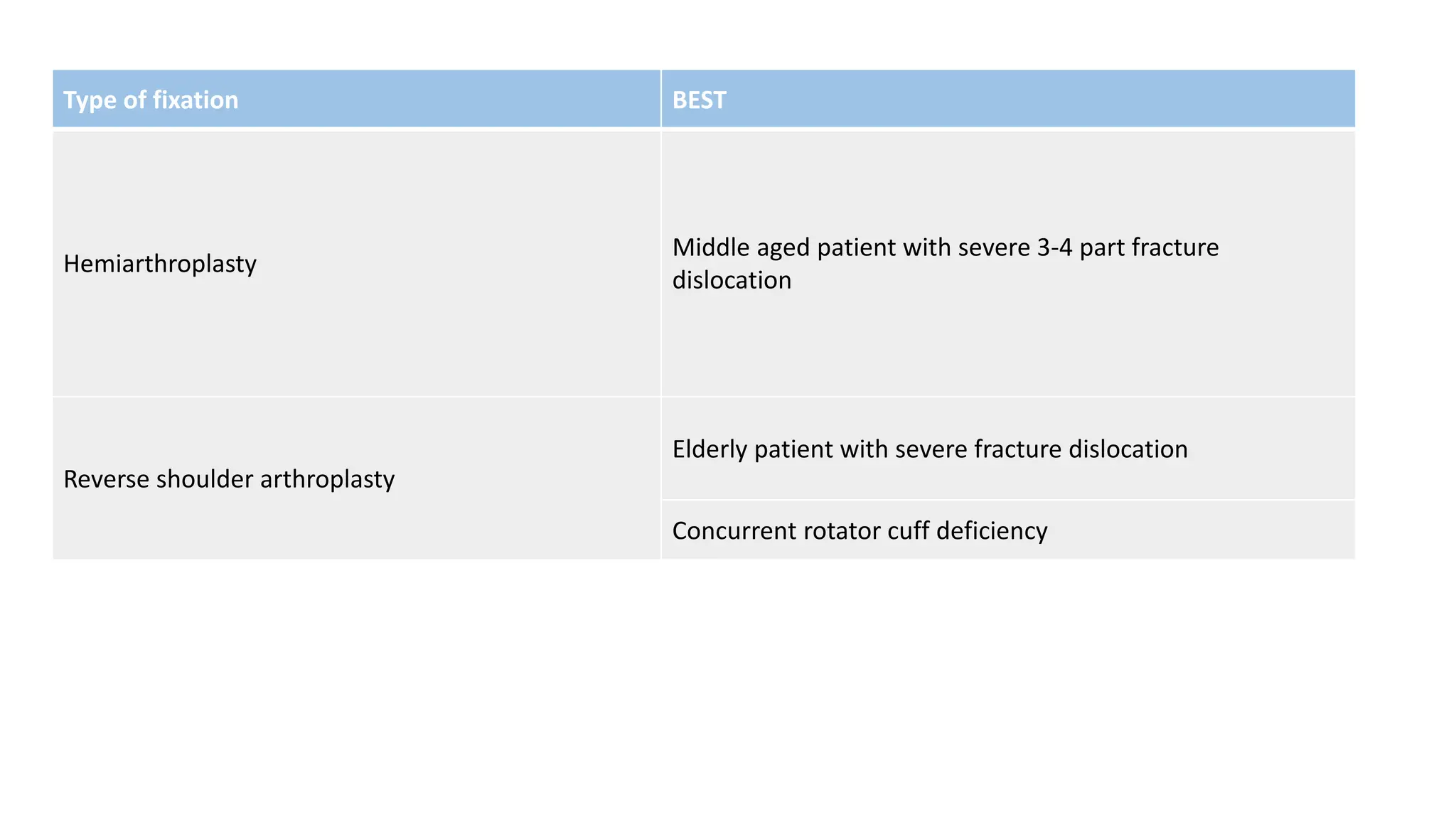
Adult: Fractures around
Shoulder Girdle (Clavicle,
Scapula, Humerus)
Presenter: Dr. Kushal Khanal
Moderator: Assoc.Prof.Dr. Rajesh B Lakhe
Guide:Dr.Jayant Bhagwan Mishra
Relevant Anatomy
• Shoulder girdle:
consists of
clavicle, scapula
and proximal
humerus and
their associated
ligaments and
muscles
The shoulder girdle
Radiographic views for shoulder girdle
Clavicle fractures
• Clavicle Fracture: 44 % to 66% of fractures about the shoulder(middle
third- 80% > lateral third-15% > medial third-5%)
• Young active individuals
• Direct blow
• Fall on to the affected shoulder leading to a bending force
• Convulsions
Clinical features
• History of fall on a shoulder
• Arm adducted to chest
• Supporting the elbow with other hand
• Clear deformity, skin tenting if displaced
• Localized tenderness
• Unwilling to move the shoulder
• Neurovascular assessment
• Possible pneumothorax and associated injuries like rib fractures
(9%)
Skin tenting
Radiological features
• Ideally taken in upright position
• AP clavicle
• 20⁰ cephalad view
• Chest X-ray showing B/L Shoulder
joints
20⁰ Cephalad view
Classification:
Neer classification
Non operative treatment
• Sling and figure of eight bandage provide
same results
• 4-6 weeks
• Active ROM of elbow,wrist and hand
Surgical indication
• Shaft fractures:
• Initial shortening of 2 cm or more is predictive of non union
• Lateral end fractures
• Mostly are undisplaced and extra-articular
• Generally progress to uncomplicated healing
• Medial end fractures:
• Usually non operative
• Posterior displacement - significant mediastinal compromise
Intramedullary fixation
Titanium Elastic Intramedullary Nails
• Advantages:
• Small skin incision
• Less periosteal stripping
• Relative stability to callus formation
• Complications:
• Intrathoracic migration
• Pin breakage
• Damage to underlying structures
• Biomechanical study: plated constructs superior in resisting displacement
Plate fixation
• Reconstruction plates:
• Facilitates contouring of implants
• Susceptible to deformation– implant
failure or malunion
• Anatomically precontoured locking
compression plate
• Further need of countouring to avoid
hardware prominence
• Calvicular hook plates:
• If distal fragment is too small
• Engage posterior aspect of acromion
Complications
• Early
• Wound complications
• Numbness- injury to supraclavicular nerves
• Hardware prominence and irritation
• Refracture
• Late:
• Non union – smoking, elderly, high energy trauma
• Malunion-corrective osteotomy with plate fixation
Acromioclavicular Joint injuries
• Diarthrodial joint between lateral end of clavicle and acromion
• Accounts 9 - 10% of traumatic injuries to shoulder girdle
• AC ligaments: anterior, posterior, superior, inferior
• Horizontal stability- AC ligaments
• Vertical stability- CC ligaments
• Deltoid and Trapezius-Secondary vertical stabilizer
• Mechanism of injury:
• Direct: fall on to shoulder with arm adducted
• Indirect: FOOSH causing force transmitted through humerus into AC joint
Clinical features
• Examination done in sitting or standing position with upper extremity
in dependent position
• Clinical deformity, focal tenderness and swelling
• Downward sag of shoulder: most characteristics anatomic deformity
• Apparent step off deformity
• Restricted range of motion
• Pain exacerbation on cross arm adduction
Radiographic evaluation
• Shoulder AP view, Axillary views
• Zanca view X-ray beam in 10◦
to 15◦ cephalad
• Stress radiographs not
recommended for routine
evaluation
Zanca view
Rockwood
Classification
Treatment:
Non-operative Operative
Type I AC joint injury Type III AC joint injury-Younger, and Labourer
using upper extremity above horizontal plane
Type II AC joint injury Type IV to VI AC joint injury
Type III AC joint injury-Inactive,Non-labouring or
recreational athletic patient
Failed non operative management
Key elements of Surgical management of ACJ
injuries
• Anatomic and accurate reduction of ACJ to correct superior
displacement and AP translation
• Direct repair or reconstruction of CC ligaments
• Supplementation of protection of CC ligament repair or
reconstruction with synthetic material (suture or tape)
• Repair of deltoid or trapezial fascia
• Distal clavicular resection in chronic ACJ injuries with clinical and
radiographical evidence of ACJ osteoarthritis
Operative treatment
• Bosworth technique of Coracoclavicular Screw with or without
primary repair of ligaments
Hook plate fixation
Arthroscopic or mini open stabilization using
suture tightropes or suture anchors
Suture or sling augmentation of CC ligaments
Sternoclavicular injuries
• Rare
• Motor vehicle accidents or sports-related
• SCJ: medial end of clavicle and manubrium
• Only true articulation between upper
extremity and axial skeleton
• Saddle type joint
• Intra articular disc ligament, extra articular
costoclavicular ligament, capsular ligament
and interclavicular ligament
Mechanism of injury
• Direct force on anteromedial aspect of clavicle-posterior SC joint dislocation
• Indirect: anterolateral force– anterior SC joint dislocation, posterolateral
force– posterior SC joint dislocation
• Associated injuries:
• Tracheal compression
• Pneumothorax
• Laceration/compression of great vessels
• Esophageal perforation
Clinical features
• Supporting the affected limb across the trunk with unaffected limb
• Unwilling to place the affected clavicle flat on table
• Swelling,tenderness and painful range of shoulder motion
• Corner of sternum is easily palpated
• Shortness of breath and dysphagia
• Neurovascular injury
Radiographic evaluation
• Hobbs view
• Serendipity view
• CT:
• Distinguishes injuries of the joint from fractures and minor subluxations
• Substantial asymmetry in joints
Hobbs view Serendipity view
Non operative treatment of SCJ injuries
• For most acute and chronic anterior subluxations and dislocations
• Acute traumatic posterior subluxations and some dislocations
• Most physeal injuries (usually in <25 yrs) heal without surgery
• Collar and cuff sling for anterior SCJ disruption
• Closed reduction techniques for posterior SCJ dislocation:
• Abduction traction technique
• Adduction traction technique
• Direct reduction with towel clip
Abduction traction technique
Buckerfield and Castle
Technique(Adduction traction
technique)
Direct reduction with towel clip
Surgical management
• Open reduction and stabilization in conjunction with vascular surgery
• Fixation of medial clavicle to sternum using fascia lata, subclavius
tendon or suture, osteotomy of medial clavicle or resection of medial
clavicle
Scapula
• Consists of
• Blade: triangular flattened region
• Spine: horizontal projection
posteriorly
• Acromion: highest and most lateral
point
• Coracoid process
• Glenoid: oriented anteriorly around
30◦
• Lateral and spinal
pillar: load bearing
part of scapula
• Spinomedial angle:
weakest area in
circumference
Anatomy of the scapula. (a) Anterior view, (b) posterior view. LP, lateral pillar;
SGN, spino-glenoidal notch; SMA, spino-medial angle; SP, spinous pillar.
Mechanism of injury
• Direct blow to the scapula
• Impact of humeral head on part of scapula
• Dislocation of humeral head
• Muscle contracture
• Associated injuries: rib fractures most common, thoracic cavity and
lungs, shoulder girdle
Clinical features
• Masked by concomitant injuries
• Upper extremity in contralateral hand in adducted and immobile
position, painful ROM especially shoulder abduction
• Suprascapular nerve risk at # of scapular neck
• Brachial plexus and axillary artery injury
Angle between line connecting the superior and inferior poles
of glenoid and line connecting the superior pole of the glenoid
and center of inferior angle of scapula
GPA <20⁰ one criteria for operative treatment
Radiographic evaluation
• AP view of entire shoulder girdle
• Neer I projection
• True AP radiograph of scapula
• Glenohumeral joint space, displacement of glenoid in relation to lateral border of scapula
• Measure glenopolar angle (GPA)
• Neer II projection
• True lateral radiograph of scapula
• translation, angulation and overlap of fragments
• CT
Classification of scapular fractures
• Anatomic Classification
• Type I: scapula body
• Type II: apophyseal fractures, including the acromion and coracoid
• Type III: Fractures of superolateral angle, including scapular neck and glenoid
Ideberg classification of intra articular glenoid
fractures
Classification of acromial fractures
Non operative treatment
• three phases:
• Immobilization (2-3 weeks)
• Passive/assisted range of motion
• Progressive resistance exercises (at 6 weeks)
Surgical indications
INDICATION CRITERION
INTRAARTICULAR FRACTURES
Articular step off ≥ 5 mm
Percentage of glenoid affected >20%
Glenohumeral articulation Unstable despite closed reduction
EXTRAARTICULAR FRACTURES
Glenopolar angle ≤ 20⁰
Angulation ≥ 45⁰
Translation ≥ 100%
Implant selection
• Glenoid rim: 4.0 mm cancellous screws
• Scapular body: reconstruction plate 3.5mm
• Acromion, acromial spine, coracoid: mini fragment plates
Fixation in scapular fractures
The standard plate for this fixation is the 2.7 or a 3.5
reconstruction plate.
Fixation with lag screw
Complications
• Shoulder Stiffness: Restricted internal rotation-most common
• Infection: deep infection rare
• Hematoma
• Suprascapular nerve palsy: decrease the range and strength of shoulder
external rotation
Floating shoulder
• Double disruptions of the Superior Shoulder Suspensory Complex
• Failure of the SSSC complex and its multiple disruptions and strut
fractures
Superior Shoulder Suspensory Complex
Treatment
• Internal fixation of clavicle alone failed to reduce the scapula
• Simultaneous fixation
- good results
- early rehabilitation
Proximal Humerus Fractures
• Most common humerus fracture
• Low energy injuries in elderly
population: fragility fracture
• Mostly- non displaced
• Mechanism:
• Elderly: fall from standing height onto
an outstretched arm
• Young: high energy trauma, more soft
tissue injury
• Seizures or electric shock
Proximal humerus
• Anatomical head: retroversion 35-40 ◦
• Greater tuberosity: ¾ rotator cuff muscle
insertion
• Lesser tuberosity: insertion of
subscapularis
• Surgical head: anatomical head and
tuberosities
• Surgical neck : junction of surgical head
and shaft
Blood supply
Blood supply of head of humerus
Clinical Examination
• Arm held close to the chest by contralateral hand
• Swelling and bruising
• Bruising may spread to dependent areas
• Restricted range of motion
• Motor and sensory function of axillary nerve
Radiographic evaluation
• AP, Lateral and axillary view of shoulder joint
• Axillary view may be impossible
• Velpeau view
AP view of shoulder joint
AP Grashey View Neer View(lateral Y View)
Axillary view Velpeau view
Neers classification
• Based on anatomical parts of the proximal humerus and their
displacement from each other
• Malposition of >1 cm or angulation of >45⁰:considered as
displacement
Classification of proximal humerus fractures
• Neer
classification
Risk of AVN
-Four part fractures and fracture dislocations-
highest risk forAVN of Humeral Head
Hertel’s Criteria:
-Metaphyseal extension of humeral head<8 mm
-Medial hinge disruption of >2 mm
-Fracture through anatomical head
The combination above factors had 97% positive
predictive value for humeral head ischaemia.
Non operative management
Indications:
• Stable non displaced or minimally displaced
• Elderly frail patients
• Patients not fit for surgery
Treatment:
• Sling immobilization for 2-3 weeks
• Early pendulum exercises or codman exercises then active ROM
Surgical indications
• Displaced fractures
• Head splitting fractures
• Neurovascular injury
• Open fractures
• Unstable fractures with disrupted medial hinge
• Floating shoulder
• Polytrauma
• Irreducible fracture dislocations
Implant selection
• Sutures, tension bands, screws: 2 part tuberosity fractures with good
bone quality
• Locking plates: displaced fractures
• MIPO: closed reduction maintained during surgery
• Arthroplasty: complex fractures in elderly osteoporotic bone
Surgical approaches
Delto pectoral approach Deltoid Splitting approach
Reduction and fixation
• Traction sutures
• Isolated 2 part fractures: suture more reliable
• Complex 3-4 part fractures: restoration of
neck shaft angle using joystick technique
• Temporary k wires- hold reduction
• Lag screws: isolated tuberosity fractures with
good bone quality
Traction sutures on tendon bone junction of
rotator cuff
Locking plates
• Correct plate position:
• 5-8 mm distal to top of greater tuberosity
• Aligned properly along the axis of the
humeral shaft
• Slightly posterior to bicipital groove
• Calcar screws maintain medial support
• MIPO
• Decrease chain of non
union and infection
• Proximal: deltoid split
• Distal: at deltoid
insertion
• Arthroplasty or
reverse shoulder
arthroplasty
Shoulder arthroplasty
Reverse Shoulder
Arthroplasty
Type of fixation
BEST
Closed reduction and percutaneous pinning
Simple # patterns in younger patients with good bone
Isolated fracture of greater tuberosity
Valgus impacted fracture pattern
IM Nailing
Proximal ring structure intact, head complex unstable
relative to shaft
Completely displaced 2 part surgical neck # in elderly
ORIF with locking plates
Both 2 and 3 part # with displacement and articular
incongruity
Excessive varus or valgus deformity
Type of fixation BEST
Hemiarthroplasty
Middle aged patient with severe 3-4 part fracture
dislocation
Reverse shoulder arthroplasty
Elderly patient with severe fracture dislocation
Concurrent rotator cuff deficiency
Complications
• Screw penetration
• Avascular necrosis:
• Length of dorsomedial metaphyseal extension
• Integrity of medial hinge
• Fracture type
• Malunion and non union: more frequently due to loss of medial
cortical buttress
• Nerve injuries
Summary
• Fractures of shoulder girdle can be associated with other serious
cardiothoracic and vascular injuries
• A simple trauma series of shoulder girdle to screen them all
• Most fractures of shoulder girdle can be managed non operatively
• Surgical indications dependent on fracture displacement and patient’s
physical demand
• Shoulder stiffness -a frequent complication
• Must not miss and must document neurovascular status
References
• Rockwood and Green’s fractures in adults
• Campbell’s operative orthopaedics
• AO principles of fracture management
• Mc Rae orthopaedic trauma management
NEXT PRESENTATION
• Journal Club(Related to Paediatric Fracture)-Dr.Arun Upreti
THANK YOU

Fracture around Shoulder Girdle Dr.Kushal

  • 1.
    Adult: Fractures around ShoulderGirdle (Clavicle, Scapula, Humerus) Presenter: Dr. Kushal Khanal Moderator: Assoc.Prof.Dr. Rajesh B Lakhe Guide:Dr.Jayant Bhagwan Mishra
  • 2.
    Relevant Anatomy • Shouldergirdle: consists of clavicle, scapula and proximal humerus and their associated ligaments and muscles The shoulder girdle
  • 3.
    Radiographic views forshoulder girdle
  • 6.
    Clavicle fractures • ClavicleFracture: 44 % to 66% of fractures about the shoulder(middle third- 80% > lateral third-15% > medial third-5%) • Young active individuals • Direct blow • Fall on to the affected shoulder leading to a bending force • Convulsions
  • 7.
    Clinical features • Historyof fall on a shoulder • Arm adducted to chest • Supporting the elbow with other hand • Clear deformity, skin tenting if displaced • Localized tenderness • Unwilling to move the shoulder • Neurovascular assessment • Possible pneumothorax and associated injuries like rib fractures (9%) Skin tenting
  • 8.
    Radiological features • Ideallytaken in upright position • AP clavicle • 20⁰ cephalad view • Chest X-ray showing B/L Shoulder joints 20⁰ Cephalad view
  • 9.
  • 10.
  • 11.
    Non operative treatment •Sling and figure of eight bandage provide same results • 4-6 weeks • Active ROM of elbow,wrist and hand
  • 12.
  • 13.
    • Shaft fractures: •Initial shortening of 2 cm or more is predictive of non union • Lateral end fractures • Mostly are undisplaced and extra-articular • Generally progress to uncomplicated healing • Medial end fractures: • Usually non operative • Posterior displacement - significant mediastinal compromise
  • 14.
    Intramedullary fixation Titanium ElasticIntramedullary Nails • Advantages: • Small skin incision • Less periosteal stripping • Relative stability to callus formation • Complications: • Intrathoracic migration • Pin breakage • Damage to underlying structures • Biomechanical study: plated constructs superior in resisting displacement
  • 15.
    Plate fixation • Reconstructionplates: • Facilitates contouring of implants • Susceptible to deformation– implant failure or malunion • Anatomically precontoured locking compression plate • Further need of countouring to avoid hardware prominence • Calvicular hook plates: • If distal fragment is too small • Engage posterior aspect of acromion
  • 16.
    Complications • Early • Woundcomplications • Numbness- injury to supraclavicular nerves • Hardware prominence and irritation • Refracture • Late: • Non union – smoking, elderly, high energy trauma • Malunion-corrective osteotomy with plate fixation
  • 17.
    Acromioclavicular Joint injuries •Diarthrodial joint between lateral end of clavicle and acromion • Accounts 9 - 10% of traumatic injuries to shoulder girdle • AC ligaments: anterior, posterior, superior, inferior • Horizontal stability- AC ligaments • Vertical stability- CC ligaments • Deltoid and Trapezius-Secondary vertical stabilizer • Mechanism of injury: • Direct: fall on to shoulder with arm adducted • Indirect: FOOSH causing force transmitted through humerus into AC joint
  • 18.
    Clinical features • Examinationdone in sitting or standing position with upper extremity in dependent position • Clinical deformity, focal tenderness and swelling • Downward sag of shoulder: most characteristics anatomic deformity • Apparent step off deformity • Restricted range of motion • Pain exacerbation on cross arm adduction
  • 19.
    Radiographic evaluation • ShoulderAP view, Axillary views • Zanca view X-ray beam in 10◦ to 15◦ cephalad • Stress radiographs not recommended for routine evaluation Zanca view
  • 20.
  • 21.
    Treatment: Non-operative Operative Type IAC joint injury Type III AC joint injury-Younger, and Labourer using upper extremity above horizontal plane Type II AC joint injury Type IV to VI AC joint injury Type III AC joint injury-Inactive,Non-labouring or recreational athletic patient Failed non operative management
  • 22.
    Key elements ofSurgical management of ACJ injuries • Anatomic and accurate reduction of ACJ to correct superior displacement and AP translation • Direct repair or reconstruction of CC ligaments • Supplementation of protection of CC ligament repair or reconstruction with synthetic material (suture or tape) • Repair of deltoid or trapezial fascia • Distal clavicular resection in chronic ACJ injuries with clinical and radiographical evidence of ACJ osteoarthritis
  • 23.
    Operative treatment • Bosworthtechnique of Coracoclavicular Screw with or without primary repair of ligaments
  • 24.
  • 25.
    Arthroscopic or miniopen stabilization using suture tightropes or suture anchors
  • 26.
    Suture or slingaugmentation of CC ligaments
  • 27.
    Sternoclavicular injuries • Rare •Motor vehicle accidents or sports-related • SCJ: medial end of clavicle and manubrium • Only true articulation between upper extremity and axial skeleton • Saddle type joint • Intra articular disc ligament, extra articular costoclavicular ligament, capsular ligament and interclavicular ligament
  • 28.
    Mechanism of injury •Direct force on anteromedial aspect of clavicle-posterior SC joint dislocation • Indirect: anterolateral force– anterior SC joint dislocation, posterolateral force– posterior SC joint dislocation • Associated injuries: • Tracheal compression • Pneumothorax • Laceration/compression of great vessels • Esophageal perforation
  • 29.
    Clinical features • Supportingthe affected limb across the trunk with unaffected limb • Unwilling to place the affected clavicle flat on table • Swelling,tenderness and painful range of shoulder motion • Corner of sternum is easily palpated • Shortness of breath and dysphagia • Neurovascular injury
  • 30.
    Radiographic evaluation • Hobbsview • Serendipity view • CT: • Distinguishes injuries of the joint from fractures and minor subluxations • Substantial asymmetry in joints Hobbs view Serendipity view
  • 32.
    Non operative treatmentof SCJ injuries • For most acute and chronic anterior subluxations and dislocations • Acute traumatic posterior subluxations and some dislocations • Most physeal injuries (usually in <25 yrs) heal without surgery • Collar and cuff sling for anterior SCJ disruption
  • 33.
    • Closed reductiontechniques for posterior SCJ dislocation: • Abduction traction technique • Adduction traction technique • Direct reduction with towel clip
  • 34.
    Abduction traction technique Buckerfieldand Castle Technique(Adduction traction technique) Direct reduction with towel clip
  • 35.
    Surgical management • Openreduction and stabilization in conjunction with vascular surgery • Fixation of medial clavicle to sternum using fascia lata, subclavius tendon or suture, osteotomy of medial clavicle or resection of medial clavicle
  • 36.
    Scapula • Consists of •Blade: triangular flattened region • Spine: horizontal projection posteriorly • Acromion: highest and most lateral point • Coracoid process • Glenoid: oriented anteriorly around 30◦
  • 37.
    • Lateral andspinal pillar: load bearing part of scapula • Spinomedial angle: weakest area in circumference Anatomy of the scapula. (a) Anterior view, (b) posterior view. LP, lateral pillar; SGN, spino-glenoidal notch; SMA, spino-medial angle; SP, spinous pillar.
  • 38.
    Mechanism of injury •Direct blow to the scapula • Impact of humeral head on part of scapula • Dislocation of humeral head • Muscle contracture • Associated injuries: rib fractures most common, thoracic cavity and lungs, shoulder girdle
  • 39.
    Clinical features • Maskedby concomitant injuries • Upper extremity in contralateral hand in adducted and immobile position, painful ROM especially shoulder abduction • Suprascapular nerve risk at # of scapular neck • Brachial plexus and axillary artery injury
  • 40.
    Angle between lineconnecting the superior and inferior poles of glenoid and line connecting the superior pole of the glenoid and center of inferior angle of scapula GPA <20⁰ one criteria for operative treatment
  • 41.
    Radiographic evaluation • APview of entire shoulder girdle • Neer I projection • True AP radiograph of scapula • Glenohumeral joint space, displacement of glenoid in relation to lateral border of scapula • Measure glenopolar angle (GPA) • Neer II projection • True lateral radiograph of scapula • translation, angulation and overlap of fragments • CT
  • 42.
    Classification of scapularfractures • Anatomic Classification • Type I: scapula body • Type II: apophyseal fractures, including the acromion and coracoid • Type III: Fractures of superolateral angle, including scapular neck and glenoid
  • 43.
    Ideberg classification ofintra articular glenoid fractures
  • 44.
  • 45.
    Non operative treatment •three phases: • Immobilization (2-3 weeks) • Passive/assisted range of motion • Progressive resistance exercises (at 6 weeks)
  • 46.
    Surgical indications INDICATION CRITERION INTRAARTICULARFRACTURES Articular step off ≥ 5 mm Percentage of glenoid affected >20% Glenohumeral articulation Unstable despite closed reduction EXTRAARTICULAR FRACTURES Glenopolar angle ≤ 20⁰ Angulation ≥ 45⁰ Translation ≥ 100%
  • 47.
    Implant selection • Glenoidrim: 4.0 mm cancellous screws • Scapular body: reconstruction plate 3.5mm • Acromion, acromial spine, coracoid: mini fragment plates
  • 48.
    Fixation in scapularfractures The standard plate for this fixation is the 2.7 or a 3.5 reconstruction plate. Fixation with lag screw
  • 49.
    Complications • Shoulder Stiffness:Restricted internal rotation-most common • Infection: deep infection rare • Hematoma • Suprascapular nerve palsy: decrease the range and strength of shoulder external rotation
  • 50.
    Floating shoulder • Doubledisruptions of the Superior Shoulder Suspensory Complex • Failure of the SSSC complex and its multiple disruptions and strut fractures
  • 51.
  • 52.
    Treatment • Internal fixationof clavicle alone failed to reduce the scapula • Simultaneous fixation - good results - early rehabilitation
  • 53.
    Proximal Humerus Fractures •Most common humerus fracture • Low energy injuries in elderly population: fragility fracture • Mostly- non displaced • Mechanism: • Elderly: fall from standing height onto an outstretched arm • Young: high energy trauma, more soft tissue injury • Seizures or electric shock
  • 54.
    Proximal humerus • Anatomicalhead: retroversion 35-40 ◦ • Greater tuberosity: ¾ rotator cuff muscle insertion • Lesser tuberosity: insertion of subscapularis • Surgical head: anatomical head and tuberosities • Surgical neck : junction of surgical head and shaft
  • 55.
    Blood supply Blood supplyof head of humerus
  • 56.
    Clinical Examination • Armheld close to the chest by contralateral hand • Swelling and bruising • Bruising may spread to dependent areas • Restricted range of motion • Motor and sensory function of axillary nerve
  • 57.
    Radiographic evaluation • AP,Lateral and axillary view of shoulder joint • Axillary view may be impossible • Velpeau view AP view of shoulder joint
  • 58.
    AP Grashey ViewNeer View(lateral Y View)
  • 59.
  • 60.
    Neers classification • Basedon anatomical parts of the proximal humerus and their displacement from each other • Malposition of >1 cm or angulation of >45⁰:considered as displacement
  • 61.
    Classification of proximalhumerus fractures • Neer classification
  • 62.
    Risk of AVN -Fourpart fractures and fracture dislocations- highest risk forAVN of Humeral Head Hertel’s Criteria: -Metaphyseal extension of humeral head<8 mm -Medial hinge disruption of >2 mm -Fracture through anatomical head The combination above factors had 97% positive predictive value for humeral head ischaemia.
  • 63.
    Non operative management Indications: •Stable non displaced or minimally displaced • Elderly frail patients • Patients not fit for surgery Treatment: • Sling immobilization for 2-3 weeks • Early pendulum exercises or codman exercises then active ROM
  • 64.
    Surgical indications • Displacedfractures • Head splitting fractures • Neurovascular injury • Open fractures • Unstable fractures with disrupted medial hinge • Floating shoulder • Polytrauma • Irreducible fracture dislocations
  • 65.
    Implant selection • Sutures,tension bands, screws: 2 part tuberosity fractures with good bone quality • Locking plates: displaced fractures • MIPO: closed reduction maintained during surgery • Arthroplasty: complex fractures in elderly osteoporotic bone
  • 66.
    Surgical approaches Delto pectoralapproach Deltoid Splitting approach
  • 67.
    Reduction and fixation •Traction sutures • Isolated 2 part fractures: suture more reliable • Complex 3-4 part fractures: restoration of neck shaft angle using joystick technique • Temporary k wires- hold reduction • Lag screws: isolated tuberosity fractures with good bone quality Traction sutures on tendon bone junction of rotator cuff
  • 68.
    Locking plates • Correctplate position: • 5-8 mm distal to top of greater tuberosity • Aligned properly along the axis of the humeral shaft • Slightly posterior to bicipital groove • Calcar screws maintain medial support
  • 69.
    • MIPO • Decreasechain of non union and infection • Proximal: deltoid split • Distal: at deltoid insertion • Arthroplasty or reverse shoulder arthroplasty Shoulder arthroplasty Reverse Shoulder Arthroplasty
  • 70.
    Type of fixation BEST Closedreduction and percutaneous pinning Simple # patterns in younger patients with good bone Isolated fracture of greater tuberosity Valgus impacted fracture pattern IM Nailing Proximal ring structure intact, head complex unstable relative to shaft Completely displaced 2 part surgical neck # in elderly ORIF with locking plates Both 2 and 3 part # with displacement and articular incongruity Excessive varus or valgus deformity
  • 71.
    Type of fixationBEST Hemiarthroplasty Middle aged patient with severe 3-4 part fracture dislocation Reverse shoulder arthroplasty Elderly patient with severe fracture dislocation Concurrent rotator cuff deficiency
  • 72.
    Complications • Screw penetration •Avascular necrosis: • Length of dorsomedial metaphyseal extension • Integrity of medial hinge • Fracture type • Malunion and non union: more frequently due to loss of medial cortical buttress • Nerve injuries
  • 73.
    Summary • Fractures ofshoulder girdle can be associated with other serious cardiothoracic and vascular injuries • A simple trauma series of shoulder girdle to screen them all • Most fractures of shoulder girdle can be managed non operatively • Surgical indications dependent on fracture displacement and patient’s physical demand • Shoulder stiffness -a frequent complication • Must not miss and must document neurovascular status
  • 74.
    References • Rockwood andGreen’s fractures in adults • Campbell’s operative orthopaedics • AO principles of fracture management • Mc Rae orthopaedic trauma management
  • 75.
    NEXT PRESENTATION • JournalClub(Related to Paediatric Fracture)-Dr.Arun Upreti THANK YOU