This document discusses ulnar nerve injuries, median nerve injuries, and radial nerve injuries. It begins by classifying ulnar nerve injuries into high ulnar nerve lesions, low ulnar nerve lesions, Guyon's tunnel syndrome, and cubital tunnel syndrome. It then describes the anatomy of the ulnar nerve, including its branches. Clinical features of different types of ulnar nerve injuries are provided, along with treatments. Carpal tunnel syndrome involving the median nerve is also described in detail. The anatomy and clinical assessments of median nerve injuries are outlined. Finally, the anatomy of the radial nerve is illustrated and different types of radial nerve lesions are classified.
paediatric injuries around the elbow
supracondylar elbow injuries
pulled elbow in paediatric age r
radiological signs around elbow in supracondylar fracture humerus
paediatric injuries around the elbow
supracondylar elbow injuries
pulled elbow in paediatric age r
radiological signs around elbow in supracondylar fracture humerus
Assessment Of Glenoid Bone Loss In Recurrent Shoulder Dislocation Samir Dwidmuthe
Bigliani coined the term glenoid rim lesions
glenoid rim erosion and
bony Bankart lesion,
Itoi et al. cadaveric study inferred that glenoid defect more than 21% produces anterior instability.
Lo and Burkhart named significant bone loss as
“inverted-pear glenoid” and
“engaging Hill-Sachs lesion”
shoulders associated with these significant bone loss are not suitable candidates for arthroscopic soft tissue stabilization
X ray
2D CT scan
3D CT scan
MRI
Arthroscopy
4th year medical student's seminar presentation under supervision of orthopedic lecturer. Reference is from Dr. Sameh Doss Textbook of upper and lower limb, and also other multiple websites.
Radial Nerve is very important topic for first year MBBS Students and as well as for day today clinical practice. This slide gives you full course & relations with clear diagrams as well as applied anatomy with clinical Co-relation.
hey this is Vedika Agrawal and this presentation is TO EXPLAIN AND HELP YOU UNDERSTAND ANATOMY OF FOREARM.
The topic is usually mixed with hand making it difficult to understand and so i seperated it to make it easy for you.
Hey this is Vedika Agrawal and my presentation explains about anatomy of forearm which covers almost every diagram and key point required to understand this topic.
This topic is usually mixed with antaomy of hand and so I separated to keep it easy for you.
reference: BD Chaurasia
This is a BASIC powerpoint focusing on the structures of the hand. Videos and pictures have been included. I trust it assists anyone who uses it. Blessings!
Injuries to the nerves of the upper limb can result from trauma, compression, lacerations, or certain medical conditions. Nerve injuries may lead to various symptoms, including pain, weakness, numbness, or loss of function in specific areas of the upper limb. Nerve injuries may range from mild to severe, and appropriate medical evaluation and treatment are essential. Physical therapy, splinting, medications, or in some cases, surgical intervention may be recommended based on the type and severity of the nerve injury. Early intervention is crucial for optimal recovery.
Ozempic: Preoperative Management of Patients on GLP-1 Receptor Agonists Saeid Safari
Preoperative Management of Patients on GLP-1 Receptor Agonists like Ozempic and Semiglutide
ASA GUIDELINE
NYSORA Guideline
2 Case Reports of Gastric Ultrasound
MANAGEMENT OF ATRIOVENTRICULAR CONDUCTION BLOCK.pdfJim Jacob Roy
Cardiac conduction defects can occur due to various causes.
Atrioventricular conduction blocks ( AV blocks ) are classified into 3 types.
This document describes the acute management of AV block.
The prostate is an exocrine gland of the male mammalian reproductive system
It is a walnut-sized gland that forms part of the male reproductive system and is located in front of the rectum and just below the urinary bladder
Function is to store and secrete a clear, slightly alkaline fluid that constitutes 10-30% of the volume of the seminal fluid that along with the spermatozoa, constitutes semen
A healthy human prostate measures (4cm-vertical, by 3cm-horizontal, 2cm ant-post ).
It surrounds the urethra just below the urinary bladder. It has anterior, median, posterior and two lateral lobes
It’s work is regulated by androgens which are responsible for male sex characteristics
Generalised disease of the prostate due to hormonal derangement which leads to non malignant enlargement of the gland (increase in the number of epithelial cells and stromal tissue)to cause compression of the urethra leading to symptoms (LUTS
Report Back from SGO 2024: What’s the Latest in Cervical Cancer?bkling
Are you curious about what’s new in cervical cancer research or unsure what the findings mean? Join Dr. Emily Ko, a gynecologic oncologist at Penn Medicine, to learn about the latest updates from the Society of Gynecologic Oncology (SGO) 2024 Annual Meeting on Women’s Cancer. Dr. Ko will discuss what the research presented at the conference means for you and answer your questions about the new developments.
micro teaching on communication m.sc nursing.pdfAnurag Sharma
Microteaching is a unique model of practice teaching. It is a viable instrument for the. desired change in the teaching behavior or the behavior potential which, in specified types of real. classroom situations, tends to facilitate the achievement of specified types of objectives.
Couples presenting to the infertility clinic- Do they really have infertility...Sujoy Dasgupta
Dr Sujoy Dasgupta presented the study on "Couples presenting to the infertility clinic- Do they really have infertility? – The unexplored stories of non-consummation" in the 13th Congress of the Asia Pacific Initiative on Reproduction (ASPIRE 2024) at Manila on 24 May, 2024.
ARTIFICIAL INTELLIGENCE IN HEALTHCARE.pdfAnujkumaranit
Artificial intelligence (AI) refers to the simulation of human intelligence processes by machines, especially computer systems. It encompasses tasks such as learning, reasoning, problem-solving, perception, and language understanding. AI technologies are revolutionizing various fields, from healthcare to finance, by enabling machines to perform tasks that typically require human intelligence.
Tom Selleck Health: A Comprehensive Look at the Iconic Actor’s Wellness Journeygreendigital
Tom Selleck, an enduring figure in Hollywood. has captivated audiences for decades with his rugged charm, iconic moustache. and memorable roles in television and film. From his breakout role as Thomas Magnum in Magnum P.I. to his current portrayal of Frank Reagan in Blue Bloods. Selleck's career has spanned over 50 years. But beyond his professional achievements. fans have often been curious about Tom Selleck Health. especially as he has aged in the public eye.
Follow us on: Pinterest
Introduction
Many have been interested in Tom Selleck health. not only because of his enduring presence on screen but also because of the challenges. and lifestyle choices he has faced and made over the years. This article delves into the various aspects of Tom Selleck health. exploring his fitness regimen, diet, mental health. and the challenges he has encountered as he ages. We'll look at how he maintains his well-being. the health issues he has faced, and his approach to ageing .
Early Life and Career
Childhood and Athletic Beginnings
Tom Selleck was born on January 29, 1945, in Detroit, Michigan, and grew up in Sherman Oaks, California. From an early age, he was involved in sports, particularly basketball. which played a significant role in his physical development. His athletic pursuits continued into college. where he attended the University of Southern California (USC) on a basketball scholarship. This early involvement in sports laid a strong foundation for his physical health and disciplined lifestyle.
Transition to Acting
Selleck's transition from an athlete to an actor came with its physical demands. His first significant role in "Magnum P.I." required him to perform various stunts and maintain a fit appearance. This role, which he played from 1980 to 1988. necessitated a rigorous fitness routine to meet the show's demands. setting the stage for his long-term commitment to health and wellness.
Fitness Regimen
Workout Routine
Tom Selleck health and fitness regimen has evolved. adapting to his changing roles and age. During his "Magnum, P.I." days. Selleck's workouts were intense and focused on building and maintaining muscle mass. His routine included weightlifting, cardiovascular exercises. and specific training for the stunts he performed on the show.
Selleck adjusted his fitness routine as he aged to suit his body's needs. Today, his workouts focus on maintaining flexibility, strength, and cardiovascular health. He incorporates low-impact exercises such as swimming, walking, and light weightlifting. This balanced approach helps him stay fit without putting undue strain on his joints and muscles.
Importance of Flexibility and Mobility
In recent years, Selleck has emphasized the importance of flexibility and mobility in his fitness regimen. Understanding the natural decline in muscle mass and joint flexibility with age. he includes stretching and yoga in his routine. These practices help prevent injuries, improve posture, and maintain mobilit
- Video recording of this lecture in English language: https://youtu.be/lK81BzxMqdo
- Video recording of this lecture in Arabic language: https://youtu.be/Ve4P0COk9OI
- Link to download the book free: https://nephrotube.blogspot.com/p/nephrotube-nephrology-books.html
- Link to NephroTube website: www.NephroTube.com
- Link to NephroTube social media accounts: https://nephrotube.blogspot.com/p/join-nephrotube-on-social-media.html
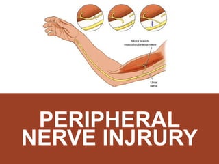
4. ANATOMY
ANATOMY OF ULNAR NERVE
Type: Mixed Never (motor & sensory)
Root Value: C7, C8 & T1
Origin: Arises in the axilla as the largest branch of
median cord of the brachial plexus, at the lower
border of pectoralis minor.
5. ANATOMY
ANATOMY OF ULNAR NERVE
Enters the forearm between 2 heads of Flexor Carpi Ulnaris
In the upper half, it rest on Flexor Digitorum Profundus &
covered by Flexor Carpi Ulnaris
In lower half, it runs lateral to Flexor Carpi Ulnaris tendon
accompanied by ulnar artery laterally
Enters palm on lateral side of pisiform bone above the flexor
retinaculum
Divides into Superficial & Deep Terminal Branches
9. ULNAR NERVE
SUMMARY OF BRANCHES OF ULNAR NERVE
MOTOR BRANCHES:
To 1 muscles only:
Flexor carpi ulnaris
Medial half of flexor digiti profundus
Superficial Terminal Branches supplies one muscle only: palmaris brevis
Deep Terminal Branch supplies:
8 interossei + medial 2 lumbricals
3 hypothenar muscle
Adductor Pollicis & may be FPB
FEMORAL
HAND
10. ULNAR NERVE
SUMMARY OF BRANCHES OF ULNAR NERVE
SENSORY BRANCHES:
Palmar cutaneous branch: Medial 1/3 of palm
Dorsal cutaneous branches: Medial 1/3 of dorsum of hand &
dorsum of medial 1
Superficial terminal branches gives 3 palmar digital nerves to the
palmar surfaces of the medial 1fingers
FEMORAL
HAND
11. 1. ABOVE ELBOW (HIGH ULNAR LESION)
ULNAR NERVE INJURY
Injuries lead to complete loss of all the functions of nerve. The
injury may be caused by:
A. Penetrating wounds
B. Gun Shots
C. Fracture of medial epicondyle
D. Cubitus Valgus (a deformity of elbow joint causing stretch of
ulnar nerve)
12. 1. ABOVE ELBOW (HIGH ULNAR LESION)
ULNAR NERVE INJURY
1. Paralysis of all muscles supplied:
1 muscles in the forearm
15 muscles in the hand
2. Weak flexion of the wrist, with radial deviation of the hand – due to
paralysis of FCU
3. Inability to flex the terminal phalanges of the medial 2 fingers – due to
paralysis of medial ½ of the FDP
4. Inability to put the hand in the writing position – due to paralysis of
interossei & medial 2 lumbricals
5. Loss of adduction of the thumb – paralysis of adductor pollicis
MOTOR AFFECTION
13. MANIFESTATIONS: ABOVE ELBOW
ULNAR NERVE INJURY
Partial Claw Hand – characterized by:
I. Extension of the metacarpophalangeal & flexion of
the interphalangeal joints – due to paralysis of
lumbricals & interossei
I. Flat hypothenar eminence – due to paralysis of it’s
muscles
Sensory loss from the skin of palmar & dorsal
surfaces of medial 1 of fingers.
DEFORMITY
SENSORY LOSS
14. 2. AT OR ABOVE THE WRIST (LOW ULNAR LESION)
ULNAR NERVE INJURY
Wrist laceration
Fracture of the carpal bones
Malunion of Colles fracture
Handcuffs
Motor:
Limited to hand muscles only. The forearm muscles are intact because they
receive supply very close to the elbow.
CAUSED BY
MANIFESTATIONS
15. 2. AT OR ABOVE THE WRIST (LOW ULNAR LESION)
ULNAR NERVE INJURY
Complete Claw Hand (in combined with median nerve injury)
More severe than in injury above the wrist, this is called the ulnar
paradox.
Sensory Loss:
Loss of sensation from the palmar surfaces of the medial 1 fingers
only – because the palmar & dorsal cutaneous branches are
intact.
DEFORMITY
16. SIGNS OF LOW LESION
ULNAR NERVE INJURY
Hypothenar muscle wasting
Claw hand
FROMENT’S SIGN POSITIVE
18. SIGNS OF LOW LESION
ULNAR NERVE INJURY
Loss of sensation over ulnar 1 ½ digits Tinel’s sign
19. COMPRESSION AT GUYON’S CANAL
ULNAR NERVE INJURY
Anatomy of Guyon’s canal :
Floor = transverse carpal ligament to
pisiform
Ulnar wall = pisiform
Radial distal wall = hook of hamate
Roof = volar carpal ligament
Contains only ulnar nerve and artery
ULNAR TUNNEL SYNDROME
20. COMPRESSION AT GUYON’S CANAL
ULNAR NERVE INJURY
Repetitive indirect trauma most common
Tumours- ganglion, lipoma
Pisiform instability
Pisotriquetral arthritis
Fractured hook of hamate / pisiform
Ulnar artery thrombosis
CAUSES
21. COMPRESSION AT GUYON’S CANAL
ULNAR NERVE INJURY
Symptoms could be:
1. Pure motor
2. Pure sensory
3. Mixed
(depending on exact site of entrapment)
CLINICAL FEATURES
22. COMPRESSION AT CUBITAL TUNNEL
ULNAR NERVE INJURY
Compression of ulnar nerve as it passes behind
elbow (behind medial epicondyle)
Intrinsic muscle wasting Waternberg’s sign
CUBITAL TUNNEL SYNDROME
23. COMPRESSION AT CUBITAL TUNNEL
ULNAR NERVE INJURY
Conservative
Modification of posture and avoidance of repetitive
trauma
Splint
Surgical
Operative decompression if symptoms persist
Tendon transfer to recover hand function
TREATMENT
26. ANATOMY
ANATOMY OF MEDIAN NERVE
Type: Mixed nerve (contains motor & sensory fibres)
Root Values: C5, 6, 7, 8 & T1
Origin: Arises in the axilla by 2 roots
Lateral root – lateral cord of the brachial plexus
Medial root – medial cord of the brachial plexus
The medial root crosses in front of the 3rd part of
axillary artery to join the lateral root.
28. ANATOMY
ANATOMY OF MEDIAN NERVE
ARM
Descends medial side at upper ½ then lateral side of brachial artery at
lower ½ arm
Cross bicipital aponeurosis then enters cubital fossa
FOREARM
Supply PT, FCR, PL, FDS
Between two head of PT, give anterior interosseous nerve branches and
supply FPL, FDP, PQ
HAND
Passes to the flexor retinaculum
Supply AbdPB, OP, FPB, 2 lumbrical and skin
32. SIGNS
Wasting of muscles of forearm
Wasting of thenar eminence
Weakness of thumb abduction and opposition
o Loss of abductor pollicis brevis + flexor pollicis brevis
The hand is held with ulnar fingers flexed and index
finger straight (pointing sign)
o Loss of FDP, FDS, FPL
Lost sensation at radial three and half digits
Weak Ok sign
Ape hand deformity
34. LOW MEDIAN NERVE LESION
MEDIAN NERVE INJURY
INJURY TO DISTAL THIRD OF THE FOREARM
I. Cuts in front of the wrist
II. Carpal dislocation
I. Wasting of thenar muscle
II. forearm muscle spared
III. Paralyzed muscle of the hand
IV. Weakness of thumb abduction and opposition
V. Loss of abductor pollicis brevis + flexor pollicis brevis
VI. Lost sensation at radial three and half digits
SIGNS
35. COMPARISON
MEDIAN NERVE INJURY
Low lesion High lesion
Wasting of thenar eminence
Weak thumb abduction
Weak thumb opposition
Loss of sensation over
lateral 3 and half digits
All the signs of low lesion
Wasting of lateral forearm
Weakened OK sign (AIN – FPL & FDP)
Pointing finger (2nd and 3rd finger remains
partially extended in an attempt to make a fist)
36. PHYSICAL EXAMINATION
MEDIAN NERVE PE
a. Thenar wasting
b. Atrophy of pulp of index, cracking of nails and other
trophic changes
c. Cigarette burns or other loss of sensory deprivation
d. Pointing finger in high nerve lesion
Flexor carpi radialis and palmaris longus. Patients hand
is placed on a flat surface, palm upwards. Ask patient
to attempt to flex the wrist with examiners hand
putting pressure on top. The (pl) and (fcr) tendon will
be prominent
INSPECTION
38. PE OF MEDIAN NERVE MUSCLE DISTRIBUTION
MEDIAN NERVE PE
Flexor pollicis longus
Flex the terminal phalanx of
the thumb against resistance
while the proximal phalanx is
kept steady by examiner
Flexor carpi radialis
The wrist deviates to the ulnar
side
39. PE OF MEDIAN NERVE MUSCLE DISTRIBUTION
MEDIAN NERVE PE
Flexor digitorum superficialis
Patient asked to clasp his hand,
the index finger will remain
straight (Pointing index)
Muscle of thenar eminence
Abductor pollicis brevis (Pen
test)
Lay the hand flat, a pen is held
above the thumb, try to touch
the pen with tip of thumb
40. PE OF MEDIAN NERVE MUSCLE DISTRIBUTION
MEDIAN NERVE PE
Opponence pollicis
Appose the tip of thumb to
other finger
44. CARPAL TUNNEL SYNDROME
MEDIAN NERVE INJURY
Common in middle age group 40-50 years
More common in female
Causes:
i. Inflammatory - RA, wrist OA,
ii. Post traumatic – Dislocation of one of the carpal
bones inside the carpal tunnel
iii. Endocrine – Myxoedema, Cushing, Acromegaly
(thickening of the tendons passing)
iv. Tumour inside the carpal tunnel pressing on the
median nerve
v. Idiopathic
45. CARPAL TUNNEL SYNDROME
MEDIAN NERVE INJURY
HISTORY
Pain and paresthesia at distribution of
median nerve in the hand
Wake up at night due to burning pain,
tingling and numbness
Relieve by shaking the arm
Dropping object
46. CARPAL TUNNEL SYNDROME
MEDIAN NERVE INJURY
PHYSICAL EXAMINATION
Hands may look normal/ wasting in
severe cases
TINEL SIGN
Percussing over the median nerve
causing sensation of current/
hyperesthesia at median nerve
distribution
PHALEN SIGN
Flexed the wrist fully for one or two
minutes causing paresthesia
48. CARPAL TUNNEL SYNDROME
MEDIAN NERVE INJURY
INVESTIGATIONS
ELECTRODIAGNOSTIC TEST
I. Nerve conduction studies
Measure Median motor and sensory latencies and
conduction velocities across the wrist
Sensory latency of greater than 3.5 millisecond or a motor
latency of greater than 4.5 millisecond is considered an
abnormal finding
Distal compound muscle action potential (CMAP) and
sensory nerve action potential (SNAP) amplitudes may be
decreased
50. CARPAL TUNNEL SYNDROME
MEDIAN NERVE INJURY
INVESTIGATIONS
ELECTRODIAGNOSTIC TEST
II. Electromyography
To determine completeness of a nerve injury
Technique:
I. Very small needle is inserted into various muscle
II. Then, the signal is magnified by high gain amplifier
III. Finally, the reading are monitored via oscilloscope and
recorded on the magnetic tape or paper recording
IV. Should performed 3-7 days after peripheral nerve injury
V. It may show low amplitude evoked compound muscle
potential (CMAP)
51. CARPAL TUNNEL SYNDROME
MEDIAN NERVE INJURY
TREATMENT
Conservatives
Painkiller - PCM,NSAIDS
Corticosteroid injection - to reduce edema
Splint - prevent wrist flexion
Physiotherapy
Modifying activities - avoid repetitive or strenuous work
Surgical
Open surgical division of transverse carpal ligament
65. RADIAL NERVE LESION
1. Very high lesion (in the axilla)
2. High lesion (humeral shaft level)
3. Low lesion(around the elbow )
4. Wartenberg’s Syndrome (at the
wrist)
66. RADIAL NERVE LESIONS
VERY HIGH LESION (IN THE AXILLA)
1. Pressure from badly fitting crutch
(‘crutch palsy’)
2. Saturday night’s palsy
3. Honeymoon palsy
CAUSES
70. RADIAL NERVE LESIONS
VERY HIGH LESION (IN THE AXILLA)
1. Weakness of wrist, fingers and thumb
extension – wrist, fingers and thumb drop
2. Weakness of elbow extension – due to
paralysis of the triceps
3. Absent of triceps reflex
4. Sensory loss in the distribution of the more
proximal cutaneous branches
CLINICAL FEATURES
72. RADIAL NERVE LESIONS
HIGH LESION (HUMERAL SHAFT)
CAUSES
1. Fracture of shaft of humerus
2. Pressure on the back of the arm on the edge of
table in an unconscious patient
3. Prolong application of tourniquet to the arm
4. Drunken man who fall asleep with the arm
dangling over the back of a chair ( Saturday night
palsy)
73. RADIAL NERVE LESIONS
HIGH LESION (HUMERAL SHAFT)
CLINICAL FEATURES
1. Wrist drop / weakness of wrist extension – due to paralysis
of the Extensor Carpi Radialis Longus and Brevis
1. Finger drop / weakness of fingers extension at the MCPJ –
due to paralysis of the Extensor Digitorum
1. Thumb drop / weakness of the whole thumb extension –
due to paralysis of the Extensor Pollicis longus and brevis)
74. RADIAL NERVE LESIONS
HIGH LESION (HUMERAL SHAFT)
CLINICAL FEATURES
4. Paralysis of the brachioradialis muscle.
The patients is asked to hold his forearm in 90 degree flexion
and midprone position. Ask him to flex the elbow
against resistance applied at the wrist. The brachio radialis
does not stand out prominently as it is paralysed.
Sensory signs: Sensory loss is minimal and is confined to a
small area in the dorsum of the hand over the metacarpal
bones of the thumb and index fingers (the distribution of the
s/f radial nerve)
76. RADIAL NERVE LESIONS
LOW LESION (AROUND THE ELBOW)
CAUSES
The posterior interosseous branch of the
radial nerve is injured in :-
o Dislocation of the head of radius
o Accidently injured during surgical excision of the
head of the radius
77. RADIAL NERVE LESIONS
LOW LESION (AROUND THE ELBOW)
CAUSES
The wrist extension is preserved
No wrist drop because branch to ECRL
arise proximal to the elbow.
Weakness of the fingers and thumb
extension at the MCPJ
No sensory loss
78. RADIAL NERVE LESIONS
WARTENBERG’S SYNDROME
Wartenberg syndrome, described in 1932, is
essentially entrapment of the superficial sensory
branch of the radial nerve.
Many factors may contribute to the development of
Wartenberg syndrome. In patients with de Quervain
tenosynovitis, secondary irritation of the RSN is
frequent. Other common causes include postoperative
injury, external compression, and trauma.
79. TREATMENT
CONSERVSTIVE TREATMENT
The wrist and fingers are splinted in a position of
extension at the wrist and M.P. Joints by ‘cock
up’ splint
To prevent overstretching of the paralysed
muscles.
Disadvantage : prevent activity of unparalysed
flexor muscles of the wrist and m.P.Joints.
80. TREATMENT
CONSERVSTIVE TREATMENT
The modern splint : Dynamic or Lively splint
Applied on the dorsal aspect
Keeps the wrist and fingers extended by elastic
bands or springs attached to it
Allows active flexion of the fingers and wrist.
81. TREATMENT
CONSERVSTIVE TREATMENT
Passive movements to the wrist and finger
joints.
Electrical stimulation is given to the
paralyzed muscles to prevent wasting of
muscles.
Progressive active exercises are given to the
muscles showing recovery. As most of the
lesions are neuoparaxia or axonotmesis,
recovery occurs in about 4 to 6 weeks.
82. TREATMENT
SURGICAL TREATMENT
When there is evidence of Neuronotmesis,
exploration and repair of the nerve gives
good results as it is mostly a motor nerve.
When the radial nerve is irreparably
damaged, tendon transfer operations are
done to restore the extensor functions at the
wrist, fingers and thumb.
86. ANATOMY
ANATOMY OF TIBIAL NERVE
The tibial nerve is derived from the L4 – S3 nerve roots as part of the
sciatica nerve.
The tibial nerve runs through the popliteal fossa to pass below the arch of
soleus.
In the popliteal fossa the nerve gives off branches to gastrocnemius,
popliteus, soleus and plantaris muscles, an articular branch to the knee
joint, and a cutaneous branch that will become the sural nerve.
After passing deep to the soleus, it continues in the posterior
compartment between the tibialis posterior and the soleus muscles.
At the medial ankle, the nerve becomes superficial, before passing into
the foot through the tarsal tunnel.
Within the tunnel it splits into the medial and lateral plantar nerves.
The medial plantar nerve divides into muscular and cutaneous branches.
The lateral plantar nerve passes between the quadrates plantae and flexor
digitorum brevis.
87. 1. TRAUMATIC
Can be direct or indirect trauma, (commonly to distal tibia, ankle ) such as
open fractures, deep laceration in the popliteal fossa, dislocation of the knee
and injuries to the ankle as well
MECHANISM OF INJURY
Can be caused by any mass, abscess, bleeding into the knee, unreleased
compartment syndrome.
2. COMPRESSION
Such as rheumatoid arthritis, diabetes or vascular disease.
Thus, any trauma or pressure will destroy the myelin sheath that protects and
insulates the nerve, or part of the nerve cell (the axon). This damage reduces
or prevents the movement of impulses through the nerve.
3. SYSTEMIC DISEASE
88. Sensation changes in the bottom of the foot and
toes, including burning sensation, numbness,
tingling, or other abnormal sensation, or pain.
Weakness of foot muscles.
Weakness of the toes or ankle.
Ankle that rolls inwards.
Muscle atrophy
SIGNS AND SYMPYTOMS
89. 1. Walking and running, ( active in sports )
2. High impact sports, ( rugby, football )
3. Hiking
4. Climbing stairs
5. Obesity
6. Diabetes and hypertension
RISK FACTORS
90. INSPECTION
PHYSICAL EXAMINATION
Look for signs of injury, wound, scars
Look for any ulcers or pressure points ( commonly in toes and medial malleolus )
Clawing of toes ( posterior tibial nerve affected )
Muscle atrophy ( calf and foot )
PALPATION
Swelling or dryness
Sensory test
Motor function test ( plantar flex and toe flex )
INVESTIGATIONS
Electromyography
Nerve conduction test
Nerve biopsy
91. MANAGEMENT
The treatment of tibial nerve injury is undertaken based on the severity of
the condition and assessment of the signs and symptoms. The condition is
often treatable without surgery.
Applying ice to the sore area. ( Cold therapy )
Taking non-steroidal anti-inflammatory medications.
Resting; avoiding running or playing high impact sports until the affected
leg heels.
If Tibial nerve injury is severe and does not resolve using the above non-
surgical treatment methods, then the following invasive surgical procedures
may be necessary:
a. Lengthening of the calf muscle.
b. Removing damaged tendon areas.
c. Osteotomy
Physiotherapy
Electric Stimulation
93. ANATOMY
The common peroneal nerve, about one-half the size of the tibial
nerve, arises from the dorsal branches of L4,L5 and S1,S2
It descends obliquely along the lateral side of the popliteal fossa to
the head of the fibula, close to the medial margin of the biceps
femoris muscle. Where the common peroneal nerve winds round
the head of the fibula, it is palpable.
Between the peroneus longus and the bone, it divides beneath the
muscle into the superficial peroneal nerve and deep peroneal
nerve.
A peroneal nerve injury (also called foot drop), is a peripheral
nerve injury that affects a patient’s ability to lift the foot at the
ankle. While foot drop injury is a neuromuscular disorder, it can
also be a symptom of a more serious injury, such as a nerve
compression or herniated disc.
95. 1. TRAUMATIC
Caused by any fractures around the knee, specifically to the head of fibula,
supracondylar even knee dislocation.
MECHANISM OF INJURY
Caused by any plaster usage or cast, swelling, mass, or abscess. Certain cases
habitual leg crossing as well.
2. COMPRESSION
Such as rheumatoid arthritis, diabetes or hypertension
3. SYSTEMIC DISEASE
96. Decreased sensation and numbness on the outer half
of the leg or dosum of the foot.
Weakness of the ankles or feet,
Foot drop
Toes drag while walking
High stepping gait
SIGNS AND SYMPYTOMS
97. INSPECTION
PHYSICAL EXAMINATION
1. High stepping gait and foot drop
2. Look for any injuries or previous scars
3. Muscle atrophy
PALPATION
1. Sensory test
2. Motor function test
INVESTIGATIONS
1. EMG
2. Nerve conduction test
3. Nerve biopsy
98. MANAGEMENT
Resting from any activities that cause the symptoms to get worse.
Applying ice to the sore area.
Use of ankle or foot braces to support the foot
Analgesics and NSAIDS
If severe, surgery is done to reduce decompression or to fix the
underlying cause. ( peroneal nerve repair