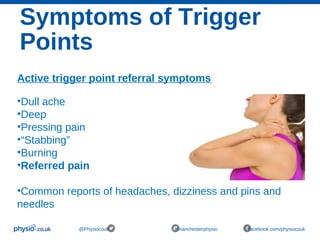
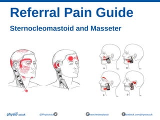
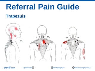
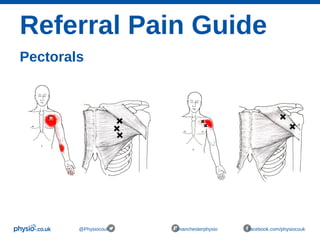
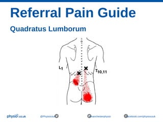
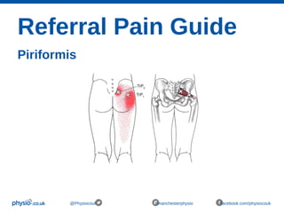
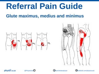
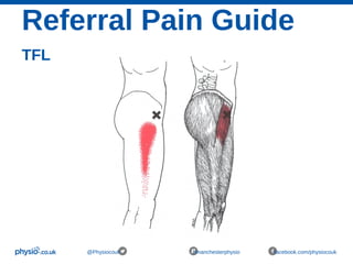
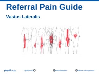
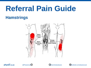
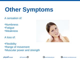
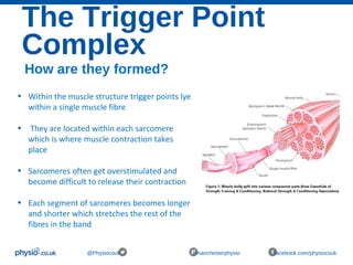
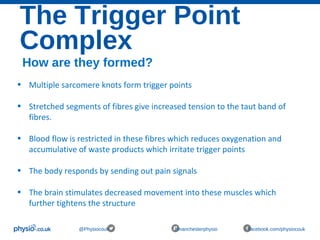
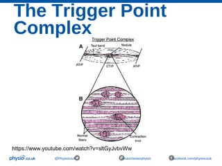
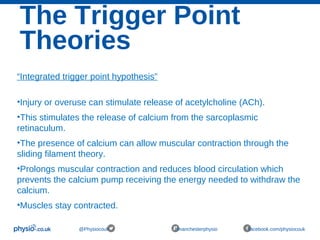
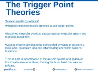
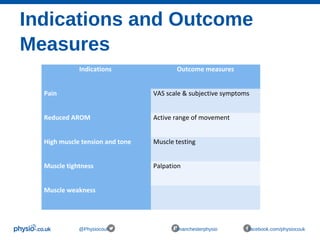
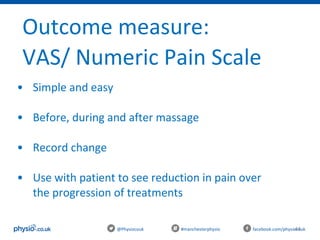
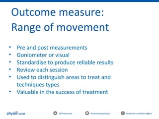
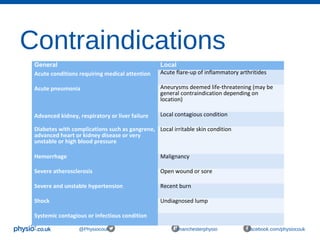
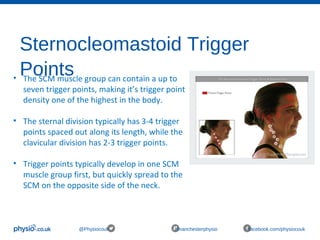
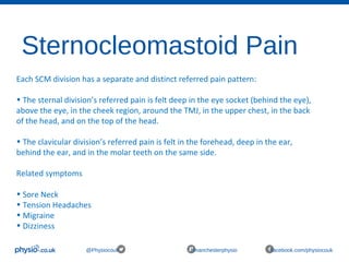
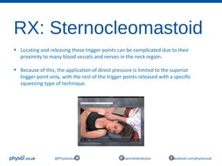
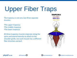
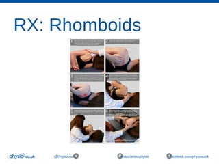
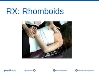
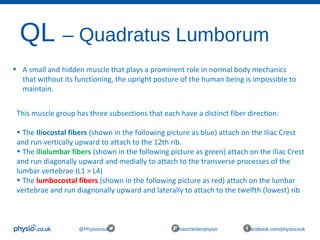
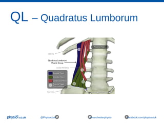
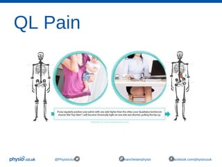
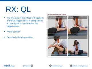
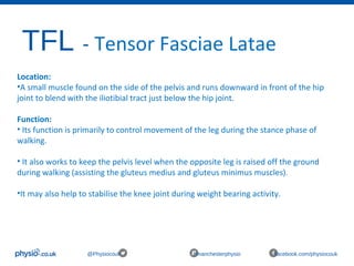
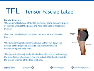
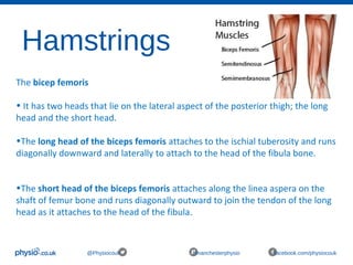
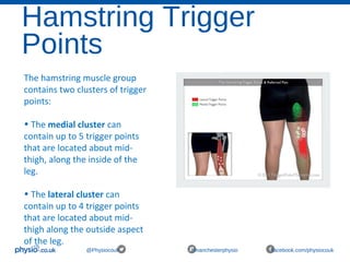
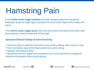
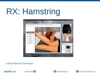
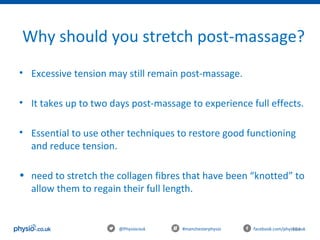
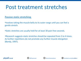
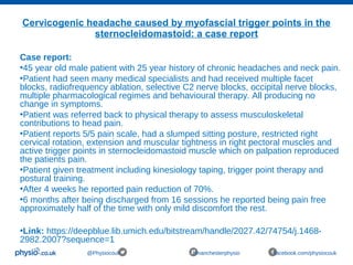
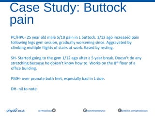
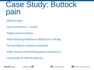
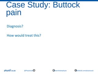
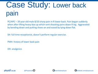
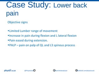
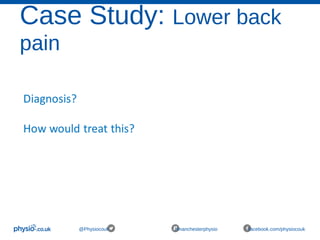
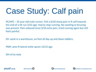
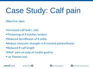
@Physiocouk #manchesterphysio facebook.com/physio
05/03/16 2
Welcome
Trigger point therapy & soft tissue release for sports
and massage therapists
With Katie Emmett & Kate Mcnally
@Physiocouk #manchesterphysio facebook.com/physiocouk
3
@Physiocouk #manchesterphysio facebook.com/physiocouk
Who are we?
Katie’s LinkedIn: www.linkedin.com/katieemmett
Twitter: @KatiePhysiocouk
Kate’s LinkedIn: www.linkedin.com/katemcnally
Twitter: @KateMcPhysiocouk
4
@Physiocouk #manchesterphysio facebook.com/physiocouk
Let’s connect
Website: www.physio.co.uk
Twitter: @physiocouk
Facebook: www.facebook.com/physiocouk
Aims of today
@Physiocouk #manchesterphysio facebook.com/physiocouk
 Learn the theory of a trigger point
 Learn the theory of trigger point therapy
 Practice the trigger point technique to muscle groups
 Use other soft tissue release techniques along side TP release
Itinerary
@Physiocouk #manchesterphysio facebook.com/physiocouk
10.00 - 10.30 - Induction / Arrival
10.30 - 10.50 - Quiz – What do you know about trigger point
therapy
10.50 -11.30 - Theory: Trigger point therapy
11.30 -12.00 - Practical: workshop
12.00 - 12.30 - Lunch
12.30 - 13.00 - Theory: Trigger pointing technique
13.00 - 14.00 - Practical: Muscle groups
14.00 - 14.30 – Practical: Tools & other STR techniques
14.30 - 15.00 - Evidence/Case Studies/Quiz answers
Quiz…
What do you know about trigger point
therapy?
@Physiocouk #manchesterphysio facebook.com/physiocouk
Question 1
@Physiocouk #manchesterphysio facebook.com/physiocouk
Name a type of Trigger Point?
Question 2
@Physiocouk #manchesterphysio facebook.com/physiocouk
How would patients describe trigger point
pain?
Question 3
@Physiocouk #manchesterphysio facebook.com/physiocouk
Name some indications for Trigger Point
Therapy?
Question 4
@Physiocouk #manchesterphysio facebook.com/physiocouk
Name 5 benefits of Trigger Point Therapy
Question 5
@Physiocouk #manchesterphysio facebook.com/physiocouk
Where are the Rhomboid muscles located?
Question 6
@Physiocouk #manchesterphysio facebook.com/physiocouk
Name the muscles in the Hamstring group
Question 7
@Physiocouk #manchesterphysio facebook.com/physiocouk
Name 5 contraindications of Trigger point
therapy
Question 8
@Physiocouk #manchesterphysio facebook.com/physiocouk
Name some related symptoms to trigger
points in the Sternocleomastoid muscle
Theory:
Trigger Point
Therapy
@Physiocouk #manchesterphysio facebook.com/physiocouk
What are trigger
points?
@Physiocouk #manchesterphysio facebook.com/physiocouk
• Trigger points are hyperirritable areas of contracted
muscle fibres that form a palatable nodule
• On a microscopic level, the contracted muscle fibres
accumulate into a small thickened area causing the
rest of the fibre to stretch
• The areas of contracted muscle restrict blood flow
within the tissue causing an accumulation of waste
products and reduced levels of nutrients available.
Brief History
@Physiocouk #manchesterphysio facebook.com/physiocouk
• 1930s -Dr Hans Lange used sclerometer to prove that tender areas in muscles
are 50% harder than surrounding areas.
• 1940s- Janet Travell developed trigger point injection therapy and termed the
ā€œtender areasā€ described by Dr Hans ā€œTrigger pointsā€.
• Travell's therapy called for the injection of saline (a salt solution) and procaine
(also known as Novocaine, an anesthetic) into the trigger point.
• Travell mapped what she termed the body's trigger points and the manner in
which pain radiates to the rest of the body.
• Travell's work came to national attention when she treated President John F.
Kennedy for his back pain.
• Travell co-authored several books with David Simons which are considered
the definitive reference for trigger point therapy.
• Travell & Simons' Myofascial Pain and Dysfunction: Upper half of body
• Travell & Simons' Myofascial Pain and Dysfunction: The Trigger Point Manual
• Myofascial Pain and Dysfunction: The Trigger Point Manual, Volume 2
Brief History
@Physiocouk #manchesterphysio facebook.com/physiocouk
• 1976- Bonnie Prudden, a physical fitness and exercise therapist
developed Travells trigger point therapy. She found that applying
sustained pressure to a trigger point using thumbs, knuckles and
elbows produced superior results to those treated with injections
when followed by corrective movements and stretching. Prudden
later went on to author two books:
• Myotherapy: Bonnie Prudden’s Complete Guide to Pain Free Living
• Pain Erasure the Bonnie Prudden Way
Different types of trigger
points
@Physiocouk #manchesterphysio facebook.com/physiocouk
• Trigger points are described according to location, tenderness and
chronicity as central (or primary), satellite (or secondary), attachment,
diffuse, inactive (or latent) and active
• The main types of trigger points are:
 Central/ primary trigger points
 Satellite/ secondary trigger points
 Active trigger points
 Latent trigger points
Central/ primary trigger
points
@Physiocouk #manchesterphysio facebook.com/physiocouk
• These are the most well-established and painful points
• Pain is felt by the individual when they are active, and are usually
what people refer to when they talk about trigger points
• Central trigger points exist at a neuromuscular point, which is the
meeting place of a nerve and muscle
Satellite/ secondary trigger
points
@Physiocouk #manchesterphysio facebook.com/physiocouk
• These trigger points are ā€œcreatedā€ as a response to the central
trigger point in neighbouring muscles that lie within the referred
pain zone.
• Form in response to central trigger points within the pain referral
patterns
• The primary trigger point is still the key to trigger pointing
intervention: the satellite trigger points often resolve once the
primary point has been effectively rendered inactive.
• Satellite points may also prove resilient to treatment until the
primary central focus is weakened; such is often the case in the
paraspinal and/or abdominal muscles.
Active trigger points
@Physiocouk #manchesterphysio facebook.com/physiocouk
• This can apply to central and satellite trigger points.
• A variety of stimulants, such as forcing muscular activity
through pain, can activate an inactive trigger point.
• This situation is common when activity is increased after trauma
i.e a road traffic accident, where multiple and diffuse trigger
points may have developed.
• This trigger point is both tender to palpation and elicits a referred
pain pattern.
• Pain can limit range of movement
Latent trigger points
@Physiocouk #manchesterphysio facebook.com/physiocouk
• This applies to lumps and nodules that feel like trigger points. These can
develop anywhere in the body and are often secondary.
• These trigger points are not painful, and do not elicit a referred pain
pathway.
• The presence of inactive trigger points within muscles may lead to
increased muscular stiffness and tension. They can build up for years.
• It has been suggested that these points are more common in those who
live a sedentary lifestyle (Starlanyl & Copeland 2001)
• These points are ā€œpotentialā€ trigger points and may reactivate if the
central or primary trigger point is (re)stimulated
• Reactivation may occur following trauma and injury
Symptoms of Trigger
Points
@Physiocouk #manchesterphysio facebook.com/physiocouk
Active trigger point referral symptoms
•Dull ache
•Deep
•Pressing pain
ā€¢ā€œStabbingā€
•Burning
•Referred pain
•Common reports of headaches, dizziness and pins and
needles
Referral Pain Guide
Sternocleomastoid and Masseter
@Physiocouk #manchesterphysio facebook.com/physiocouk
Referral Pain Guide
Trapezuis
@Physiocouk #manchesterphysio facebook.com/physiocouk
Referral Pain Guide
Pectorals
@Physiocouk #manchesterphysio facebook.com/physiocouk
Referral Pain Guide
Quadratus Lumborum
@Physiocouk #manchesterphysio facebook.com/physiocouk
Referral Pain Guide
Piriformis
@Physiocouk #manchesterphysio facebook.com/physiocouk
Referral Pain Guide
Glute maximus, medius and minimus
@Physiocouk #manchesterphysio facebook.com/physiocouk
Referral Pain Guide
TFL
@Physiocouk #manchesterphysio facebook.com/physiocouk
Referral Pain Guide
Vastus Lateralis
@Physiocouk #manchesterphysio facebook.com/physiocouk
Referral Pain Guide
Hamstrings
@Physiocouk #manchesterphysio facebook.com/physiocouk
Other Symptoms
@Physiocouk #manchesterphysio facebook.com/physiocouk
A sensation of:
•Numbness
•Fatigue
•Weakness
A loss of:
•Flexibility
•Range of movement
•Muscular power and strength
Why are they
present?
@Physiocouk #manchesterphysio facebook.com/physiocouk
• Repetitive overuse injuries (using the same body parts
in the same way hundreds of times on a daily basis) from
activities such as typing/mousing, handheld electronics,
gardening, home improvement projects, work environments,
etc.
• Sustained loading e.g heavy lifting, carrying babies,
briefcases, boxes or lifting bedridden patients.
Why are they
present?
@Physiocouk #manchesterphysio facebook.com/physiocouk
•Poor posture due to our sedentary lifestyles, de-
conditioning, poorly designed furniture and technology.
•Muscle clenching and tensing due to mental/emotional
stress.
•Direct injury such as a strain, break, twist or tear e.g car
accidents, sports injuries, falling down stairs.
•Trigger points can even develop due to inactivity such as
prolonged bed rest or sitting.
The Trigger Point
Complex
@Physiocouk #manchesterphysio facebook.com/physiocouk
How are they formed?
• Within the muscle structure trigger points lye
within a single muscle fibre
• They are located within each sarcomere
which is where muscle contraction takes
place
• Sarcomeres often get overstimulated and
become difficult to release their contraction
• Each segment of sarcomeres becomes longer
and shorter which stretches the rest of the
fibres in the band
The Trigger Point
Complex
@Physiocouk #manchesterphysio facebook.com/physiocouk
How are they formed?
• Multiple sarcomere knots form trigger points
• Stretched segments of fibres give increased tension to the taut band of
fibres.
• Blood flow is restricted in these fibres which reduces oxygenation and
accumulative of waste products which irritate trigger points
• The body responds by sending out pain signals
• The brain stimulates decreased movement into these muscles which
further tightens the structure
The Trigger Point
Complex
@Physiocouk #manchesterphysio facebook.com/physiocouk
https://www.youtube.com/watch?v=sltGyJvbvWw
The Trigger Point
Theories
@Physiocouk #manchesterphysio facebook.com/physiocouk
ā€œIntegrated trigger point hypothesisā€
•Injury or overuse can stimulate release of acetylcholine (ACh).
•This stimulates the release of calcium from the sarcoplasmic
retinaculum.
•The presence of calcium can allow muscular contraction through the
sliding filament theory.
•Prolongs muscular contraction and reduces blood circulation which
prevents the calcium pump receiving the energy needed to withdraw the
calcium.
•Muscles stay contracted.
The Trigger Point
Theories
@Physiocouk #manchesterphysio facebook.com/physiocouk
ā€œMuscle spindle hypothesisā€
•Proposes inflamed muscle spindles cause trigger points.
•Sustained muscular overload causes fatigue, muscular spasm and
restricted blood flow.
•Causes muscle spindles to be surrounded by waste products e.g.
lactic acid, potassium ions and inflammatory chemicals such as
histamine.
•This results in inflammation of the muscle spindle and spasm of
the extrafusal muscle fibres, forming the taunt band that we can
palpate.
Indications and Outcome
Measures
@Physiocouk #manchesterphysio facebook.com/physiocouk
Indications Outcome measures
Pain VAS scale & subjective symptoms
Reduced AROM Active range of movement
High muscle tension and tone Muscle testing
Muscle tightness Palpation
Muscle weakness
Ā Ā 
44@Physiocouk #manchesterphysio facebook.com/physiocouk
Outcome measure:
VAS/ Numeric Pain Scale
• Simple and easy
• Before, during and after massage
• Record change
• Use with patient to see reduction in pain over
the progression of treatments
45@Physiocouk #manchesterphysio facebook.com/physiocouk
Outcome measure:
Range of movement
• Pre and post measurements
• Goniometer or visual
• Standardise to produce reliable results
• Review each session
• Used to distinguish areas to treat and
techniques types
• Valuable in the success of treatment
46@Physiocouk #manchesterphysio facebook.com/physiocouk
Outcome measure:
Muscle testing
• Measure nerve conduction, muscle recruitment
to determine a deficit
• Test uninjured side for norm
• Patient will see and feel a progression
• Strengthening exercises needs to be used along
side massage
47@Physiocouk #manchesterphysio facebook.com/physiocouk
Outcome measure:
Palpation
• Use palpation as a measure
• ā€œthe four T’sā€
Temperature
Tissue may be hot or cold, indicating inflammation or ischaemia
Texture
Swelling (acute-hard, chronic – ā€œboggyā€, congested)
healthy tissues should have an even texture
Adhesions feel like tissues are ā€œstuckā€ and less mobile
ā€œaudible crunchingā€
48@Physiocouk #manchesterphysio facebook.com/physiocouk
Outcome measure:
Palpation
Tenderness
Pain can be indicated through response/ use vas scores
Structures that are too painful to palpate
Tone
Tissues may be hypertonic or hypotonic
Use to compare
Practical:
Trigger point
workshop:
Symptoms
@Physiocouk #manchesterphysio facebook.com/physiocouk
Lunch
@Physiocouk #manchesterphysio facebook.com/physiocouk
Theory:
Trigger Pointing
Therapy
@Physiocouk #manchesterphysio facebook.com/physiocouk
How to treat a Trigger
Point
@Physiocouk #manchesterphysio facebook.com/physiocouk
Assessment
•Find the most painful TP using patient response and Numeric Rating Scale or (VAS)
•Treat the highest rated point and radiate out from this point
•Once the points are found – a good amount of pressure is applied (perform with
precaution - keep communication with patient)
•Initial pain is stimulated and you hold the pressure until the pain has eased completely or
in some cases reduced slightly
•Reapply pressure onto the same point until the pain eases off quicker or it isn’t felt
anymore
•Thumbs/elbows or tools can be used
How to treat a Trigger
Point
@Physiocouk #manchesterphysio facebook.com/physiocouk
Guidelines
Application of direct pressure onto the trigger points for around 30 seconds or until
the patient’s pain has decreased to at least 3/10 VAS score.
The applied pressure help breakup the adhesive fibre connections within the
trigger points and push out blood containing waste products and toxins.
After 30 seconds the pressure is released allowing a rush of fresh blood containing
nutrients to circulate the trigger point.
Repeat 3 times in conjunction with deep massage strokes.
• This can depend on the severity of pain/ how deep or superficial the TP is –
subjective and variable to each patient
The Benefits
@Physiocouk #manchesterphysio facebook.com/physiocouk
• Reduced pain
• Increased range of motion
• Decreased muscle stiffness and tension
• Reduction in headaches
• Improved flexibility
• Improved circulation
• Fewer muscle spasms
Precautions
@Physiocouk #manchesterphysio facebook.com/physiocouk
• High pain scales
• Patient Anxiety
• Acute/ Inflammatory stage of healing
• Hypersensitivity
• Pregnancy
• Epilepsy
• Asthma
• Hypertension
• Prescribed medication
Contraindications
@Physiocouk #manchesterphysio facebook.com/physiocouk
General Local
Acute conditions requiring medical attention Acute flare-up of inflammatory arthritides
Acute pneumonia Aneurysms deemed life-threatening (may be
general contraindication depending on
location)
Advanced kidney, respiratory or liver failure Local contagious condition
Diabetes with complications such as gangrene,
advanced heart or kidney disease or very
unstable or high blood pressure
Local irritable skin condition
Hemorrhage Malignancy
Severe atherosclerosis Open wound or sore
Severe and unstable hypertension Recent burn
Shock Undiagnosed lump
Systemic contagious or infectious condition
Manual Handling and
Body Position
@Physiocouk #manchesterphysio facebook.com/physiocouk
• Posture
– Bed height
– Stance
– Patient position
• Use different parts of your hands/ arms to apply pressure
• Keep arms straight to utilise body weight when applying
pressure/resistance.
• Move from the hips and knees as much as possible
• Oil (or cream)- only needs to be a little bit, if any.
Look after yourself before you look after the patient!
Post Treatment Irritation
@Physiocouk #manchesterphysio facebook.com/physiocouk
Very common for people to experience irritation for up to 72
hours after treatment.
Side effects can include:
• Bruising
• Redness
• Tenderness/Increased Sensitivity
• Increased symptoms
• Aching similar to DOMS
Post Treatment Irritation
@Physiocouk #manchesterphysio facebook.com/physiocouk
Causes
• The release of toxins/waste products from muscular tissue
• Neurological sensitisation
• Increased blood flow and micro trauma can lead to bruising and
redness
Advice
•Reassure the patient it's a normal response to be
sore after soft tissue treatment
•Recommend they drink water to keep hydrated
Practical:
Trigger pointing
muscles
@Physiocouk #manchesterphysio facebook.com/physiocouk
• Sternocleomastoid
• UFT
• Rhomboids
• QL
• TFL
• Vastus Lateralis
• Hamstrings
Sternocleomastoid
@Physiocouk #manchesterphysio facebook.com/physiocouk
Anatomical Highlights:
• Each SCM group has two divisions that originate off the mastoid process behind the ear.
The sternal division runs diagonally downward to attach to the sternum, while the clavicular
division attaches right behind it on the medial clavicle.
• Acting unilaterally, contraction of the SCM muscle turns the head towards the opposite
side, while bilateral contraction flexes the neck and head forward.
• The most important function of the SCM is to control and monitor the head’s position in
space. Proprioceptive feedback from the SCM is essential to being able to maintain one’s
balance, and is also important for interpreting visual information.
Sternocleomastoid Trigger
Points
@Physiocouk #manchesterphysio facebook.com/physiocouk
• The SCM muscle group can contain a up to
seven trigger points, making it’s trigger point
density one of the highest in the body.
• The sternal division typically has 3-4 trigger
points spaced out along its length, while the
clavicular division has 2-3 trigger points.
• Trigger points typically develop in one SCM
muscle group first, but quickly spread to the
SCM on the opposite side of the neck.
Sternocleomastoid Pain
@Physiocouk #manchesterphysio facebook.com/physiocouk
Each SCM division has a separate and distinct referred pain pattern:
• The sternal division’s referred pain is felt deep in the eye socket (behind the eye),
above the eye, in the cheek region, around the TMJ, in the upper chest, in the back
of the head, and on the top of the head.
• The clavicular division’s referred pain is felt in the forehead, deep in the ear,
behind the ear, and in the molar teeth on the same side.
Related symptoms
• Sore Neck
• Tension Headaches
• Migraine
• Dizziness
RX: Sternocleomastoid
@Physiocouk #manchesterphysio facebook.com/physiocouk
• Locating and releasing these trigger points can be complicated due to their
proximity to many blood vessels and nerves in the neck region.
• Because of this, the application of direct pressure is limited to the superior
trigger point only, with the rest of the trigger points released with a specific
squeezing-type of technique.
Upper Fiber Traps
@Physiocouk #manchesterphysio facebook.com/physiocouk
The trapezius is not one, but three separate
muscles:
•The upper trapezius
•The middle trapezius
•The lower trapezius
All three trapezius muscles originate along the
spine and extend laterally to attach to the
shoulder girdle, but each muscle has a different
fiber direction and pull.
Upper Fiber Traps
@Physiocouk #manchesterphysio facebook.com/physiocouk
The whole trapezius muscle creates various movements of the shoulder blade, neck,
and head.
An example, the simple act of flexing the head to the right requires:
•Contraction of the lower trapezius on the right side to fix the right shoulder blade in
place.
•Contraction of the right upper trapezius to pull the neck and head to the right.
•Relaxation of the left lower trapezius to allow the left shoulder blade to rise.
•Relaxation of the left upper trapezius to allow the neck and head to move to the right.
This type of complexity makes it easy for trigger point activity to spread quickly
through the muscle group as a whole.
UFT Trigger Points
@Physiocouk #manchesterphysio facebook.com/physiocouk
Four primary trigger points in the
trapezius muscle group; two trigger
points in the upper fibers, and one each
in the middle and lower fibers.
• The anterior trapezius trigger point
• The upper trapezius trigger point
• The middle trapezius trigger point
• The lower trapezius trigger point
UFT Pain
@Physiocouk #manchesterphysio facebook.com/physiocouk
• ā€œPain in the neckā€
• The mental and emotional stress of modern day life often takes physical form as trigger
points in the lower and upper trapezius muscles.
• The lower trapezius trigger point is the most sensitive to psychological and projects
pain and tenderness upward into the neck and shoulder region.
• The anterior trigger point refers pain to the side of the neck, jaw, and face, but it is
notorious for producing a throbbing headache in the temple region. This headache
pain may also be described as ā€œbehind the eye.ā€
• Middle trapezius trigger point, which produces a localised burning-type pain along the
spine. It will often recruit the rhomboid trigger points as they share a similar intra-
scapular pain pattern.
RX: UFT
@Physiocouk #manchesterphysio facebook.com/physiocouk
• The anterior trapezius trigger point
• The upper trapezius trigger point
• The middle trapezius trigger point
• The lower trapezius trigger point
Rhomboids
@Physiocouk #manchesterphysio facebook.com/physiocouk
ā€œThat Nagging Pain Between the Shoulder Bladesā€
• Location: The rhomboid muscle group is found between the spine and the scapula
in the mid- back region. It lies deep to the Trapezius muscle and is composed of
the rhomboid major and rhomboid minor muscles.
• Structure: The rhomboid minor is smaller than and lies above (superior to) the
rhomboid major. Both muscles originate along the thoracic spine with their fibers
running diagonally downward and outward to attach along the inside border of the
scapula.
• Function: In everyday life, the rhomboid muscles function to position the scapula
during various movements of the shoulder and arm.
Rhomboids
@Physiocouk #manchesterphysio facebook.com/physiocouk
ā€œThat Nagging Pain Between the Shoulder Bladesā€
•TheĀ rhomboid minorĀ originatesĀ onĀ theĀ spinousĀ processesĀ ofĀ C7Ā andĀ T1Ā andĀ attachesĀ toĀ 
theĀ medialĀ borderĀ ofĀ theĀ scapulaĀ nearĀ theĀ rootĀ ofĀ scapularĀ spine.
•TheĀ rhomboid majorĀ originatesĀ fromĀ theĀ spinousĀ processesĀ ofĀ T2Ā toĀ T5Ā andĀ attachesĀ 
alongĀ theĀ lowerĀ halfĀ ofĀ theĀ scapularĀ border.
Rhomboid Trigger
Points
@Physiocouk #manchesterphysio facebook.com/physiocouk
Ā 3Ā primaryĀ triggerĀ points
• TheĀ rhomboid minor trigger pointĀ liesĀ justĀ medialĀ toĀ theĀ insideĀ edgeĀ ofĀ theĀ scapula,Ā 
levelĀ withĀ theĀ scapularĀ spine.
• TheĀ rhomboid major trigger pointsĀ lieĀ oneĀ aboveĀ theĀ other,Ā alongĀ theĀ lowerĀ partĀ ofĀ theĀ 
scapularĀ border.
ItĀ shouldĀ beĀ notedĀ thatĀ allĀ threeĀ ofĀ theĀ rhomboidĀ triggerĀ pointsĀ lieĀ beneathĀ theĀ trapeziusĀ muscleĀ andĀ 
mayĀ beĀ difficultĀ toĀ palpateĀ ifĀ thereĀ isĀ tensionĀ orĀ triggerĀ pointĀ activityĀ inĀ theĀ trapezius.
Rhomboid Pain
@Physiocouk #manchesterphysio facebook.com/physiocouk
Referred Pain: TheĀ painĀ concentratesĀ inĀ theĀ regionĀ betweenĀ theĀ spineĀ andĀ theĀ shoulderĀ 
blade.Ā ItĀ mayĀ alsoĀ extendĀ toĀ theĀ regionĀ aboveĀ theĀ shoulderĀ bladeĀ asĀ well.
TheĀ rhomboidĀ andĀ levatorĀ scapulaeĀ triggerĀ pointĀ painĀ patternsĀ areĀ veryĀ similarĀ exceptĀ 
thatĀ theĀ rhomboidĀ patternĀ doesĀ notĀ involveĀ theĀ neck.
Symptoms/ Clinical Findings
•PainĀ BetweenĀ theĀ ShoulderĀ Blades:Ā anĀ achingĀ (butĀ notĀ deep)Ā painĀ thatĀ isĀ feltĀ alongĀ theĀ 
insideĀ ofĀ theĀ shoulderĀ blade.
•PainĀ isĀ usuallyĀ feltĀ atĀ restĀ andĀ notĀ typicallyĀ affectedĀ myĀ movement.
•AĀ patientĀ willĀ typicallyĀ presentĀ withĀ rounded-shoulder,Ā sunkenĀ chestĀ postureĀ whereĀ tightĀ 
pectoralisĀ musclesĀ pullĀ theĀ shoulderĀ forward,Ā producingĀ aĀ chronicĀ strainĀ andĀ stretchĀ onĀ 
theĀ rhomboidsĀ andĀ middleĀ trapeziusĀ muscles.
•RhomboidĀ weaknessĀ 
•PatientsĀ mayĀ hearĀ snappingĀ orĀ grindingĀ noisesĀ fromĀ theĀ regionĀ aroundĀ theĀ shoulderĀ 
bladeĀ duringĀ movementsĀ ofĀ theĀ arm.
RX: Rhomboids
@Physiocouk #manchesterphysio facebook.com/physiocouk
• MakeĀ sureĀ thatĀ youĀ haveĀ releasedĀ anyĀ trapeziusĀ triggerĀ pointsĀ first.Ā 
• IfĀ youĀ don’t,Ā youĀ willĀ neverĀ beĀ ableĀ toĀ accuratelyĀ locateĀ theĀ rhomboidĀ 
triggerĀ pointsĀ byĀ palpation.Ā EvenĀ withĀ aĀ relaxedĀ trapeziusĀ muscles,Ā theseĀ 
triggerĀ pointsĀ willĀ feelĀ ratherĀ deepĀ toĀ yourĀ touchĀ (evenĀ thoughĀ theyĀ reallyĀ 
aren’tĀ thatĀ deep)
Positions:
• Side-lyingĀ positionĀ toĀ allowĀ moreĀ forwardĀ movementĀ ofĀ theirĀ shoulder
• ProneĀ toĀ allowĀ moreĀ pressureĀ toĀ beĀ appliedĀ 
RX: Rhomboids
@Physiocouk #manchesterphysio facebook.com/physiocouk
RX: Rhomboids
@Physiocouk #manchesterphysio facebook.com/physiocouk
Have a go!
@Physiocouk #manchesterphysio facebook.com/physiocouk
QL – Quadratus Lumborum
@Physiocouk #manchesterphysio facebook.com/physiocouk
• AĀ smallĀ andĀ hiddenĀ muscleĀ thatĀ playsĀ aĀ prominentĀ roleĀ inĀ normalĀ bodyĀ mechanicsĀ 
thatĀ withoutĀ itsĀ functioning,Ā theĀ uprightĀ postureĀ ofĀ theĀ humanĀ beingĀ isĀ impossibleĀ toĀ 
maintain.
ThisĀ muscleĀ groupĀ hasĀ threeĀ subsectionsĀ thatĀ eachĀ haveĀ aĀ distinctĀ fiberĀ direction:
• TheĀ Iliocostal fibersĀ (shownĀ inĀ theĀ followingĀ pictureĀ asĀ blue)Ā attachĀ onĀ theĀ IliacĀ CrestĀ 
andĀ runĀ verticallyĀ upwardĀ toĀ attachĀ toĀ theĀ 12thĀ rib.
• TheĀ iliolumbar fibersĀ (shownĀ inĀ theĀ followingĀ pictureĀ asĀ green)Ā attachĀ onĀ theĀ IliacĀ CrestĀ 
andĀ runĀ diagonallyĀ upwardĀ andĀ mediallyĀ toĀ attachĀ toĀ theĀ transverseĀ processesĀ ofĀ theĀ 
lumbarĀ vertebraeĀ (L1Ā >Ā L4)
• TheĀ lumbocostal fibersĀ (shownĀ inĀ theĀ followingĀ pictureĀ asĀ red)Ā attachĀ onĀ theĀ lumbarĀ 
vertebraeĀ andĀ runĀ diagnonallyĀ upwardĀ andĀ laterallyĀ toĀ attachĀ toĀ theĀ twelfthĀ (lowest)Ā rib
QL – Quadratus Lumborum
@Physiocouk #manchesterphysio facebook.com/physiocouk
QL Trigger points
@Physiocouk #manchesterphysio facebook.com/physiocouk
• TheĀ primaryĀ antagonistĀ toĀ eachĀ QLĀ muscleĀ isĀ theĀ opposingĀ QLĀ muscleĀ onĀ theĀ 
otherĀ sideĀ ofĀ theĀ body.Ā 
• IfĀ oneĀ muscleĀ developsĀ triggerĀ pointĀ activity,Ā theĀ muscleĀ onĀ theĀ otherĀ sideĀ willĀ 
becomeĀ overloadedĀ andĀ developĀ triggerĀ pointsĀ asĀ well.Ā 
• FromĀ aĀ clinicalĀ perspective,Ā youĀ shouldĀ alwaysĀ addressĀ theĀ triggerĀ pointsĀ inĀ bothĀ 
theĀ leftĀ andĀ rightĀ QLĀ muscles,Ā evenĀ ifĀ theĀ painĀ isĀ limitedĀ onlyĀ toĀ oneĀ side.
QL Trigger points
@Physiocouk #manchesterphysio facebook.com/physiocouk
ThereĀ areĀ fourĀ potentialĀ triggerĀ pointsĀ inĀ theĀ 
QLĀ muscle:
• TheĀ upper QL trigger pointĀ isĀ foundĀ justĀ 
lateralĀ toĀ whereĀ theĀ lumbarĀ paraspinalĀ musclesĀ 
andĀ theĀ twelfthĀ ribĀ meet.Ā 
•TheĀ lower QL trigger point liesĀ deepĀ inĀ theĀ 
regionĀ whereĀ theĀ paraspinalĀ musclesĀ meetĀ theĀ 
hipĀ crestĀ (iliacĀ crest).
•TheĀ middle or deep QL trigger pointsĀ lieĀ 
closerĀ toĀ theĀ spineĀ thanĀ theĀ superiorĀ orĀ lowerĀ 
triggerĀ points,Ā nextĀ toĀ theĀ thirdĀ andĀ fourthĀ 
lumbarĀ vertebrae.
QL Pain
@Physiocouk #manchesterphysio facebook.com/physiocouk
• UsuallyĀ describedĀ asĀ anĀ intense,Ā deepĀ acheĀ butĀ occasionallyĀ canĀ initiateĀ aĀ sharp,Ā 
knifelikeĀ symptom,Ā particularlyĀ duringĀ movement.Ā 
TheĀ distributionĀ ofĀ theĀ referredĀ painĀ fromĀ eachĀ TPĀ is:
• TheĀ upper trigger point refersĀ painĀ toĀ theĀ flankĀ regionĀ ofĀ theĀ lowĀ back,Ā alongĀ theĀ 
crestĀ ofĀ theĀ hip,Ā andĀ aroundĀ theĀ frontĀ toĀ theĀ upperĀ groinĀ region.
• Ā TheĀ lower trigger point refersĀ painĀ andĀ tendernessĀ toĀ theĀ hipĀ jointĀ region,Ā makingĀ 
layingĀ onĀ thatĀ sideĀ tooĀ painfulĀ duringĀ sleep.
• TheĀ middle trigger pointsĀ referĀ painĀ andĀ tendernessĀ stronglyĀ toĀ theĀ S.I.Ā jointĀ andĀ 
lowerĀ buttockĀ regions.Ā Occasionally,Ā theseĀ triggerĀ pointsĀ mayĀ referĀ aĀ sharp,Ā 
ā€œlighteningĀ boltā€Ā ofĀ painĀ toĀ theĀ frontĀ ofĀ theĀ thigh.
QL Pain
@Physiocouk #manchesterphysio facebook.com/physiocouk
RX: QL
@Physiocouk #manchesterphysio facebook.com/physiocouk
• TheĀ firstĀ stepĀ inĀ theĀ effectiveĀ treatmentĀ 
ofĀ theĀ QLĀ triggerĀ pointsĀ isĀ beingĀ ableĀ toĀ 
accuratelyĀ locateĀ andĀ contactĀ theĀ 
triggerĀ points.
• ProneĀ positionĀ 
• ExtendedĀ side-lyingĀ positionĀ 
TFL - TensorĀ FasciaeĀ Latae
@Physiocouk #manchesterphysio facebook.com/physiocouk
Location:
•A smallĀ muscleĀ foundĀ onĀ theĀ sideĀ ofĀ theĀ pelvisĀ andĀ runsĀ downwardĀ inĀ frontĀ ofĀ theĀ hipĀ 
jointĀ toĀ blendĀ withĀ theĀ iliotibialĀ tractĀ justĀ belowĀ theĀ hipĀ joint.
Function:
• Its functionĀ isĀ primarilyĀ toĀ controlĀ movementĀ ofĀ theĀ legĀ duringĀ theĀ stanceĀ phaseĀ ofĀ 
walking.
• ItĀ alsoĀ worksĀ toĀ keepĀ theĀ pelvisĀ levelĀ whenĀ theĀ oppositeĀ legĀ isĀ raisedĀ offĀ theĀ groundĀ 
duringĀ walkingĀ (assistingĀ theĀ gluteusĀ mediusĀ andĀ gluteusĀ minimusĀ muscles).Ā 
•ItĀ mayĀ alsoĀ helpĀ toĀ stabiliseĀ theĀ kneeĀ jointĀ duringĀ weightĀ bearingĀ activity.
TFL - TensorĀ FasciaeĀ Latae
@Physiocouk #manchesterphysio facebook.com/physiocouk
Muscle Structure:
•TheĀ upperĀ attachmentĀ ofĀ theĀ TFLĀ originatesĀ alongĀ theĀ outerĀ aspectĀ 
ofĀ theĀ IliacĀ CrestĀ (ofĀ theĀ pelvis)Ā andĀ AnteriorĀ SuperiorĀ IliacĀ SpineĀ 
(A.S.I.S).
•TwoĀ functionallyĀ distinctĀ sections,Ā theĀ anteriorĀ andĀ posteriorĀ 
fibers.Ā 
•TheĀ anterior fibersĀ becomeĀ tendinousĀ asĀ theyĀ runĀ downĀ theĀ 
outsideĀ ofĀ theĀ thighĀ andĀ attachĀ toĀ theĀ connectiveĀ tissueĀ 
encapsulatingĀ theĀ kneeĀ joint.Ā 
•TheĀ posteriorĀ fibersĀ joinĀ theĀ iliotibialĀ tractĀ (aĀ centralĀ thickeningĀ ofĀ 
theĀ largeĀ fascialĀ Ā sheathĀ coveringĀ theĀ outsideĀ thigh)Ā andĀ attachĀ toĀ 
theĀ lateralĀ tubercleĀ ofĀ theĀ tibiaĀ legĀ bone.
TFL Trigger Point
@Physiocouk #manchesterphysio facebook.com/physiocouk
• ThereĀ isĀ onlyĀ oneĀ triggerĀ pointĀ foundĀ inĀ theĀ TFLĀ andĀ itĀ isĀ locatedĀ inĀ theĀ upperĀ 
regionĀ ofĀ theĀ muscleĀ justĀ belowĀ whereĀ itĀ attachesĀ toĀ theĀ A.S.I.S.
TFL Pain
@Physiocouk #manchesterphysio facebook.com/physiocouk
• The referred pain pattern associated with this trigger point covers the entire
hip joint and extends down the outside aspect of the thigh, sometimes nearly
to the knee joint. Tenderness to touch may also be prominent in the hip joint
and down the thigh
Symptoms/Clinical Findings
• PainĀ and/orĀ sorenessĀ inĀ theĀ hipĀ jointĀ (greaterĀ trochanter)Ā andĀ downĀ theĀ outsideĀ 
thighĀ duringĀ movementĀ ofĀ theĀ hip.
• PainĀ preventsĀ themĀ fromĀ walkingĀ quickly.
• UnableĀ toĀ sitĀ inĀ aĀ deepĀ (orĀ low)Ā chairĀ orĀ flexĀ theirĀ hipĀ moreĀ thanĀ 90°.
• UnableĀ toĀ lieĀ onĀ theĀ affectedĀ hipĀ duringĀ sleepĀ andĀ unableĀ toĀ lieĀ onĀ theĀ unaffectedĀ 
sideĀ duringĀ sleepĀ withoutĀ aĀ pillowĀ betweenĀ theirĀ knees.
• AdductionĀ ofĀ theĀ thighĀ atĀ theĀ hipĀ isĀ limitedĀ toĀ 15° orĀ less.
• SwingingĀ theĀ legĀ onĀ theĀ affectedĀ sideĀ upĀ andĀ toĀ theĀ sideĀ (hipĀ abduction)Ā mayĀ beĀ 
painful.
RX: TFL
@Physiocouk #manchesterphysio facebook.com/physiocouk
Vastus Lateralis
@Physiocouk #manchesterphysio facebook.com/physiocouk
Location:Ā TheĀ quadricepsĀ femorisĀ muscleĀ groupĀ 
formĀ theĀ thighĀ musculatureĀ foundĀ onĀ theĀ frontĀ ofĀ 
theĀ upperĀ leg.Ā TheĀ groupĀ isĀ comprisedĀ ofĀ fourĀ 
muscles:
• TheĀ Vastus LateralisĀ 
• TheĀ Rectus FemorisĀ 
• TheĀ Vastus MedialisĀ 
• TheĀ Vastus IntermediusĀ 
Vastus Lateralis
@Physiocouk #manchesterphysio facebook.com/physiocouk
Function
•TheĀ quadricepĀ muscleĀ groupĀ asĀ aĀ wholeĀ functionsĀ toĀ allowĀ aĀ personĀ toĀ squat,Ā bendĀ 
backwards,Ā walkĀ upĀ orĀ downĀ stairs,Ā andĀ moveĀ fromĀ aĀ standingĀ toĀ aĀ seatedĀ positionĀ (orĀ vice-
versa).Ā 
•TheseĀ musclesĀ areĀ notĀ activeĀ whileĀ standingĀ withĀ theĀ kneesĀ locked,Ā butĀ becomeĀ activeĀ 
duringĀ theĀ heel-strikeĀ andĀ toe-offĀ phasesĀ ofĀ walking.
Muscle Structure and Actions
•TheĀ vastusĀ lateralisĀ isĀ theĀ largestĀ muscleĀ inĀ theĀ group.
•ItĀ originatesĀ alongĀ theĀ posterior-lateralĀ aspectĀ ofĀ theĀ femurĀ boneĀ andĀ runsĀ downĀ theĀ 
outsideĀ ofĀ theĀ thighĀ toĀ attachĀ toĀ theĀ lateralĀ aspectĀ ofĀ theĀ patellaĀ bone.
•ContractionĀ ofĀ thisĀ muscleĀ producesĀ extensionĀ ofĀ theĀ lowerĀ legĀ atĀ theĀ knee.
Vastus Lateralis Trigger
Points
@Physiocouk #manchesterphysio facebook.com/physiocouk
ThereĀ areĀ twoĀ setsĀ ofĀ triggerĀ pointsĀ inĀ theĀ vastusĀ lateralisĀ muscle:
• TheĀ upper vastus lateralis trigger pointsĀ areĀ locatedĀ inĀ mid-thighĀ regionĀ onĀ theĀ 
outsideĀ aspectĀ ofĀ theĀ leg.Ā 
• TheyĀ referĀ painĀ allĀ alongĀ theĀ outsideĀ ofĀ theĀ thighĀ andĀ knee,Ā fromĀ theĀ pelvicĀ crestĀ 
downĀ toĀ theĀ lowerĀ legĀ regionĀ justĀ belowĀ theĀ knee.
• TheĀ lower vastus lateralis trigger pointsĀ areĀ foundĀ justĀ aboveĀ andĀ toĀ theĀ outsideĀ ofĀ 
theĀ kneeĀ joint.Ā TheyĀ referĀ painĀ aroundĀ theĀ outsideĀ aspectĀ ofĀ theĀ kneeĀ jointĀ andĀ belowĀ 
it,Ā sometimesĀ extendingĀ upĀ intoĀ theĀ lowerĀ lateralĀ thighĀ region.Ā 
• TheĀ painĀ mayĀ alsoĀ beĀ experiencedĀ asĀ goingĀ ā€œthroughĀ theĀ kneeā€Ā andĀ intoĀ theĀ backĀ ofĀ 
theĀ knee,Ā especiallyĀ ifĀ itĀ occursĀ inĀ children.
Vastus Lateralis Trigger
Points
@Physiocouk #manchesterphysio facebook.com/physiocouk
RX: Vastus Lateralis
@Physiocouk #manchesterphysio facebook.com/physiocouk
Hamstrings
@Physiocouk #manchesterphysio facebook.com/physiocouk
Muscle Structure & Attachments: TheĀ fourĀ componentsĀ ofĀ theĀ hamstringĀ muscleĀ groupĀ 
areĀ detailedĀ below:
TheĀ semitendinosusĀ 
•MedialĀ aspectĀ ofĀ theĀ posteriorĀ thigh
•OriginatesĀ onĀ theĀ ischialĀ tuberosityĀ ofĀ theĀ pelvisĀ andĀ runsĀ downĀ theĀ legĀ toĀ attachĀ belowĀ 
theĀ medialĀ condyleĀ onĀ theĀ tibia.Ā 
•TheĀ bellyĀ ofĀ thisĀ muscleĀ isĀ foundĀ inĀ theĀ topĀ portionĀ ofĀ theĀ posteriorĀ thigh.
TheĀ semimembranosus
•AlsoĀ liesĀ onĀ theĀ medialĀ aspectĀ ofĀ theĀ posteriorĀ thigh
•ItĀ attachesĀ toĀ theĀ ischialĀ tuberosityĀ ofĀ theĀ pelvisĀ andĀ runsĀ deepĀ toĀ theĀ otherĀ hamstringĀ 
musclesĀ toĀ attachĀ toĀ theĀ medialĀ condyleĀ ofĀ theĀ tibiaĀ justĀ belowĀ theĀ kneeĀ jointĀ capsule.
Hamstrings
@Physiocouk #manchesterphysio facebook.com/physiocouk
TheĀ bicep femoris
• ItĀ hasĀ twoĀ headsĀ thatĀ lieĀ onĀ theĀ lateralĀ aspectĀ ofĀ theĀ posteriorĀ thigh;Ā theĀ longĀ 
headĀ andĀ theĀ shortĀ head.Ā 
•TheĀ long head of the biceps femorisĀ attachesĀ toĀ theĀ ischialĀ tuberosityĀ andĀ runsĀ 
diagonallyĀ downwardĀ andĀ laterallyĀ toĀ attachĀ toĀ theĀ headĀ ofĀ theĀ fibulaĀ bone.
•TheĀ short head of the biceps femorisĀ attachesĀ alongĀ theĀ lineaĀ asperaĀ onĀ theĀ 
shaftĀ ofĀ femurĀ boneĀ andĀ runsĀ diagonallyĀ outwardĀ toĀ joinĀ theĀ tendonĀ ofĀ theĀ longĀ 
headĀ asĀ itĀ attachesĀ toĀ theĀ headĀ ofĀ theĀ fibula.
Hamstring Trigger
Points
@Physiocouk #manchesterphysio facebook.com/physiocouk
TheĀ hamstringĀ muscleĀ groupĀ 
containsĀ twoĀ clustersĀ ofĀ triggerĀ 
points:
• TheĀ medial clusterĀ canĀ 
containĀ upĀ toĀ 5Ā triggerĀ pointsĀ 
thatĀ areĀ locatedĀ aboutĀ mid-
thigh,Ā alongĀ theĀ insideĀ ofĀ theĀ 
leg.
• TheĀ lateral clusterĀ canĀ 
containĀ upĀ toĀ 4Ā triggerĀ pointsĀ 
thatĀ areĀ locatedĀ aboutĀ mid-
thighĀ alongĀ theĀ outsideĀ aspectĀ 
ofĀ theĀ leg.
Hamstring Pain
@Physiocouk #manchesterphysio facebook.com/physiocouk
• TheĀ medial cluster trigger point(s)Ā referĀ painĀ stronglyĀ upwardĀ toĀ theĀ glutealĀ 
fold/upperĀ posteriorĀ thighĀ regionĀ andĀ downĀ theĀ backĀ ofĀ theĀ thighĀ toĀ theĀ medialĀ calfĀ 
region.
• TheĀ lateral cluster trigger pointsĀ referĀ painĀ primarilyĀ toĀ theĀ backĀ ofĀ theĀ knee,Ā withĀ 
someĀ spilloverĀ referralĀ toĀ theĀ backĀ ofĀ theĀ thigh.
Symptoms/Clinical Findings of active hamstring
• PosteriorĀ thighĀ orĀ posteriorĀ kneeĀ pain,Ā worseĀ whenĀ walking,Ā oftenĀ causesĀ aĀ limp.
• PainĀ inĀ buttocks,Ā backĀ ofĀ theĀ thighĀ and/orĀ kneeĀ whileĀ sitting
• LegĀ painĀ thatĀ disturbsĀ sleep
• QuadricepsĀ femorisĀ triggerĀ pointĀ symptomsĀ dueĀ toĀ theĀ prominentĀ antagonisticĀ 
relationshipĀ betweenĀ theseĀ muscleĀ groups.
RX: Hamstring
@Physiocouk #manchesterphysio facebook.com/physiocouk
+Ā ActiveĀ ReleaseĀ TechniqueĀ 
Have a go!
@Physiocouk #manchesterphysio facebook.com/physiocouk
The use of other STR
@Physiocouk #manchesterphysio facebook.com/physiocouk
•HelpsĀ warmĀ upĀ anĀ area
•RemovesĀ wasteĀ productsĀ 
•IncreasesĀ oxygenationĀ 
•IncreasesĀ newĀ bloodĀ flowĀ 
•FurtherĀ breaksĀ downĀ collagen
•HelpsĀ soothĀ anĀ areaĀ afterĀ deepĀ pressureĀ hasĀ beenĀ appliedĀ 
•Nice,Ā relaxingĀ endĀ toĀ aĀ treatmentĀ Ā 
102@PhysiocoukĀ Ā Ā Ā Ā Ā Ā Ā Ā Ā Ā Ā Ā Ā Ā Ā Ā Ā Ā Ā Ā Ā Ā Ā Ā Ā Ā Ā Ā Ā Ā  #manchesterphysio Ā Ā Ā Ā Ā Ā Ā facebook.com/physiocouk
EffleurageĀ Ā 
• TechniqueĀ usedĀ toĀ warmĀ upĀ orĀ warmĀ downĀ theĀ tissues
• TensileĀ force,Ā worksĀ asĀ aĀ mechanicalĀ pump
• IncreasesĀ fluidĀ flowĀ encouragesĀ venousĀ andĀ lymphaticĀ return
• IncreasesĀ tissueĀ mobility
• DilationĀ ofĀ capillaries
• CanĀ increaseĀ orĀ decreaseĀ toneĀ dependingĀ uponĀ speed
103@PhysiocoukĀ Ā Ā Ā Ā Ā Ā Ā Ā Ā Ā Ā Ā Ā Ā Ā Ā Ā Ā Ā Ā Ā Ā Ā Ā Ā Ā Ā Ā Ā Ā  #manchesterphysio Ā Ā Ā Ā Ā Ā Ā facebook.com/physiocouk
PetrissageĀ 
• ExamplesĀ ofĀ petrissage-Ā Kneading,Ā wringingĀ &Ā skinĀ rolling
• AĀ groupĀ ofĀ techniquesĀ thatĀ areĀ appliedĀ withĀ pressureĀ andĀ 
areĀ deepĀ andĀ compressĀ theĀ underlyingĀ muscles
• MovementsĀ shouldĀ beĀ slowĀ andĀ repetitiveĀ withĀ pressureĀ inĀ 
orderĀ toĀ loosenĀ theĀ musclesĀ andĀ increaseĀ bloodĀ flowĀ toĀ theĀ 
area
• PromotesĀ relaxationĀ 
• IncreasesĀ fluidĀ flow
• IncreasesĀ mobilityĀ ofĀ fibrousĀ tissue
• DecreasesĀ tone
104@PhysiocoukĀ Ā Ā Ā Ā Ā Ā Ā Ā Ā Ā Ā Ā Ā Ā Ā Ā Ā Ā Ā Ā Ā Ā Ā Ā Ā Ā Ā Ā Ā Ā  #manchesterphysio Ā Ā Ā Ā Ā Ā Ā facebook.com/physiocouk
WhyĀ shouldĀ youĀ stretchĀ post-massage?
• ExcessiveĀ tensionĀ mayĀ stillĀ remainĀ post-massage.
• ItĀ takesĀ upĀ toĀ twoĀ daysĀ post-massageĀ toĀ experienceĀ fullĀ effects.
• EssentialĀ toĀ useĀ otherĀ techniquesĀ toĀ restoreĀ goodĀ functioningĀ 
andĀ reduceĀ tension.
• needĀ toĀ stretchĀ theĀ collagenĀ fibresĀ thatĀ haveĀ beenĀ ā€œknottedā€Ā toĀ 
allowĀ themĀ toĀ regainĀ theirĀ fullĀ length.
105@PhysiocoukĀ Ā Ā Ā Ā Ā Ā Ā Ā Ā Ā Ā Ā Ā Ā Ā Ā Ā Ā Ā Ā Ā Ā Ā Ā Ā Ā Ā Ā Ā Ā  #manchesterphysio Ā Ā Ā Ā Ā Ā Ā facebook.com/physiocouk
PostĀ treatmentĀ stretchesĀ 
PassiveĀ staticĀ stretchingĀ 
•InvolvesĀ takingĀ theĀ muscleĀ bellyĀ toĀ itsĀ outerĀ rangeĀ untilĀ youĀ canĀ feelĀ aĀ 
gentleĀ stretch.
•StaticĀ stretchesĀ areĀ usuallyĀ heldĀ forĀ atĀ leastĀ 30Ā painĀ freeĀ seconds.
•ResearchĀ suggestsĀ staticĀ stretchesĀ shouldĀ beĀ repeatedĀ fromĀ 2Ā toĀ 4Ā times.Ā 
AsĀ furtherĀ repetitionsĀ doĀ notĀ promoteĀ anyĀ furtherĀ muscleĀ elongationĀ 
(Bandy,Ā 1997).
Ā 
Practical:
Tool and other STR
techniques
@Physiocouk #manchesterphysio facebook.com/physiocouk
Supporting
Evidence
@Physiocouk #manchesterphysio facebook.com/physiocouk
Myofascial trigger points in subjects presenting with
mechanical neck pain: a blinded, controlled study
@Physiocouk #manchesterphysio facebook.com/physiocouk
Fernandez-de-las-penas, 2006
•Aim: To highlight the presence of trigger points in subjects complaining of
mechanical neck pain within the upper trapezius, sternocleidomastoid, levator
scapulae and suboccipital muscles.
•Method: 20 subjects with mechanical neck pain matched with 20 healthy
subjects. TrPs were identified, by an assessor blinded to the subjects' condition,
when there was a hypersensible tender spot in a palpable taut band, local twitch
response elicited by the snapping palpation of the taut band, and reproduction of
the referred pain typical of each TrP.
•Results: the mean number of TrPs present on each neck pain patient was 4.3
(SD: 0.9), of which 2.5 (SD: 1.3) were latent and 1.8 (SD: 0.8) were active TrPs. All
the examined muscles evoked referred pain patterns contributing to patients'
symptoms. Active TrPs were more frequent in patients presenting with mechanical
neck pain than in healthy subjects.
•Link: http://www.manualtherapyjournal.com/article/S1356-689X(06)00031-
2/fulltext?refuid=S1479-2354(07)00108-3&refissn=1479-2354
Effectiveness of Myofascial Trigger Point Manual Therapy Combined
With a Self-Stretching Protocol for the Management of Plantar Heel Pain:
A Randomized Controlled Trial
@Physiocouk #manchesterphysio facebook.com/physiocouk
Renan-Ordine et al, (2011)
•Aim: to assess the effect of trigger point therapy and stretching or stretching alone in the treatment
for plantar heel pain.
•Method: 60 patients with plantar heel pain were divided into 2 groups a)self-stretching b) self-
stretching and trigger point therapy.
•Outcome measures: assessed at baseline and at a 1-month follow up.
– Physical function and bodily pains assessed through a quality of life questionnaire.
– pressure pain thresholds were assessed over affected gastroc, soleus muscles and over the
calcaneus using a mechanical pressure algometer.
•Results: trigger point therapy and self-stretching is superior to stretching alone in the treatment of
patients with plantar heel pain.
•Link: http://www.jospt.org/doi/full/10.2519/jospt.2011.3504
Comparative study on effects of manipulation treatment and
transcutaneous electrical nerve stimulation on patients with cervicogenic
headache
@Physiocouk #manchesterphysio facebook.com/physiocouk
Li et al, (2007)
•Aim: To compare the effects of trigger pointing and transcutaneous electrical nerve
stimulation (TENS) on patients with cervicogenic headache.
•Method: 70 patients with cervicoigenic headaches were randomly allocated to receive
trigger pointing or TENS every other day for 40 days.
•Outcome measures: taken 2 weeks pre-treatment and 4 weeks post-treatment.
– headache degree, frequency and lasting time using a numeric rating scale
– ROM of cervical spine.
•Results: Trigger pointing was superior to TENS in headache frequency, lasting time and
ROM scores. Response rate of trigger pointing treatment was 94.5%, significantly higher
than 64.5% of TENS treatment.
•Link: http://europepmc.org/abstract/med/17631795
Immediate effect of activator trigger point therapy and myofascial band
therapy on non-specific neck pain in patients with upper trapezius trigger
points compared to sham ultrasound: A randomised controlled trial
@Physiocouk #manchesterphysio facebook.com/physiocouk
Blikstad and Gemmell, (2007)
•Aim: To determine the immediate effect of activator trigger point therapy and myofascial
band therapy compared to sham ultrasound on non-specific neck pain
•Method: 45 patients with non-specific neck pain of at least 4 on an 11-point numerical
rating scale and upper trap trigger points, decreased cervical lateral flexion away from the
active trigger points participated. Participants were assigned to one of three treatment
groups; trigger point therapy, myofascial band therapy or sham ultrasound.
•Outcome measures: assessed before and 5 min after treatment
– pain levels assessed using numerical scale
– cervical ROM using goniometer
– pain perceived thresholds using pain pressure algometer.
•Results: For the primary outcome measure of pain reduction the odds of a patient
improving with activator trigger point therapy was 7 times higher than a patient treated with
myofascial band therapy or sham ultrasound.
•Link: http://www.sciencedirect.com/science/article/pii/S1479235407001083
Cervicogenic headache caused by myofascial trigger points in the
sternocleidomastoid: a case report
@Physiocouk #manchesterphysio facebook.com/physiocouk
Case report:
•45 year old male patient with 25 year history of chronic headaches and neck pain.
•Patient had seen many medical specialists and had received multiple facet
blocks, radiofrequency ablation, selective C2 nerve blocks, occipital nerve blocks,
multiple pharmacological regimes and behavioural therapy. All producing no
change in symptoms.
•Patient was referred back to physical therapy to assess musculoskeletal
contributions to head pain.
•Patient reports 5/5 pain scale, had a slumped sitting posture, restricted right
cervical rotation, extension and muscular tightness in right pectoral muscles and
active trigger points in sternocleidomastoid muscle which on palpation reproduced
the patients pain.
•Patient given treatment including kinesiology taping, trigger point therapy and
postural training.
•After 4 weeks he reported pain reduction of 70%.
•6 months after being discharged from 16 sessions he reported being pain free
approximately half of the time with only mild discomfort the rest.
•Link: https://deepblue.lib.umich.edu/bitstream/handle/2027.42/74754/j.1468-
2982.2007?sequence=1
Supporting
Evidence:
Other STR techniques
@Physiocouk #manchesterphysio facebook.com/physiocouk
Therapeutic evaluation of lumbar tender point deep
massage for chronic non-specific low back pain
@Physiocouk #manchesterphysio facebook.com/physiocouk
Zheng et al, 2012
•Aim: To investigate the effects of lumber traction along and in combination with
deep tissue massage in patients with chronic low back pain.
•Method: 64 patients with LBP were divided to two groups A) lumber traction and
deep tissue massage or B) lumber traction who both received treatment twice a
week for 3 weeks.
•Outcome measures: tissue hardness meter/algometer and VAS pain scores.
•Results: Patients receiving deep tissue massage and traction experienced
significant decreases in muscle hardness and pain intensity when compared to
those who received lumber traction alone.
•Link: http://www.sciencedirect.com/science/article/pii/S0254627213600667
Massage therapy as an effective treatment for carpal
tunnel syndrome
@Physiocouk #manchesterphysio facebook.com/physiocouk
Elliott and Burkett, 2013
•Aim: To investigate the effects of massage therapy as the
treatment for carpal tunnel syndrome.
•Method: 21 participants received 30 min of massage
including trigger point therapy twice a week for 6 weeks.
•Outcome measures: Carpel tunnel questionnaires, Phalen
and Tinel test assessment.
•Results: Participants experienced a significant reduction in
symptom severity and improvements in physical function.
•Link:
http://www.sciencedirect.com/science/article/pii/S1360859212002434
Case Studies
@Physiocouk #manchesterphysio facebook.com/physiocouk
Case Study: Shoulder
pain
@Physiocouk #manchesterphysio facebook.com/physiocouk
PC/HPC -21 year old female with an gradual onset of ache pain in shoulders over
past 1/12 rating 4/10 on VAS scale. The pain is aggravated by sitting at a desk for
long hours and eased with the application of heat.
SH- final year art student with a sudden increase in workload as final project is
due in 2/12. Carry heavy art portfolio to and from university. Attends a LBP class
at the gym 1 x a month.
PMH- nil to note
DH- paracetamol when needed
Case Study: Shoulder
pain
@Physiocouk #manchesterphysio facebook.com/physiocouk
Objective signs
• Increased UFT tone
• Reduced cervical lateral flexion due to UFT tightness
• TOP of L and R UFT and Rhomboids
• Active Trigger points in R and L Rhomboids
• No neurological symptoms
Case Study: Shoulder
pain
@Physiocouk #manchesterphysio facebook.com/physiocouk
Case Study: Buttock
pain
@Physiocouk #manchesterphysio facebook.com/physiocouk
PC/HPC- 25 year old male 5/10 pain in L buttock. 1/12 ago increased pain
following legs gym session, gradually worsening since. Aggravated by
climbing multiple flights of stairs at work. Eased by resting.
SH- Started going to the gym 1/12 ago after a 5 year break. Doesn’t do any
stretching because he doesn’t know how to. Works on the 8th
floor of a
office building.
PMH- over pronate both feet, especially bad in L side.
DH- nil to note
Case Study: Buttock
pain
@Physiocouk #manchesterphysio facebook.com/physiocouk
Objective signs
•Over pronation in L > R foot
•Valgus position of knees
•Poor hamstring flexibility on 90/90 test in L>R legs
•No neurological symptoms during SLR
•PALP: tension L>R hamstring, glutes and piriformis
•Very tender on PALP of piriformis
Case Study: Buttock
pain
@Physiocouk #manchesterphysio facebook.com/physiocouk
Diagnosis?
How would treat this?
Case Study: Lower back
pain
@Physiocouk #manchesterphysio facebook.com/physiocouk
PC/HPC – 39 year old male 8/10 sharp pain in R lower back. Pain began suddenly
when after lifting heavy box up which sent shooting pains down R leg. Aggravated
by bending down and putting shoes on and eased by lying down flat.
SH- full time receptionist, doesn’t perform regular exercise.
PMH- history of lower back pain
DH- analgesics
Case Study: Lower back
pain
@Physiocouk #manchesterphysio facebook.com/physiocouk
Objective signs
•Limited Lumber range of movement
•Increase in pain during flexion and L lateral flexion
•Pain eased during extension.
•PALP – pain on palp of QL and L3 spinous process
Case Study: Lower back
pain
@Physiocouk #manchesterphysio facebook.com/physiocouk
Case Study: Calf pain
@Physiocouk #manchesterphysio facebook.com/physiocouk
PC/HPC – 35 year old male runner. Felt a 6/10 sharp pain in R calf towards
the end of a 5K run 2/52 ago. Had to stop running. No swelling or bruising
was present. Pain reduced since 3/10 ache pain, tried running again but still
feels painful.
SH- work in a warehouse, on feet all day up and down ladders.
PMH- prev R lateral ankle sprain 12/12 ago
DH-nil to note
Case Study: Calf pain
@Physiocouk #manchesterphysio facebook.com/physiocouk
Objective signs
•Increased calf bulk L side
•Thickening of R Achilles tendon
•Reduced dorsiflexion of R ankle
•Reduce muscular strength in R resisted plantarflexion
•Reduced R calf length
•PALP- pain on palp of medial gastroc
•-ve Thomas test
Case Study: Calf pain
@Physiocouk #manchesterphysio facebook.com/physiocouk
Quiz…
Answers
@Physiocouk #manchesterphysio facebook.com/physiocouk
Question 1
@Physiocouk #manchesterphysio facebook.com/physiocouk
• Central/ Primary
• Satellite/Secondary
• Active
• Latent/potential
Question 2
@Physiocouk #manchesterphysio facebook.com/physiocouk
• Dull ache
• Deep
• Sharp
• Pressing pain
• Stabbing
• Burning
• Travelling pain
• Head pain
Question 3
@Physiocouk #manchesterphysio facebook.com/physiocouk
• Pain
• Reduced AROM
• High muscle tension or tone
• Muscle tightness
Question 4
@Physiocouk #manchesterphysio facebook.com/physiocouk
• Reduced pain
• Increased range of motion
• Decreased muscle stiffness and tension
• Reduction in headaches
• Improved flexibility
• Improved circulation
• Fewer muscle spasms
Question 5
@Physiocouk #manchesterphysio facebook.com/physiocouk
• The rhomboid muscle group is found between the spine and the scapula in the
mid- back region. It lies deep to the Trapezius muscle and is composed of
the rhomboid major and rhomboid minor muscles.
• The rhomboid minor originates on the spinous processes of C7 and T1 and
attaches to the medial border of the scapula near the root of scapular spine.
• The rhomboid major originates from the spinous processes of T2 to T5 and
attaches along the lower half of the scapular border
Question 6
@Physiocouk #manchesterphysio facebook.com/physiocouk
The semitendinosus
•Medial aspect of the posterior thigh
•Originates on the ischial tuberosity of the pelvis and runs down the leg to attach below the
medial condyle on the tibia.
•The belly of this muscle is found in the top portion of the posterior thigh.
The semimembranosus
•Also lies on the medial aspect of the posterior thigh
•It attaches to the ischial tuberosity of the pelvis and runs deep to the other hamstring muscles to
attach to the medial condyle of the tibia just below the knee joint capsule.
• The long head of the biceps femoris attaches to the ischial tuberosity and runs diagonally
downward and laterally to attach to the head of the fibula bone.
• The short head of the biceps femoris attaches along the linea aspera on the shaft of femur
bone and runs diagonally outward to join the tendon of the long head as it attaches to the head
of the fibula.
Question 7
@Physiocouk #manchesterphysio facebook.com/physiocouk
General Local
Acute conditions requiring medical attention Acute flare-up of inflammatory arthritides
Acute pneumonia Aneurysms deemed life-threatening (may be general
contraindication depending on location)
Advanced kidney, respiratory or liver failure Local contagious condition
Diabetes with complications such as gangrene,
advanced heart or kidney disease or very unstable or
high blood pressure
Local irritable skin condition
Hemorrhage Malignancy
Severe atherosclerosis Open wound or sore
Severe and unstable hypertension Recent burn
Shock Undiagnosed lump
Systemic contagious or infectious condition
Question 8
@Physiocouk #manchesterphysio facebook.com/physiocouk
• The sternal division’s referred pain is felt deep in the eye socket (behind the eye),
above the eye, in the cheek region, around the TMJ, in the upper chest, in the back
of the head, and on the top of the head.
• The clavicular division’s referred pain is felt in the forehead, deep in the ear,
behind the ear, and in the molar teeth on the same side.
Related symptoms
• Sore Neck
• Tension Headaches
• Migraine
• Dizziness
138
Thanks for coming!
Don’t forget to follow us on Twitter: @physiocouk
@Physiocouk #manchesterphysio facebook.com/physiocouk

Trigger Point Therapy Slides

  • 1.
  • 2.
    05/03/16 2 Welcome Trigger pointtherapy & soft tissue release for sports and massage therapists With Katie Emmett & Kate Mcnally @Physiocouk #manchesterphysio facebook.com/physiocouk
  • 3.
    3 @Physiocouk #manchesterphysio facebook.com/physiocouk Whoare we? Katie’s LinkedIn: www.linkedin.com/katieemmett Twitter: @KatiePhysiocouk Kate’s LinkedIn: www.linkedin.com/katemcnally Twitter: @KateMcPhysiocouk
  • 4.
    4 @Physiocouk #manchesterphysio facebook.com/physiocouk Let’sconnect Website: www.physio.co.uk Twitter: @physiocouk Facebook: www.facebook.com/physiocouk
  • 5.
    Aims of today @Physiocouk#manchesterphysio facebook.com/physiocouk  Learn the theory of a trigger point  Learn the theory of trigger point therapy  Practice the trigger point technique to muscle groups  Use other soft tissue release techniques along side TP release
  • 6.
    Itinerary @Physiocouk #manchesterphysio facebook.com/physiocouk 10.00- 10.30 - Induction / Arrival 10.30 - 10.50 - Quiz – What do you know about trigger point therapy 10.50 -11.30 - Theory: Trigger point therapy 11.30 -12.00 - Practical: workshop 12.00 - 12.30 - Lunch 12.30 - 13.00 - Theory: Trigger pointing technique 13.00 - 14.00 - Practical: Muscle groups 14.00 - 14.30 – Practical: Tools & other STR techniques 14.30 - 15.00 - Evidence/Case Studies/Quiz answers
  • 7.
    Quiz… What do youknow about trigger point therapy? @Physiocouk #manchesterphysio facebook.com/physiocouk
  • 8.
    Question 1 @Physiocouk #manchesterphysiofacebook.com/physiocouk Name a type of Trigger Point?
  • 9.
    Question 2 @Physiocouk #manchesterphysiofacebook.com/physiocouk How would patients describe trigger point pain?
  • 10.
    Question 3 @Physiocouk #manchesterphysiofacebook.com/physiocouk Name some indications for Trigger Point Therapy?
  • 11.
    Question 4 @Physiocouk #manchesterphysiofacebook.com/physiocouk Name 5 benefits of Trigger Point Therapy
  • 12.
    Question 5 @Physiocouk #manchesterphysiofacebook.com/physiocouk Where are the Rhomboid muscles located?
  • 13.
    Question 6 @Physiocouk #manchesterphysiofacebook.com/physiocouk Name the muscles in the Hamstring group
  • 14.
    Question 7 @Physiocouk #manchesterphysiofacebook.com/physiocouk Name 5 contraindications of Trigger point therapy
  • 15.
    Question 8 @Physiocouk #manchesterphysiofacebook.com/physiocouk Name some related symptoms to trigger points in the Sternocleomastoid muscle
  • 16.
  • 17.
    What are trigger points? @Physiocouk#manchesterphysio facebook.com/physiocouk • Trigger points are hyperirritable areas of contracted muscle fibres that form a palatable nodule • On a microscopic level, the contracted muscle fibres accumulate into a small thickened area causing the rest of the fibre to stretch • The areas of contracted muscle restrict blood flow within the tissue causing an accumulation of waste products and reduced levels of nutrients available.
  • 18.
    Brief History @Physiocouk #manchesterphysiofacebook.com/physiocouk • 1930s -Dr Hans Lange used sclerometer to prove that tender areas in muscles are 50% harder than surrounding areas. • 1940s- Janet Travell developed trigger point injection therapy and termed the ā€œtender areasā€ described by Dr Hans ā€œTrigger pointsā€. • Travell's therapy called for the injection of saline (a salt solution) and procaine (also known as Novocaine, an anesthetic) into the trigger point. • Travell mapped what she termed the body's trigger points and the manner in which pain radiates to the rest of the body. • Travell's work came to national attention when she treated President John F. Kennedy for his back pain. • Travell co-authored several books with David Simons which are considered the definitive reference for trigger point therapy. • Travell & Simons' Myofascial Pain and Dysfunction: Upper half of body • Travell & Simons' Myofascial Pain and Dysfunction: The Trigger Point Manual • Myofascial Pain and Dysfunction: The Trigger Point Manual, Volume 2
  • 19.
    Brief History @Physiocouk #manchesterphysiofacebook.com/physiocouk • 1976- Bonnie Prudden, a physical fitness and exercise therapist developed Travells trigger point therapy. She found that applying sustained pressure to a trigger point using thumbs, knuckles and elbows produced superior results to those treated with injections when followed by corrective movements and stretching. Prudden later went on to author two books: • Myotherapy: Bonnie Prudden’s Complete Guide to Pain Free Living • Pain Erasure the Bonnie Prudden Way
  • 20.
    Different types oftrigger points @Physiocouk #manchesterphysio facebook.com/physiocouk • Trigger points are described according to location, tenderness and chronicity as central (or primary), satellite (or secondary), attachment, diffuse, inactive (or latent) and active • The main types of trigger points are:  Central/ primary trigger points  Satellite/ secondary trigger points  Active trigger points  Latent trigger points
  • 21.
    Central/ primary trigger points @Physiocouk#manchesterphysio facebook.com/physiocouk • These are the most well-established and painful points • Pain is felt by the individual when they are active, and are usually what people refer to when they talk about trigger points • Central trigger points exist at a neuromuscular point, which is the meeting place of a nerve and muscle
  • 22.
    Satellite/ secondary trigger points @Physiocouk#manchesterphysio facebook.com/physiocouk • These trigger points are ā€œcreatedā€ as a response to the central trigger point in neighbouring muscles that lie within the referred pain zone. • Form in response to central trigger points within the pain referral patterns • The primary trigger point is still the key to trigger pointing intervention: the satellite trigger points often resolve once the primary point has been effectively rendered inactive. • Satellite points may also prove resilient to treatment until the primary central focus is weakened; such is often the case in the paraspinal and/or abdominal muscles.
  • 23.
    Active trigger points @Physiocouk#manchesterphysio facebook.com/physiocouk • This can apply to central and satellite trigger points. • A variety of stimulants, such as forcing muscular activity through pain, can activate an inactive trigger point. • This situation is common when activity is increased after trauma i.e a road traffic accident, where multiple and diffuse trigger points may have developed. • This trigger point is both tender to palpation and elicits a referred pain pattern. • Pain can limit range of movement
  • 24.
    Latent trigger points @Physiocouk#manchesterphysio facebook.com/physiocouk • This applies to lumps and nodules that feel like trigger points. These can develop anywhere in the body and are often secondary. • These trigger points are not painful, and do not elicit a referred pain pathway. • The presence of inactive trigger points within muscles may lead to increased muscular stiffness and tension. They can build up for years. • It has been suggested that these points are more common in those who live a sedentary lifestyle (Starlanyl & Copeland 2001) • These points are ā€œpotentialā€ trigger points and may reactivate if the central or primary trigger point is (re)stimulated • Reactivation may occur following trauma and injury
  • 25.
    Symptoms of Trigger Points @Physiocouk#manchesterphysio facebook.com/physiocouk Active trigger point referral symptoms •Dull ache •Deep •Pressing pain ā€¢ā€œStabbingā€ •Burning •Referred pain •Common reports of headaches, dizziness and pins and needles
  • 26.
    Referral Pain Guide Sternocleomastoidand Masseter @Physiocouk #manchesterphysio facebook.com/physiocouk
  • 27.
    Referral Pain Guide Trapezuis @Physiocouk#manchesterphysio facebook.com/physiocouk
  • 28.
    Referral Pain Guide Pectorals @Physiocouk#manchesterphysio facebook.com/physiocouk
  • 29.
    Referral Pain Guide QuadratusLumborum @Physiocouk #manchesterphysio facebook.com/physiocouk
  • 30.
    Referral Pain Guide Piriformis @Physiocouk#manchesterphysio facebook.com/physiocouk
  • 31.
    Referral Pain Guide Glutemaximus, medius and minimus @Physiocouk #manchesterphysio facebook.com/physiocouk
  • 32.
    Referral Pain Guide TFL @Physiocouk#manchesterphysio facebook.com/physiocouk
  • 33.
    Referral Pain Guide VastusLateralis @Physiocouk #manchesterphysio facebook.com/physiocouk
  • 34.
    Referral Pain Guide Hamstrings @Physiocouk#manchesterphysio facebook.com/physiocouk
  • 35.
    Other Symptoms @Physiocouk #manchesterphysiofacebook.com/physiocouk A sensation of: •Numbness •Fatigue •Weakness A loss of: •Flexibility •Range of movement •Muscular power and strength
  • 36.
    Why are they present? @Physiocouk#manchesterphysio facebook.com/physiocouk • Repetitive overuse injuries (using the same body parts in the same way hundreds of times on a daily basis) from activities such as typing/mousing, handheld electronics, gardening, home improvement projects, work environments, etc. • Sustained loading e.g heavy lifting, carrying babies, briefcases, boxes or lifting bedridden patients.
  • 37.
    Why are they present? @Physiocouk#manchesterphysio facebook.com/physiocouk •Poor posture due to our sedentary lifestyles, de- conditioning, poorly designed furniture and technology. •Muscle clenching and tensing due to mental/emotional stress. •Direct injury such as a strain, break, twist or tear e.g car accidents, sports injuries, falling down stairs. •Trigger points can even develop due to inactivity such as prolonged bed rest or sitting.
  • 38.
    The Trigger Point Complex @Physiocouk#manchesterphysio facebook.com/physiocouk How are they formed? • Within the muscle structure trigger points lye within a single muscle fibre • They are located within each sarcomere which is where muscle contraction takes place • Sarcomeres often get overstimulated and become difficult to release their contraction • Each segment of sarcomeres becomes longer and shorter which stretches the rest of the fibres in the band
  • 39.
    The Trigger Point Complex @Physiocouk#manchesterphysio facebook.com/physiocouk How are they formed? • Multiple sarcomere knots form trigger points • Stretched segments of fibres give increased tension to the taut band of fibres. • Blood flow is restricted in these fibres which reduces oxygenation and accumulative of waste products which irritate trigger points • The body responds by sending out pain signals • The brain stimulates decreased movement into these muscles which further tightens the structure
  • 40.
    The Trigger Point Complex @Physiocouk#manchesterphysio facebook.com/physiocouk https://www.youtube.com/watch?v=sltGyJvbvWw
  • 41.
    The Trigger Point Theories @Physiocouk#manchesterphysio facebook.com/physiocouk ā€œIntegrated trigger point hypothesisā€ •Injury or overuse can stimulate release of acetylcholine (ACh). •This stimulates the release of calcium from the sarcoplasmic retinaculum. •The presence of calcium can allow muscular contraction through the sliding filament theory. •Prolongs muscular contraction and reduces blood circulation which prevents the calcium pump receiving the energy needed to withdraw the calcium. •Muscles stay contracted.
  • 42.
    The Trigger Point Theories @Physiocouk#manchesterphysio facebook.com/physiocouk ā€œMuscle spindle hypothesisā€ •Proposes inflamed muscle spindles cause trigger points. •Sustained muscular overload causes fatigue, muscular spasm and restricted blood flow. •Causes muscle spindles to be surrounded by waste products e.g. lactic acid, potassium ions and inflammatory chemicals such as histamine. •This results in inflammation of the muscle spindle and spasm of the extrafusal muscle fibres, forming the taunt band that we can palpate.
  • 43.
    Indications and Outcome Measures @Physiocouk#manchesterphysio facebook.com/physiocouk Indications Outcome measures Pain VAS scale & subjective symptoms Reduced AROM Active range of movement High muscle tension and tone Muscle testing Muscle tightness Palpation Muscle weakness Ā Ā 
  • 44.
    44@Physiocouk #manchesterphysio facebook.com/physiocouk Outcomemeasure: VAS/ Numeric Pain Scale • Simple and easy • Before, during and after massage • Record change • Use with patient to see reduction in pain over the progression of treatments
  • 45.
    45@Physiocouk #manchesterphysio facebook.com/physiocouk Outcomemeasure: Range of movement • Pre and post measurements • Goniometer or visual • Standardise to produce reliable results • Review each session • Used to distinguish areas to treat and techniques types • Valuable in the success of treatment
  • 46.
    46@Physiocouk #manchesterphysio facebook.com/physiocouk Outcomemeasure: Muscle testing • Measure nerve conduction, muscle recruitment to determine a deficit • Test uninjured side for norm • Patient will see and feel a progression • Strengthening exercises needs to be used along side massage
  • 47.
    47@Physiocouk #manchesterphysio facebook.com/physiocouk Outcomemeasure: Palpation • Use palpation as a measure • ā€œthe four T’sā€ Temperature Tissue may be hot or cold, indicating inflammation or ischaemia Texture Swelling (acute-hard, chronic – ā€œboggyā€, congested) healthy tissues should have an even texture Adhesions feel like tissues are ā€œstuckā€ and less mobile ā€œaudible crunchingā€
  • 48.
    48@Physiocouk #manchesterphysio facebook.com/physiocouk Outcomemeasure: Palpation Tenderness Pain can be indicated through response/ use vas scores Structures that are too painful to palpate Tone Tissues may be hypertonic or hypotonic Use to compare
  • 49.
  • 50.
  • 51.
  • 52.
    How to treata Trigger Point @Physiocouk #manchesterphysio facebook.com/physiocouk Assessment •Find the most painful TP using patient response and Numeric Rating Scale or (VAS) •Treat the highest rated point and radiate out from this point •Once the points are found – a good amount of pressure is applied (perform with precaution - keep communication with patient) •Initial pain is stimulated and you hold the pressure until the pain has eased completely or in some cases reduced slightly •Reapply pressure onto the same point until the pain eases off quicker or it isn’t felt anymore •Thumbs/elbows or tools can be used
  • 53.
    How to treata Trigger Point @Physiocouk #manchesterphysio facebook.com/physiocouk Guidelines Application of direct pressure onto the trigger points for around 30 seconds or until the patient’s pain has decreased to at least 3/10 VAS score. The applied pressure help breakup the adhesive fibre connections within the trigger points and push out blood containing waste products and toxins. After 30 seconds the pressure is released allowing a rush of fresh blood containing nutrients to circulate the trigger point. Repeat 3 times in conjunction with deep massage strokes. • This can depend on the severity of pain/ how deep or superficial the TP is – subjective and variable to each patient
  • 54.
    The Benefits @Physiocouk #manchesterphysiofacebook.com/physiocouk • Reduced pain • Increased range of motion • Decreased muscle stiffness and tension • Reduction in headaches • Improved flexibility • Improved circulation • Fewer muscle spasms
  • 55.
    Precautions @Physiocouk #manchesterphysio facebook.com/physiocouk •High pain scales • Patient Anxiety • Acute/ Inflammatory stage of healing • Hypersensitivity • Pregnancy • Epilepsy • Asthma • Hypertension • Prescribed medication
  • 56.
    Contraindications @Physiocouk #manchesterphysio facebook.com/physiocouk GeneralLocal Acute conditions requiring medical attention Acute flare-up of inflammatory arthritides Acute pneumonia Aneurysms deemed life-threatening (may be general contraindication depending on location) Advanced kidney, respiratory or liver failure Local contagious condition Diabetes with complications such as gangrene, advanced heart or kidney disease or very unstable or high blood pressure Local irritable skin condition Hemorrhage Malignancy Severe atherosclerosis Open wound or sore Severe and unstable hypertension Recent burn Shock Undiagnosed lump Systemic contagious or infectious condition
  • 57.
    Manual Handling and BodyPosition @Physiocouk #manchesterphysio facebook.com/physiocouk • Posture – Bed height – Stance – Patient position • Use different parts of your hands/ arms to apply pressure • Keep arms straight to utilise body weight when applying pressure/resistance. • Move from the hips and knees as much as possible • Oil (or cream)- only needs to be a little bit, if any. Look after yourself before you look after the patient!
  • 58.
    Post Treatment Irritation @Physiocouk#manchesterphysio facebook.com/physiocouk Very common for people to experience irritation for up to 72 hours after treatment. Side effects can include: • Bruising • Redness • Tenderness/Increased Sensitivity • Increased symptoms • Aching similar to DOMS
  • 59.
    Post Treatment Irritation @Physiocouk#manchesterphysio facebook.com/physiocouk Causes • The release of toxins/waste products from muscular tissue • Neurological sensitisation • Increased blood flow and micro trauma can lead to bruising and redness Advice •Reassure the patient it's a normal response to be sore after soft tissue treatment •Recommend they drink water to keep hydrated
  • 60.
    Practical: Trigger pointing muscles @Physiocouk #manchesterphysiofacebook.com/physiocouk • Sternocleomastoid • UFT • Rhomboids • QL • TFL • Vastus Lateralis • Hamstrings
  • 61.
    Sternocleomastoid @Physiocouk #manchesterphysio facebook.com/physiocouk AnatomicalHighlights: • Each SCM group has two divisions that originate off the mastoid process behind the ear. The sternal division runs diagonally downward to attach to the sternum, while the clavicular division attaches right behind it on the medial clavicle. • Acting unilaterally, contraction of the SCM muscle turns the head towards the opposite side, while bilateral contraction flexes the neck and head forward. • The most important function of the SCM is to control and monitor the head’s position in space. Proprioceptive feedback from the SCM is essential to being able to maintain one’s balance, and is also important for interpreting visual information.
  • 62.
    Sternocleomastoid Trigger Points @Physiocouk #manchesterphysiofacebook.com/physiocouk • The SCM muscle group can contain a up to seven trigger points, making it’s trigger point density one of the highest in the body. • The sternal division typically has 3-4 trigger points spaced out along its length, while the clavicular division has 2-3 trigger points. • Trigger points typically develop in one SCM muscle group first, but quickly spread to the SCM on the opposite side of the neck.
  • 63.
    Sternocleomastoid Pain @Physiocouk #manchesterphysiofacebook.com/physiocouk Each SCM division has a separate and distinct referred pain pattern: • The sternal division’s referred pain is felt deep in the eye socket (behind the eye), above the eye, in the cheek region, around the TMJ, in the upper chest, in the back of the head, and on the top of the head. • The clavicular division’s referred pain is felt in the forehead, deep in the ear, behind the ear, and in the molar teeth on the same side. Related symptoms • Sore Neck • Tension Headaches • Migraine • Dizziness
  • 64.
    RX: Sternocleomastoid @Physiocouk #manchesterphysiofacebook.com/physiocouk • Locating and releasing these trigger points can be complicated due to their proximity to many blood vessels and nerves in the neck region. • Because of this, the application of direct pressure is limited to the superior trigger point only, with the rest of the trigger points released with a specific squeezing-type of technique.
  • 65.
    Upper Fiber Traps @Physiocouk#manchesterphysio facebook.com/physiocouk The trapezius is not one, but three separate muscles: •The upper trapezius •The middle trapezius •The lower trapezius All three trapezius muscles originate along the spine and extend laterally to attach to the shoulder girdle, but each muscle has a different fiber direction and pull.
  • 66.
    Upper Fiber Traps @Physiocouk#manchesterphysio facebook.com/physiocouk The whole trapezius muscle creates various movements of the shoulder blade, neck, and head. An example, the simple act of flexing the head to the right requires: •Contraction of the lower trapezius on the right side to fix the right shoulder blade in place. •Contraction of the right upper trapezius to pull the neck and head to the right. •Relaxation of the left lower trapezius to allow the left shoulder blade to rise. •Relaxation of the left upper trapezius to allow the neck and head to move to the right. This type of complexity makes it easy for trigger point activity to spread quickly through the muscle group as a whole.
  • 67.
    UFT Trigger Points @Physiocouk#manchesterphysio facebook.com/physiocouk Four primary trigger points in the trapezius muscle group; two trigger points in the upper fibers, and one each in the middle and lower fibers. • The anterior trapezius trigger point • The upper trapezius trigger point • The middle trapezius trigger point • The lower trapezius trigger point
  • 68.
    UFT Pain @Physiocouk #manchesterphysiofacebook.com/physiocouk • ā€œPain in the neckā€ • The mental and emotional stress of modern day life often takes physical form as trigger points in the lower and upper trapezius muscles. • The lower trapezius trigger point is the most sensitive to psychological and projects pain and tenderness upward into the neck and shoulder region. • The anterior trigger point refers pain to the side of the neck, jaw, and face, but it is notorious for producing a throbbing headache in the temple region. This headache pain may also be described as ā€œbehind the eye.ā€ • Middle trapezius trigger point, which produces a localised burning-type pain along the spine. It will often recruit the rhomboid trigger points as they share a similar intra- scapular pain pattern.
  • 69.
    RX: UFT @Physiocouk #manchesterphysiofacebook.com/physiocouk • The anterior trapezius trigger point • The upper trapezius trigger point • The middle trapezius trigger point • The lower trapezius trigger point
  • 70.
    Rhomboids @Physiocouk #manchesterphysio facebook.com/physiocouk ā€œThatNagging Pain Between the Shoulder Bladesā€ • Location: The rhomboid muscle group is found between the spine and the scapula in the mid- back region. It lies deep to the Trapezius muscle and is composed of the rhomboid major and rhomboid minor muscles. • Structure: The rhomboid minor is smaller than and lies above (superior to) the rhomboid major. Both muscles originate along the thoracic spine with their fibers running diagonally downward and outward to attach along the inside border of the scapula. • Function: In everyday life, the rhomboid muscles function to position the scapula during various movements of the shoulder and arm.
  • 71.
    Rhomboids @Physiocouk #manchesterphysio facebook.com/physiocouk ā€œThatNagging Pain Between the Shoulder Bladesā€ •TheĀ rhomboid minorĀ originatesĀ onĀ theĀ spinousĀ processesĀ ofĀ C7Ā andĀ T1Ā andĀ attachesĀ toĀ  theĀ medialĀ borderĀ ofĀ theĀ scapulaĀ nearĀ theĀ rootĀ ofĀ scapularĀ spine. •TheĀ rhomboid majorĀ originatesĀ fromĀ theĀ spinousĀ processesĀ ofĀ T2Ā toĀ T5Ā andĀ attachesĀ  alongĀ theĀ lowerĀ halfĀ ofĀ theĀ scapularĀ border.
  • 72.
    Rhomboid Trigger Points @Physiocouk #manchesterphysiofacebook.com/physiocouk Ā 3Ā primaryĀ triggerĀ points • TheĀ rhomboid minor trigger pointĀ liesĀ justĀ medialĀ toĀ theĀ insideĀ edgeĀ ofĀ theĀ scapula,Ā  levelĀ withĀ theĀ scapularĀ spine. • TheĀ rhomboid major trigger pointsĀ lieĀ oneĀ aboveĀ theĀ other,Ā alongĀ theĀ lowerĀ partĀ ofĀ theĀ  scapularĀ border. ItĀ shouldĀ beĀ notedĀ thatĀ allĀ threeĀ ofĀ theĀ rhomboidĀ triggerĀ pointsĀ lieĀ beneathĀ theĀ trapeziusĀ muscleĀ andĀ  mayĀ beĀ difficultĀ toĀ palpateĀ ifĀ thereĀ isĀ tensionĀ orĀ triggerĀ pointĀ activityĀ inĀ theĀ trapezius.
  • 73.
    Rhomboid Pain @Physiocouk #manchesterphysiofacebook.com/physiocouk Referred Pain: TheĀ painĀ concentratesĀ inĀ theĀ regionĀ betweenĀ theĀ spineĀ andĀ theĀ shoulderĀ  blade.Ā ItĀ mayĀ alsoĀ extendĀ toĀ theĀ regionĀ aboveĀ theĀ shoulderĀ bladeĀ asĀ well. TheĀ rhomboidĀ andĀ levatorĀ scapulaeĀ triggerĀ pointĀ painĀ patternsĀ areĀ veryĀ similarĀ exceptĀ  thatĀ theĀ rhomboidĀ patternĀ doesĀ notĀ involveĀ theĀ neck. Symptoms/ Clinical Findings •PainĀ BetweenĀ theĀ ShoulderĀ Blades:Ā anĀ achingĀ (butĀ notĀ deep)Ā painĀ thatĀ isĀ feltĀ alongĀ theĀ  insideĀ ofĀ theĀ shoulderĀ blade. •PainĀ isĀ usuallyĀ feltĀ atĀ restĀ andĀ notĀ typicallyĀ affectedĀ myĀ movement. •AĀ patientĀ willĀ typicallyĀ presentĀ withĀ rounded-shoulder,Ā sunkenĀ chestĀ postureĀ whereĀ tightĀ  pectoralisĀ musclesĀ pullĀ theĀ shoulderĀ forward,Ā producingĀ aĀ chronicĀ strainĀ andĀ stretchĀ onĀ  theĀ rhomboidsĀ andĀ middleĀ trapeziusĀ muscles. •RhomboidĀ weaknessĀ  •PatientsĀ mayĀ hearĀ snappingĀ orĀ grindingĀ noisesĀ fromĀ theĀ regionĀ aroundĀ theĀ shoulderĀ  bladeĀ duringĀ movementsĀ ofĀ theĀ arm.
  • 74.
    RX: Rhomboids @Physiocouk #manchesterphysiofacebook.com/physiocouk • MakeĀ sureĀ thatĀ youĀ haveĀ releasedĀ anyĀ trapeziusĀ triggerĀ pointsĀ first.Ā  • IfĀ youĀ don’t,Ā youĀ willĀ neverĀ beĀ ableĀ toĀ accuratelyĀ locateĀ theĀ rhomboidĀ  triggerĀ pointsĀ byĀ palpation.Ā EvenĀ withĀ aĀ relaxedĀ trapeziusĀ muscles,Ā theseĀ  triggerĀ pointsĀ willĀ feelĀ ratherĀ deepĀ toĀ yourĀ touchĀ (evenĀ thoughĀ theyĀ reallyĀ  aren’tĀ thatĀ deep) Positions: • Side-lyingĀ positionĀ toĀ allowĀ moreĀ forwardĀ movementĀ ofĀ theirĀ shoulder • ProneĀ toĀ allowĀ moreĀ pressureĀ toĀ beĀ appliedĀ 
  • 75.
  • 76.
  • 77.
    Have a go! @Physiocouk#manchesterphysio facebook.com/physiocouk
  • 78.
    QL – QuadratusLumborum @Physiocouk #manchesterphysio facebook.com/physiocouk • AĀ smallĀ andĀ hiddenĀ muscleĀ thatĀ playsĀ aĀ prominentĀ roleĀ inĀ normalĀ bodyĀ mechanicsĀ  thatĀ withoutĀ itsĀ functioning,Ā theĀ uprightĀ postureĀ ofĀ theĀ humanĀ beingĀ isĀ impossibleĀ toĀ  maintain. ThisĀ muscleĀ groupĀ hasĀ threeĀ subsectionsĀ thatĀ eachĀ haveĀ aĀ distinctĀ fiberĀ direction: • TheĀ Iliocostal fibersĀ (shownĀ inĀ theĀ followingĀ pictureĀ asĀ blue)Ā attachĀ onĀ theĀ IliacĀ CrestĀ  andĀ runĀ verticallyĀ upwardĀ toĀ attachĀ toĀ theĀ 12thĀ rib. • TheĀ iliolumbar fibersĀ (shownĀ inĀ theĀ followingĀ pictureĀ asĀ green)Ā attachĀ onĀ theĀ IliacĀ CrestĀ  andĀ runĀ diagonallyĀ upwardĀ andĀ mediallyĀ toĀ attachĀ toĀ theĀ transverseĀ processesĀ ofĀ theĀ  lumbarĀ vertebraeĀ (L1Ā >Ā L4) • TheĀ lumbocostal fibersĀ (shownĀ inĀ theĀ followingĀ pictureĀ asĀ red)Ā attachĀ onĀ theĀ lumbarĀ  vertebraeĀ andĀ runĀ diagnonallyĀ upwardĀ andĀ laterallyĀ toĀ attachĀ toĀ theĀ twelfthĀ (lowest)Ā rib
  • 79.
    QL – QuadratusLumborum @Physiocouk #manchesterphysio facebook.com/physiocouk
  • 80.
    QL Trigger points @Physiocouk#manchesterphysio facebook.com/physiocouk • TheĀ primaryĀ antagonistĀ toĀ eachĀ QLĀ muscleĀ isĀ theĀ opposingĀ QLĀ muscleĀ onĀ theĀ  otherĀ sideĀ ofĀ theĀ body.Ā  • IfĀ oneĀ muscleĀ developsĀ triggerĀ pointĀ activity,Ā theĀ muscleĀ onĀ theĀ otherĀ sideĀ willĀ  becomeĀ overloadedĀ andĀ developĀ triggerĀ pointsĀ asĀ well.Ā  • FromĀ aĀ clinicalĀ perspective,Ā youĀ shouldĀ alwaysĀ addressĀ theĀ triggerĀ pointsĀ inĀ bothĀ  theĀ leftĀ andĀ rightĀ QLĀ muscles,Ā evenĀ ifĀ theĀ painĀ isĀ limitedĀ onlyĀ toĀ oneĀ side.
  • 81.
    QL Trigger points @Physiocouk#manchesterphysio facebook.com/physiocouk ThereĀ areĀ fourĀ potentialĀ triggerĀ pointsĀ inĀ theĀ  QLĀ muscle: • TheĀ upper QL trigger pointĀ isĀ foundĀ justĀ  lateralĀ toĀ whereĀ theĀ lumbarĀ paraspinalĀ musclesĀ  andĀ theĀ twelfthĀ ribĀ meet.Ā  •TheĀ lower QL trigger point liesĀ deepĀ inĀ theĀ  regionĀ whereĀ theĀ paraspinalĀ musclesĀ meetĀ theĀ  hipĀ crestĀ (iliacĀ crest). •TheĀ middle or deep QL trigger pointsĀ lieĀ  closerĀ toĀ theĀ spineĀ thanĀ theĀ superiorĀ orĀ lowerĀ  triggerĀ points,Ā nextĀ toĀ theĀ thirdĀ andĀ fourthĀ  lumbarĀ vertebrae.
  • 82.
    QL Pain @Physiocouk #manchesterphysiofacebook.com/physiocouk • UsuallyĀ describedĀ asĀ anĀ intense,Ā deepĀ acheĀ butĀ occasionallyĀ canĀ initiateĀ aĀ sharp,Ā  knifelikeĀ symptom,Ā particularlyĀ duringĀ movement.Ā  TheĀ distributionĀ ofĀ theĀ referredĀ painĀ fromĀ eachĀ TPĀ is: • TheĀ upper trigger point refersĀ painĀ toĀ theĀ flankĀ regionĀ ofĀ theĀ lowĀ back,Ā alongĀ theĀ  crestĀ ofĀ theĀ hip,Ā andĀ aroundĀ theĀ frontĀ toĀ theĀ upperĀ groinĀ region. • Ā TheĀ lower trigger point refersĀ painĀ andĀ tendernessĀ toĀ theĀ hipĀ jointĀ region,Ā makingĀ  layingĀ onĀ thatĀ sideĀ tooĀ painfulĀ duringĀ sleep. • TheĀ middle trigger pointsĀ referĀ painĀ andĀ tendernessĀ stronglyĀ toĀ theĀ S.I.Ā jointĀ andĀ  lowerĀ buttockĀ regions.Ā Occasionally,Ā theseĀ triggerĀ pointsĀ mayĀ referĀ aĀ sharp,Ā  ā€œlighteningĀ boltā€Ā ofĀ painĀ toĀ theĀ frontĀ ofĀ theĀ thigh.
  • 83.
  • 84.
    RX: QL @Physiocouk #manchesterphysiofacebook.com/physiocouk • TheĀ firstĀ stepĀ inĀ theĀ effectiveĀ treatmentĀ  ofĀ theĀ QLĀ triggerĀ pointsĀ isĀ beingĀ ableĀ toĀ  accuratelyĀ locateĀ andĀ contactĀ theĀ  triggerĀ points. • ProneĀ positionĀ  • ExtendedĀ side-lyingĀ positionĀ 
  • 85.
    TFL - TensorĀ FasciaeĀ Latae @Physiocouk#manchesterphysio facebook.com/physiocouk Location: •A smallĀ muscleĀ foundĀ onĀ theĀ sideĀ ofĀ theĀ pelvisĀ andĀ runsĀ downwardĀ inĀ frontĀ ofĀ theĀ hipĀ  jointĀ toĀ blendĀ withĀ theĀ iliotibialĀ tractĀ justĀ belowĀ theĀ hipĀ joint. Function: • Its functionĀ isĀ primarilyĀ toĀ controlĀ movementĀ ofĀ theĀ legĀ duringĀ theĀ stanceĀ phaseĀ ofĀ  walking. • ItĀ alsoĀ worksĀ toĀ keepĀ theĀ pelvisĀ levelĀ whenĀ theĀ oppositeĀ legĀ isĀ raisedĀ offĀ theĀ groundĀ  duringĀ walkingĀ (assistingĀ theĀ gluteusĀ mediusĀ andĀ gluteusĀ minimusĀ muscles).Ā  •ItĀ mayĀ alsoĀ helpĀ toĀ stabiliseĀ theĀ kneeĀ jointĀ duringĀ weightĀ bearingĀ activity.
  • 86.
    TFL - TensorĀ FasciaeĀ Latae @Physiocouk#manchesterphysio facebook.com/physiocouk Muscle Structure: •TheĀ upperĀ attachmentĀ ofĀ theĀ TFLĀ originatesĀ alongĀ theĀ outerĀ aspectĀ  ofĀ theĀ IliacĀ CrestĀ (ofĀ theĀ pelvis)Ā andĀ AnteriorĀ SuperiorĀ IliacĀ SpineĀ  (A.S.I.S). •TwoĀ functionallyĀ distinctĀ sections,Ā theĀ anteriorĀ andĀ posteriorĀ  fibers.Ā  •TheĀ anterior fibersĀ becomeĀ tendinousĀ asĀ theyĀ runĀ downĀ theĀ  outsideĀ ofĀ theĀ thighĀ andĀ attachĀ toĀ theĀ connectiveĀ tissueĀ  encapsulatingĀ theĀ kneeĀ joint.Ā  •TheĀ posteriorĀ fibersĀ joinĀ theĀ iliotibialĀ tractĀ (aĀ centralĀ thickeningĀ ofĀ  theĀ largeĀ fascialĀ Ā sheathĀ coveringĀ theĀ outsideĀ thigh)Ā andĀ attachĀ toĀ  theĀ lateralĀ tubercleĀ ofĀ theĀ tibiaĀ legĀ bone.
  • 87.
    TFL Trigger Point @Physiocouk#manchesterphysio facebook.com/physiocouk • ThereĀ isĀ onlyĀ oneĀ triggerĀ pointĀ foundĀ inĀ theĀ TFLĀ andĀ itĀ isĀ locatedĀ inĀ theĀ upperĀ  regionĀ ofĀ theĀ muscleĀ justĀ belowĀ whereĀ itĀ attachesĀ toĀ theĀ A.S.I.S.
  • 88.
    TFL Pain @Physiocouk #manchesterphysiofacebook.com/physiocouk • The referred pain pattern associated with this trigger point covers the entire hip joint and extends down the outside aspect of the thigh, sometimes nearly to the knee joint. Tenderness to touch may also be prominent in the hip joint and down the thigh Symptoms/Clinical Findings • PainĀ and/orĀ sorenessĀ inĀ theĀ hipĀ jointĀ (greaterĀ trochanter)Ā andĀ downĀ theĀ outsideĀ  thighĀ duringĀ movementĀ ofĀ theĀ hip. • PainĀ preventsĀ themĀ fromĀ walkingĀ quickly. • UnableĀ toĀ sitĀ inĀ aĀ deepĀ (orĀ low)Ā chairĀ orĀ flexĀ theirĀ hipĀ moreĀ thanĀ 90°. • UnableĀ toĀ lieĀ onĀ theĀ affectedĀ hipĀ duringĀ sleepĀ andĀ unableĀ toĀ lieĀ onĀ theĀ unaffectedĀ  sideĀ duringĀ sleepĀ withoutĀ aĀ pillowĀ betweenĀ theirĀ knees. • AdductionĀ ofĀ theĀ thighĀ atĀ theĀ hipĀ isĀ limitedĀ toĀ 15° orĀ less. • SwingingĀ theĀ legĀ onĀ theĀ affectedĀ sideĀ upĀ andĀ toĀ theĀ sideĀ (hipĀ abduction)Ā mayĀ beĀ  painful.
  • 89.
  • 90.
    Vastus Lateralis @Physiocouk #manchesterphysiofacebook.com/physiocouk Location:Ā TheĀ quadricepsĀ femorisĀ muscleĀ groupĀ  formĀ theĀ thighĀ musculatureĀ foundĀ onĀ theĀ frontĀ ofĀ  theĀ upperĀ leg.Ā TheĀ groupĀ isĀ comprisedĀ ofĀ fourĀ  muscles: • TheĀ Vastus LateralisĀ  • TheĀ Rectus FemorisĀ  • TheĀ Vastus MedialisĀ  • TheĀ Vastus IntermediusĀ 
  • 91.
    Vastus Lateralis @Physiocouk #manchesterphysiofacebook.com/physiocouk Function •TheĀ quadricepĀ muscleĀ groupĀ asĀ aĀ wholeĀ functionsĀ toĀ allowĀ aĀ personĀ toĀ squat,Ā bendĀ  backwards,Ā walkĀ upĀ orĀ downĀ stairs,Ā andĀ moveĀ fromĀ aĀ standingĀ toĀ aĀ seatedĀ positionĀ (orĀ vice- versa).Ā  •TheseĀ musclesĀ areĀ notĀ activeĀ whileĀ standingĀ withĀ theĀ kneesĀ locked,Ā butĀ becomeĀ activeĀ  duringĀ theĀ heel-strikeĀ andĀ toe-offĀ phasesĀ ofĀ walking. Muscle Structure and Actions •TheĀ vastusĀ lateralisĀ isĀ theĀ largestĀ muscleĀ inĀ theĀ group. •ItĀ originatesĀ alongĀ theĀ posterior-lateralĀ aspectĀ ofĀ theĀ femurĀ boneĀ andĀ runsĀ downĀ theĀ  outsideĀ ofĀ theĀ thighĀ toĀ attachĀ toĀ theĀ lateralĀ aspectĀ ofĀ theĀ patellaĀ bone. •ContractionĀ ofĀ thisĀ muscleĀ producesĀ extensionĀ ofĀ theĀ lowerĀ legĀ atĀ theĀ knee.
  • 92.
    Vastus Lateralis Trigger Points @Physiocouk#manchesterphysio facebook.com/physiocouk ThereĀ areĀ twoĀ setsĀ ofĀ triggerĀ pointsĀ inĀ theĀ vastusĀ lateralisĀ muscle: • TheĀ upper vastus lateralis trigger pointsĀ areĀ locatedĀ inĀ mid-thighĀ regionĀ onĀ theĀ  outsideĀ aspectĀ ofĀ theĀ leg.Ā  • TheyĀ referĀ painĀ allĀ alongĀ theĀ outsideĀ ofĀ theĀ thighĀ andĀ knee,Ā fromĀ theĀ pelvicĀ crestĀ  downĀ toĀ theĀ lowerĀ legĀ regionĀ justĀ belowĀ theĀ knee. • TheĀ lower vastus lateralis trigger pointsĀ areĀ foundĀ justĀ aboveĀ andĀ toĀ theĀ outsideĀ ofĀ  theĀ kneeĀ joint.Ā TheyĀ referĀ painĀ aroundĀ theĀ outsideĀ aspectĀ ofĀ theĀ kneeĀ jointĀ andĀ belowĀ  it,Ā sometimesĀ extendingĀ upĀ intoĀ theĀ lowerĀ lateralĀ thighĀ region.Ā  • TheĀ painĀ mayĀ alsoĀ beĀ experiencedĀ asĀ goingĀ ā€œthroughĀ theĀ kneeā€Ā andĀ intoĀ theĀ backĀ ofĀ  theĀ knee,Ā especiallyĀ ifĀ itĀ occursĀ inĀ children.
  • 93.
    Vastus Lateralis Trigger Points @Physiocouk#manchesterphysio facebook.com/physiocouk
  • 94.
    RX: Vastus Lateralis @Physiocouk#manchesterphysio facebook.com/physiocouk
  • 95.
    Hamstrings @Physiocouk #manchesterphysio facebook.com/physiocouk MuscleStructure & Attachments: TheĀ fourĀ componentsĀ ofĀ theĀ hamstringĀ muscleĀ groupĀ  areĀ detailedĀ below: TheĀ semitendinosusĀ  •MedialĀ aspectĀ ofĀ theĀ posteriorĀ thigh •OriginatesĀ onĀ theĀ ischialĀ tuberosityĀ ofĀ theĀ pelvisĀ andĀ runsĀ downĀ theĀ legĀ toĀ attachĀ belowĀ  theĀ medialĀ condyleĀ onĀ theĀ tibia.Ā  •TheĀ bellyĀ ofĀ thisĀ muscleĀ isĀ foundĀ inĀ theĀ topĀ portionĀ ofĀ theĀ posteriorĀ thigh. TheĀ semimembranosus •AlsoĀ liesĀ onĀ theĀ medialĀ aspectĀ ofĀ theĀ posteriorĀ thigh •ItĀ attachesĀ toĀ theĀ ischialĀ tuberosityĀ ofĀ theĀ pelvisĀ andĀ runsĀ deepĀ toĀ theĀ otherĀ hamstringĀ  musclesĀ toĀ attachĀ toĀ theĀ medialĀ condyleĀ ofĀ theĀ tibiaĀ justĀ belowĀ theĀ kneeĀ jointĀ capsule.
  • 96.
    Hamstrings @Physiocouk #manchesterphysio facebook.com/physiocouk TheĀ bicepfemoris • ItĀ hasĀ twoĀ headsĀ thatĀ lieĀ onĀ theĀ lateralĀ aspectĀ ofĀ theĀ posteriorĀ thigh;Ā theĀ longĀ  headĀ andĀ theĀ shortĀ head.Ā  •TheĀ long head of the biceps femorisĀ attachesĀ toĀ theĀ ischialĀ tuberosityĀ andĀ runsĀ  diagonallyĀ downwardĀ andĀ laterallyĀ toĀ attachĀ toĀ theĀ headĀ ofĀ theĀ fibulaĀ bone. •TheĀ short head of the biceps femorisĀ attachesĀ alongĀ theĀ lineaĀ asperaĀ onĀ theĀ  shaftĀ ofĀ femurĀ boneĀ andĀ runsĀ diagonallyĀ outwardĀ toĀ joinĀ theĀ tendonĀ ofĀ theĀ longĀ  headĀ asĀ itĀ attachesĀ toĀ theĀ headĀ ofĀ theĀ fibula.
  • 97.
    Hamstring Trigger Points @Physiocouk #manchesterphysiofacebook.com/physiocouk TheĀ hamstringĀ muscleĀ groupĀ  containsĀ twoĀ clustersĀ ofĀ triggerĀ  points: • TheĀ medial clusterĀ canĀ  containĀ upĀ toĀ 5Ā triggerĀ pointsĀ  thatĀ areĀ locatedĀ aboutĀ mid- thigh,Ā alongĀ theĀ insideĀ ofĀ theĀ  leg. • TheĀ lateral clusterĀ canĀ  containĀ upĀ toĀ 4Ā triggerĀ pointsĀ  thatĀ areĀ locatedĀ aboutĀ mid- thighĀ alongĀ theĀ outsideĀ aspectĀ  ofĀ theĀ leg.
  • 98.
    Hamstring Pain @Physiocouk #manchesterphysiofacebook.com/physiocouk • TheĀ medial cluster trigger point(s)Ā referĀ painĀ stronglyĀ upwardĀ toĀ theĀ glutealĀ  fold/upperĀ posteriorĀ thighĀ regionĀ andĀ downĀ theĀ backĀ ofĀ theĀ thighĀ toĀ theĀ medialĀ calfĀ  region. • TheĀ lateral cluster trigger pointsĀ referĀ painĀ primarilyĀ toĀ theĀ backĀ ofĀ theĀ knee,Ā withĀ  someĀ spilloverĀ referralĀ toĀ theĀ backĀ ofĀ theĀ thigh. Symptoms/Clinical Findings of active hamstring • PosteriorĀ thighĀ orĀ posteriorĀ kneeĀ pain,Ā worseĀ whenĀ walking,Ā oftenĀ causesĀ aĀ limp. • PainĀ inĀ buttocks,Ā backĀ ofĀ theĀ thighĀ and/orĀ kneeĀ whileĀ sitting • LegĀ painĀ thatĀ disturbsĀ sleep • QuadricepsĀ femorisĀ triggerĀ pointĀ symptomsĀ dueĀ toĀ theĀ prominentĀ antagonisticĀ  relationshipĀ betweenĀ theseĀ muscleĀ groups.
  • 99.
    RX: Hamstring @Physiocouk #manchesterphysiofacebook.com/physiocouk +Ā ActiveĀ ReleaseĀ TechniqueĀ 
  • 100.
    Have a go! @Physiocouk#manchesterphysio facebook.com/physiocouk
  • 101.
    The use ofother STR @Physiocouk #manchesterphysio facebook.com/physiocouk •HelpsĀ warmĀ upĀ anĀ area •RemovesĀ wasteĀ productsĀ  •IncreasesĀ oxygenationĀ  •IncreasesĀ newĀ bloodĀ flowĀ  •FurtherĀ breaksĀ downĀ collagen •HelpsĀ soothĀ anĀ areaĀ afterĀ deepĀ pressureĀ hasĀ beenĀ appliedĀ  •Nice,Ā relaxingĀ endĀ toĀ aĀ treatmentĀ Ā 
  • 102.
    102@PhysiocoukĀ Ā Ā Ā Ā Ā Ā Ā Ā Ā Ā Ā Ā Ā Ā Ā Ā Ā Ā Ā Ā Ā Ā Ā Ā Ā Ā Ā Ā Ā Ā  #manchesterphysio Ā Ā Ā Ā Ā Ā Ā facebook.com/physiocouk EffleurageĀ Ā  •TechniqueĀ usedĀ toĀ warmĀ upĀ orĀ warmĀ downĀ theĀ tissues • TensileĀ force,Ā worksĀ asĀ aĀ mechanicalĀ pump • IncreasesĀ fluidĀ flowĀ encouragesĀ venousĀ andĀ lymphaticĀ return • IncreasesĀ tissueĀ mobility • DilationĀ ofĀ capillaries • CanĀ increaseĀ orĀ decreaseĀ toneĀ dependingĀ uponĀ speed
  • 103.
    103@PhysiocoukĀ Ā Ā Ā Ā Ā Ā Ā Ā Ā Ā Ā Ā Ā Ā Ā Ā Ā Ā Ā Ā Ā Ā Ā Ā Ā Ā Ā Ā Ā Ā  #manchesterphysio Ā Ā Ā Ā Ā Ā Ā facebook.com/physiocouk PetrissageĀ  •ExamplesĀ ofĀ petrissage-Ā Kneading,Ā wringingĀ &Ā skinĀ rolling • AĀ groupĀ ofĀ techniquesĀ thatĀ areĀ appliedĀ withĀ pressureĀ andĀ  areĀ deepĀ andĀ compressĀ theĀ underlyingĀ muscles • MovementsĀ shouldĀ beĀ slowĀ andĀ repetitiveĀ withĀ pressureĀ inĀ  orderĀ toĀ loosenĀ theĀ musclesĀ andĀ increaseĀ bloodĀ flowĀ toĀ theĀ  area • PromotesĀ relaxationĀ  • IncreasesĀ fluidĀ flow • IncreasesĀ mobilityĀ ofĀ fibrousĀ tissue • DecreasesĀ tone
  • 104.
    104@PhysiocoukĀ Ā Ā Ā Ā Ā Ā Ā Ā Ā Ā Ā Ā Ā Ā Ā Ā Ā Ā Ā Ā Ā Ā Ā Ā Ā Ā Ā Ā Ā Ā  #manchesterphysio Ā Ā Ā Ā Ā Ā Ā facebook.com/physiocouk WhyĀ shouldĀ youĀ stretchĀ post-massage? •ExcessiveĀ tensionĀ mayĀ stillĀ remainĀ post-massage. • ItĀ takesĀ upĀ toĀ twoĀ daysĀ post-massageĀ toĀ experienceĀ fullĀ effects. • EssentialĀ toĀ useĀ otherĀ techniquesĀ toĀ restoreĀ goodĀ functioningĀ  andĀ reduceĀ tension. • needĀ toĀ stretchĀ theĀ collagenĀ fibresĀ thatĀ haveĀ beenĀ ā€œknottedā€Ā toĀ  allowĀ themĀ toĀ regainĀ theirĀ fullĀ length.
  • 105.
  • 106.
    Practical: Tool and otherSTR techniques @Physiocouk #manchesterphysio facebook.com/physiocouk
  • 107.
  • 108.
    Myofascial trigger pointsin subjects presenting with mechanical neck pain: a blinded, controlled study @Physiocouk #manchesterphysio facebook.com/physiocouk Fernandez-de-las-penas, 2006 •Aim: To highlight the presence of trigger points in subjects complaining of mechanical neck pain within the upper trapezius, sternocleidomastoid, levator scapulae and suboccipital muscles. •Method: 20 subjects with mechanical neck pain matched with 20 healthy subjects. TrPs were identified, by an assessor blinded to the subjects' condition, when there was a hypersensible tender spot in a palpable taut band, local twitch response elicited by the snapping palpation of the taut band, and reproduction of the referred pain typical of each TrP. •Results: the mean number of TrPs present on each neck pain patient was 4.3 (SD: 0.9), of which 2.5 (SD: 1.3) were latent and 1.8 (SD: 0.8) were active TrPs. All the examined muscles evoked referred pain patterns contributing to patients' symptoms. Active TrPs were more frequent in patients presenting with mechanical neck pain than in healthy subjects. •Link: http://www.manualtherapyjournal.com/article/S1356-689X(06)00031- 2/fulltext?refuid=S1479-2354(07)00108-3&refissn=1479-2354
  • 109.
    Effectiveness of MyofascialTrigger Point Manual Therapy Combined With a Self-Stretching Protocol for the Management of Plantar Heel Pain: A Randomized Controlled Trial @Physiocouk #manchesterphysio facebook.com/physiocouk Renan-Ordine et al, (2011) •Aim: to assess the effect of trigger point therapy and stretching or stretching alone in the treatment for plantar heel pain. •Method: 60 patients with plantar heel pain were divided into 2 groups a)self-stretching b) self- stretching and trigger point therapy. •Outcome measures: assessed at baseline and at a 1-month follow up. – Physical function and bodily pains assessed through a quality of life questionnaire. – pressure pain thresholds were assessed over affected gastroc, soleus muscles and over the calcaneus using a mechanical pressure algometer. •Results: trigger point therapy and self-stretching is superior to stretching alone in the treatment of patients with plantar heel pain. •Link: http://www.jospt.org/doi/full/10.2519/jospt.2011.3504
  • 110.
    Comparative study oneffects of manipulation treatment and transcutaneous electrical nerve stimulation on patients with cervicogenic headache @Physiocouk #manchesterphysio facebook.com/physiocouk Li et al, (2007) •Aim: To compare the effects of trigger pointing and transcutaneous electrical nerve stimulation (TENS) on patients with cervicogenic headache. •Method: 70 patients with cervicoigenic headaches were randomly allocated to receive trigger pointing or TENS every other day for 40 days. •Outcome measures: taken 2 weeks pre-treatment and 4 weeks post-treatment. – headache degree, frequency and lasting time using a numeric rating scale – ROM of cervical spine. •Results: Trigger pointing was superior to TENS in headache frequency, lasting time and ROM scores. Response rate of trigger pointing treatment was 94.5%, significantly higher than 64.5% of TENS treatment. •Link: http://europepmc.org/abstract/med/17631795
  • 111.
    Immediate effect ofactivator trigger point therapy and myofascial band therapy on non-specific neck pain in patients with upper trapezius trigger points compared to sham ultrasound: A randomised controlled trial @Physiocouk #manchesterphysio facebook.com/physiocouk Blikstad and Gemmell, (2007) •Aim: To determine the immediate effect of activator trigger point therapy and myofascial band therapy compared to sham ultrasound on non-specific neck pain •Method: 45 patients with non-specific neck pain of at least 4 on an 11-point numerical rating scale and upper trap trigger points, decreased cervical lateral flexion away from the active trigger points participated. Participants were assigned to one of three treatment groups; trigger point therapy, myofascial band therapy or sham ultrasound. •Outcome measures: assessed before and 5 min after treatment – pain levels assessed using numerical scale – cervical ROM using goniometer – pain perceived thresholds using pain pressure algometer. •Results: For the primary outcome measure of pain reduction the odds of a patient improving with activator trigger point therapy was 7 times higher than a patient treated with myofascial band therapy or sham ultrasound. •Link: http://www.sciencedirect.com/science/article/pii/S1479235407001083
  • 112.
    Cervicogenic headache causedby myofascial trigger points in the sternocleidomastoid: a case report @Physiocouk #manchesterphysio facebook.com/physiocouk Case report: •45 year old male patient with 25 year history of chronic headaches and neck pain. •Patient had seen many medical specialists and had received multiple facet blocks, radiofrequency ablation, selective C2 nerve blocks, occipital nerve blocks, multiple pharmacological regimes and behavioural therapy. All producing no change in symptoms. •Patient was referred back to physical therapy to assess musculoskeletal contributions to head pain. •Patient reports 5/5 pain scale, had a slumped sitting posture, restricted right cervical rotation, extension and muscular tightness in right pectoral muscles and active trigger points in sternocleidomastoid muscle which on palpation reproduced the patients pain. •Patient given treatment including kinesiology taping, trigger point therapy and postural training. •After 4 weeks he reported pain reduction of 70%. •6 months after being discharged from 16 sessions he reported being pain free approximately half of the time with only mild discomfort the rest. •Link: https://deepblue.lib.umich.edu/bitstream/handle/2027.42/74754/j.1468- 2982.2007?sequence=1
  • 113.
    Supporting Evidence: Other STR techniques @Physiocouk#manchesterphysio facebook.com/physiocouk
  • 114.
    Therapeutic evaluation oflumbar tender point deep massage for chronic non-specific low back pain @Physiocouk #manchesterphysio facebook.com/physiocouk Zheng et al, 2012 •Aim: To investigate the effects of lumber traction along and in combination with deep tissue massage in patients with chronic low back pain. •Method: 64 patients with LBP were divided to two groups A) lumber traction and deep tissue massage or B) lumber traction who both received treatment twice a week for 3 weeks. •Outcome measures: tissue hardness meter/algometer and VAS pain scores. •Results: Patients receiving deep tissue massage and traction experienced significant decreases in muscle hardness and pain intensity when compared to those who received lumber traction alone. •Link: http://www.sciencedirect.com/science/article/pii/S0254627213600667
  • 115.
    Massage therapy asan effective treatment for carpal tunnel syndrome @Physiocouk #manchesterphysio facebook.com/physiocouk Elliott and Burkett, 2013 •Aim: To investigate the effects of massage therapy as the treatment for carpal tunnel syndrome. •Method: 21 participants received 30 min of massage including trigger point therapy twice a week for 6 weeks. •Outcome measures: Carpel tunnel questionnaires, Phalen and Tinel test assessment. •Results: Participants experienced a significant reduction in symptom severity and improvements in physical function. •Link: http://www.sciencedirect.com/science/article/pii/S1360859212002434
  • 116.
  • 117.
    Case Study: Shoulder pain @Physiocouk#manchesterphysio facebook.com/physiocouk PC/HPC -21 year old female with an gradual onset of ache pain in shoulders over past 1/12 rating 4/10 on VAS scale. The pain is aggravated by sitting at a desk for long hours and eased with the application of heat. SH- final year art student with a sudden increase in workload as final project is due in 2/12. Carry heavy art portfolio to and from university. Attends a LBP class at the gym 1 x a month. PMH- nil to note DH- paracetamol when needed
  • 118.
    Case Study: Shoulder pain @Physiocouk#manchesterphysio facebook.com/physiocouk Objective signs • Increased UFT tone • Reduced cervical lateral flexion due to UFT tightness • TOP of L and R UFT and Rhomboids • Active Trigger points in R and L Rhomboids • No neurological symptoms
  • 119.
    Case Study: Shoulder pain @Physiocouk#manchesterphysio facebook.com/physiocouk
  • 120.
    Case Study: Buttock pain @Physiocouk#manchesterphysio facebook.com/physiocouk PC/HPC- 25 year old male 5/10 pain in L buttock. 1/12 ago increased pain following legs gym session, gradually worsening since. Aggravated by climbing multiple flights of stairs at work. Eased by resting. SH- Started going to the gym 1/12 ago after a 5 year break. Doesn’t do any stretching because he doesn’t know how to. Works on the 8th floor of a office building. PMH- over pronate both feet, especially bad in L side. DH- nil to note
  • 121.
    Case Study: Buttock pain @Physiocouk#manchesterphysio facebook.com/physiocouk Objective signs •Over pronation in L > R foot •Valgus position of knees •Poor hamstring flexibility on 90/90 test in L>R legs •No neurological symptoms during SLR •PALP: tension L>R hamstring, glutes and piriformis •Very tender on PALP of piriformis
  • 122.
    Case Study: Buttock pain @Physiocouk#manchesterphysio facebook.com/physiocouk Diagnosis? How would treat this?
  • 123.
    Case Study: Lowerback pain @Physiocouk #manchesterphysio facebook.com/physiocouk PC/HPC – 39 year old male 8/10 sharp pain in R lower back. Pain began suddenly when after lifting heavy box up which sent shooting pains down R leg. Aggravated by bending down and putting shoes on and eased by lying down flat. SH- full time receptionist, doesn’t perform regular exercise. PMH- history of lower back pain DH- analgesics
  • 124.
    Case Study: Lowerback pain @Physiocouk #manchesterphysio facebook.com/physiocouk Objective signs •Limited Lumber range of movement •Increase in pain during flexion and L lateral flexion •Pain eased during extension. •PALP – pain on palp of QL and L3 spinous process
  • 125.
    Case Study: Lowerback pain @Physiocouk #manchesterphysio facebook.com/physiocouk
  • 126.
    Case Study: Calfpain @Physiocouk #manchesterphysio facebook.com/physiocouk PC/HPC – 35 year old male runner. Felt a 6/10 sharp pain in R calf towards the end of a 5K run 2/52 ago. Had to stop running. No swelling or bruising was present. Pain reduced since 3/10 ache pain, tried running again but still feels painful. SH- work in a warehouse, on feet all day up and down ladders. PMH- prev R lateral ankle sprain 12/12 ago DH-nil to note
  • 127.
    Case Study: Calfpain @Physiocouk #manchesterphysio facebook.com/physiocouk Objective signs •Increased calf bulk L side •Thickening of R Achilles tendon •Reduced dorsiflexion of R ankle •Reduce muscular strength in R resisted plantarflexion •Reduced R calf length •PALP- pain on palp of medial gastroc •-ve Thomas test
  • 128.
    Case Study: Calfpain @Physiocouk #manchesterphysio facebook.com/physiocouk
  • 129.
  • 130.
    Question 1 @Physiocouk #manchesterphysiofacebook.com/physiocouk • Central/ Primary • Satellite/Secondary • Active • Latent/potential
  • 131.
    Question 2 @Physiocouk #manchesterphysiofacebook.com/physiocouk • Dull ache • Deep • Sharp • Pressing pain • Stabbing • Burning • Travelling pain • Head pain
  • 132.
    Question 3 @Physiocouk #manchesterphysiofacebook.com/physiocouk • Pain • Reduced AROM • High muscle tension or tone • Muscle tightness
  • 133.
    Question 4 @Physiocouk #manchesterphysiofacebook.com/physiocouk • Reduced pain • Increased range of motion • Decreased muscle stiffness and tension • Reduction in headaches • Improved flexibility • Improved circulation • Fewer muscle spasms
  • 134.
    Question 5 @Physiocouk #manchesterphysiofacebook.com/physiocouk • The rhomboid muscle group is found between the spine and the scapula in the mid- back region. It lies deep to the Trapezius muscle and is composed of the rhomboid major and rhomboid minor muscles. • The rhomboid minor originates on the spinous processes of C7 and T1 and attaches to the medial border of the scapula near the root of scapular spine. • The rhomboid major originates from the spinous processes of T2 to T5 and attaches along the lower half of the scapular border
  • 135.
    Question 6 @Physiocouk #manchesterphysiofacebook.com/physiocouk The semitendinosus •Medial aspect of the posterior thigh •Originates on the ischial tuberosity of the pelvis and runs down the leg to attach below the medial condyle on the tibia. •The belly of this muscle is found in the top portion of the posterior thigh. The semimembranosus •Also lies on the medial aspect of the posterior thigh •It attaches to the ischial tuberosity of the pelvis and runs deep to the other hamstring muscles to attach to the medial condyle of the tibia just below the knee joint capsule. • The long head of the biceps femoris attaches to the ischial tuberosity and runs diagonally downward and laterally to attach to the head of the fibula bone. • The short head of the biceps femoris attaches along the linea aspera on the shaft of femur bone and runs diagonally outward to join the tendon of the long head as it attaches to the head of the fibula.
  • 136.
    Question 7 @Physiocouk #manchesterphysiofacebook.com/physiocouk General Local Acute conditions requiring medical attention Acute flare-up of inflammatory arthritides Acute pneumonia Aneurysms deemed life-threatening (may be general contraindication depending on location) Advanced kidney, respiratory or liver failure Local contagious condition Diabetes with complications such as gangrene, advanced heart or kidney disease or very unstable or high blood pressure Local irritable skin condition Hemorrhage Malignancy Severe atherosclerosis Open wound or sore Severe and unstable hypertension Recent burn Shock Undiagnosed lump Systemic contagious or infectious condition
  • 137.
    Question 8 @Physiocouk #manchesterphysiofacebook.com/physiocouk • The sternal division’s referred pain is felt deep in the eye socket (behind the eye), above the eye, in the cheek region, around the TMJ, in the upper chest, in the back of the head, and on the top of the head. • The clavicular division’s referred pain is felt in the forehead, deep in the ear, behind the ear, and in the molar teeth on the same side. Related symptoms • Sore Neck • Tension Headaches • Migraine • Dizziness
  • 138.
    138 Thanks for coming! Don’tforget to follow us on Twitter: @physiocouk @Physiocouk #manchesterphysio facebook.com/physiocouk