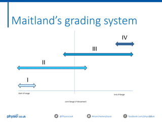
This document outlines an introduction to basic joint mobilizations for sports and massage therapists. It includes the itinerary for the workshop, which involves learning the theory of joint mobilizations, practicing assessments, and practicing different joint mobilization techniques. The document covers topics such as the definition of a joint mobilization, anatomy of synovial joints, types of synovial joints, physiological and accessory joint movements, assessment of range of motion and end feels, contraindications to joint mobilizations, and Maitland's grading system for joint mobilizations.
2. Welcome
An introduction to basic joint mobilisations for sports and
massage therapists
With Kevin Parry
@Physiocouk #manchesterphysio facebook.com/physiocouk
5. 5
Aims of today
@Physiocouk #manchesterphysio facebook.com/physiocouk
Learn the theory of joint mobilisations
Learn how to assess a joint before mobilising
Practice different joint mobilisations
Learn the evidence and research behind joint mobilisations
6. 6
Itinerary
10.00 - 10.30: Induction/arrival
10.30 – 11.30: Theory: Mobilisations and Manual therapy
11.30 – 12.00: Assessment Practical
12.00 – 12.30: Lunch
12.30 - 13.30 : Practical: Mobilisations and Manual therapy
13.30 - 14.00: Evidence and recent research
14.00 – 15.00: Case studies and Practical
@Physiocouk #manchesterphysio facebook.com/physiocouk
8. 8
Definition of a joint
mobilisation
@Physiocouk #manchesterphysio facebook.com/physiocouk
A skilled passive movement of the articular surfaces
performed by a physical therapist to decrease pain
or increase joint mobility.
Edward P. Mulligan, 2001
9. 9@Physiocouk #manchesterphysio facebook.com/physiocouk
Anatomy of a synovial joint
• The synovial joint is the most common type of joint found in the
body
• Most evolved and therefore most mobile type of joints
• Articular surfaces are covered with hyaline cartilage
10. 10@Physiocouk #manchesterphysio facebook.com/physiocouk
Anatomy of a synovial joint
• Between the articular surfaces there is a joint cavity filled with
synovial fluid
• The joint is surrounded by an articular capsule which is fibrous
in nature and is lined by synovial membrane
• The synovial membrane lines the entire joint except the
articular surfaces covered by hyaline cartilage
16. 16
Accessory
@Physiocouk #manchesterphysio facebook.com/physiocouk
• Known as Arthrokinematic joint movements
• Articular movements between two joint surfaces:
• Roll
• Glide
• Spin
• Occur with all active/passive physiological joint movement
.
• Necessary for full, pain-free range of movement
• Movements that we FEEL
17. 17
Arthrokinematic Roll
@Physiocouk #manchesterphysio facebook.com/physiocouk
• New points of one surface come into contact with
the other surface
• This can only occur when the two joint surfaces are
incongruent
• Analogy: wheel
18. 18
Arthrokinematic Glide
@Physiocouk #manchesterphysio facebook.com/physiocouk
• One joint surfaces slides or translates over the other
• Occurs when two surfaces are congruent and flat, or
congruent and curved
• Analogy: An ice-skater’s blade (one point) sliding
across the ice surface (many points)
19. 19
Arthrokinematic Spin
@Physiocouk #manchesterphysio facebook.com/physiocouk
• Rotation around a longitudinal axis
• One joint surface rotates around another
• Analogy: a top spinning on the table (if it
were to remain upright and in one place)
20. 20
Joint Morphology
@Physiocouk #manchesterphysio facebook.com/physiocouk
Joint surfaces can be described as
either:
1. Convex: Male, Arched, Rounded
2. Concave: Female, Shallow, Hollowed
Knowing that a joint surface is concave
or convex is important because shape
determines motion
21. 21
Convex on Concave
@Physiocouk #manchesterphysio facebook.com/physiocouk
• Concave surface is fixed and
the convex surface moves
over it.
• Physiological and accessory
joint movements occur in the
opposite direction
• Glide and Roll are in opposite
directions
22. 22
Concave on Convex
@Physiocouk #manchesterphysio facebook.com/physiocouk
• Convex segment is static
with the concave surface
moving over it
• Physiological and accessory
joint motions are in the
same direction
• Roll and glide are in the
same direction
24. 24
Assessing physiological joint
movements
@Physiocouk #manchesterphysio facebook.com/physiocouk
The therapist passively takes joints through their
available range.
Used to assess:
1. Available range of movement at a joint
2. Presence/absence of a capsular pattern
3. End-feel
4. Pain
25. 25
Capsular Patterns – Cyriax (1982)
@Physiocouk #manchesterphysio facebook.com/physiocouk
• A series of limitations of joint movement when the
joint capsule is a limiting structure.
• Usually represents pathology/restriction from within
the joint or capsule itself.
• Unique pattern to each synovial joint
• Assessed by evaluating the available ROM and ‘end-
feel’ in joints passively
26. 26@Physiocouk #manchesterphysio facebook.com/physiocouk
Joint Capsular Pattern (in order of most
limited)
Cervical Spine Side flexion & rotations equally limited,
extension
Thoracic Spine Side flexion & rotation equally limited,
extension
Lumbar Spine Extension, Side flexion & rotation equally
limited.
Shoulder (Glenohumeral) Lateral rotation, abduction, medial
rotation
Elbow (Humeroulnar) Flexion, extension
Wrist Flexion & extension equally limited
Hip Medial rotation, flexion, abduction
Knee Flexion, extension
Ankle (Talocrural) Plantar flexion, dorsi flexion
28. 28
Normal End-feels
@Physiocouk #manchesterphysio facebook.com/physiocouk
1. Hard
Bone-to-bone approximation
E.g. extension of the elbow
2. Soft
Characteristic of a stop to the movement due to
approximation of tissue
E.g. Knee flexion
3. Elastic
Felt when tissues are placed on a passive stretch causing an
elastic resistance
E.g. Lateral rotation of the hip or shoulder
29. 29
Abnormal End-feels
@Physiocouk #manchesterphysio facebook.com/physiocouk
1. Hard
Different from that of ‘normal’ hard end-feel
Often felt in early OA
Involuntary muscle spasm causes provides a break to movement
Also due to capsular contracture
2. Springy
Associated with mechanical joint displacement, usually a loose body
Feels like the joint springs or bounces back just before end range
3. Empty
Examiner does not have the opportunity to appreciate true end-feel
Due to pain or apprehension
36. @Physiocouk #manchesterphysio facebook.com/physiocouk
Restoring normal range of
movement
• Reduces pain (PGT)
• Enables normal biomechanics
• Functional movement
• Indication of proper muscle tone and balance around a joint
• Abnormal joint function are secondary to abnormal postures,
injury and stress
37. @Physiocouk #manchesterphysio facebook.com/physiocouk
Pain gate theory (PGT)
• Proposed in 1965 by Melzack and Wall
• Commonly used explanation of pain transmission
• 3 types of sensory nerves involved in the
transmission:
• A- Beta fibres
• A- Delta fibres
• C-fibres
38. 38
Pain gate theory (PGT)
@Physiocouk #manchesterphysio facebook.com/physiocouk
- a-beta fibres
Responsible for “sharp” pain, large diameter and
myelinated, fast transmission fibre
- a-Delta fibres
Small diameter and myelinated, responsive to vibration
and light touch – fast reactive
- C – fibres
Small diameter and un-myelinated, throbbing or burning,
slow
• Size = bigger a nerve, the quicker its conduction
• Speed = increased with myelin sheath
39. @Physiocouk #manchesterphysio facebook.com/physiocouk
Pain gate theory (PGT)
• All nerves synapse onto projection cells and travel up the CNS
to the brain
• Spinal cord has inhibitory interneurons acts as “gate keeper”
• When there is no sensation from the nerves the inhibitory
interneurons stop signals – no need for brain response (“gate
closed”)
• When smaller fibres are stimulated the inhibitory interneurons
do not act – “gate open”
• Pain is sensed
41. @Physiocouk #manchesterphysio facebook.com/physiocouk
Descending inhibition
• Mobilisations have shown to stimulate areas if the brain,
instrumental in experience of pain
• These areas include:
- Anterior cingulate cortex (ACC)
- Amygdyla
- Periaqueductal Gray (PAG)
- Rostral Ventromedial Medualla (RVM)
• The doral area of PAG and RVM, have been shown to selective
produce analgesia to cause sympatho-excitation and the
release of endorphins
42. @Physiocouk #manchesterphysio facebook.com/physiocouk
Increased local blood flow
Increased nutrition supply
Remove inflammatory exudate
Produces movement so that blood/fluid can move in and out of
articular cartilage within joints
Maintenance of healthy articular cartilage and proper joint
function.
Stimulates repair of cellular damage
Enhances the healing process
44. @Physiocouk #manchesterphysio facebook.com/physiocouk
Synovial sweep
• An oscillation/movement increases lubrication of
cartilage
• Provides nutrients to maintain healthy joints
• Elasticity increases range of movement
• Synovial fluid is found in the cavities of synovial
joints
• Egg white–like consistency, with the principal role of
reducing friction between the articulating surfaces
during movement.
• Lack of lubricated synovial fluid causes poor joint
dysfunction and secondary injuries
48. 48@Physiocouk #manchesterphysio facebook.com/physiocouk
Treatment Principles
Need to consider the following:
1. The Desired Effect - what effect of the mobilisation is the
therapist wanting? Relieve pain or stretch tissues?
2. The Starting Position - of patient and therapist to make the
treatment effective and comfortable.
3. The Direction - AP/PA
49. 49@Physiocouk #manchesterphysio facebook.com/physiocouk
4. The Method of Application - The
position, grade, amplitude, rhythm and
duration of the technique.
5. The Expected Response - Should the
patient be pain-free, have an increased
range or have reduced soreness?
(Hengeveld and Banks, 2005)
Treatment Principles
1 oscillation per second = 30 oscillations if high SIN factor /
60 if low SIN factor (Donatelli, 2001)
50. 50@Physiocouk #manchesterphysio facebook.com/physiocouk
Tractions
• Traction is the force applied to separate joint surfaces – gap/widen the
joint space
Aims:
• Relief pain
• Create space
• Produce negative pressure
• Tighten ligaments
• Reduce loose body.
Indications: OA, loose body, disc, stenosis
52. 52@Physiocouk #manchesterphysio facebook.com/physiocouk
Small amplitude movement at the beginning of the
available ROM
Clinical Reasoning: Donatelli (2001)
• 7-10/10 VAS pain rating
• Pain before resistance upon palpation
• Acute phase of injury
• Inflammatory phase of healing
• Aim to reduce pain and neutralise joint pressures
Grade 1
53. 53@Physiocouk #manchesterphysio facebook.com/physiocouk
Grade 2
Large amplitude movement at within the available ROM
Clinical Reasoning: Donatelli (2001)
• 5-7/10 VAS pain rating
• Pain and resistance occur simultaneously upon
palpation
• Proliferation stage of recovery
• Aim to reduce pain and neutralise joint pressures
54. 54@Physiocouk #manchesterphysio facebook.com/physiocouk
Grade 3
Large amplitude movement that reaches the end ROM
Clinical Reasoning: Donatelli (2001)
• 3-5/10 VAS pain rating
• Resistance before pain
• Scar maturation/remodelling phase of healing
• Aim to treat stiffness/hypomobility
55. 55@Physiocouk #manchesterphysio facebook.com/physiocouk
Grade 4
Small amplitude movement at the very end range of motion
Clinical Reasoning: Donatelli (2001)
• 1-3/10 VAS pain rating
• Increase ROM through promotion of capsular mobility
and plastic deformation
56. 56@Physiocouk #manchesterphysio facebook.com/physiocouk
Grading mobilisations
(Maitland)
The rate of mobilisation should be thought of as an
oscillation in a rhythmical fashion at:
2Hz - 120 movements per minute
For 30 seconds - 1 minute
Kessler RM, Hertling (1983)
57. @Physiocouk #manchesterphysio facebook.com/physiocouk
Manual Handling and Body
Positioning
• Posture
• Bed height
• Stance
• Patient position
• Use different parts of your hands/ arms to apply pressure
• Keep arms straight to utilise body weight when applying
pressure/resistance.
• Move from the hips and knees as much as possible
• Oil (or cream)- only needs to be a little bit.
• Look after yourself before you look after the patient!
65. Lumbar spine:
Case study 1
@Physiocouk #manchesterphysio facebook.com/physiocouk
33 year old women who works as a social worker. Reports a
lifting and twisting injury 2 days ago. Immediate pain into lumbar
spine and referred unilateral leg sensations.
Aggravating factors are bending forwards and prolonged sitting.
Finds walking and bending backwards easing. She rates her pain
score 8/10 on the VAS scale.
• Diagnosis?
• What mobilisations would you perform to relieve symptoms?
• What grade would you perform?
66. @Physiocouk #manchesterphysio facebook.com/physiocouk
55 year old taxi driver involved in a RTC 2 weeks ago. Reports
instant pain and reduced range of movement and now struggles
to check blind spot during driving. His current VAS score is 7/10.
Objective findings of limitation in bilateral side flexion and
rotation.
• Diagnosis?
• What mobilisations would you perform to relieve symptoms?
• What would you reassess after treatment?
Neck:
Case study 2
67. @Physiocouk #manchesterphysio facebook.com/physiocouk
Hip:
Case study 3
60 year old female reports gradual onset of pain into right hip
(5/10 VAS). The main aggravating factors are prolonged standing
and walking and she also reports morning stiffness. Objective
findings of a mild capsular pattern are found. Diagnosis is OA of
the R hip.
• What is the capsular pattern of the hip?
• What mobilisations would you perform to relieve symptoms?
• What grade would you perform?
• What else would you advise to this patient?
69. 69@Physiocouk #manchesterphysio facebook.com/physiocouk
Upper Extremity
Comparison of Supervised Exercise With and Without Manual Physical
Therapy for Patients With Shoulder Impingement Syndrome (Bang et al,
2000):
Manual therapy combined with supervised clinical exercise resulted in
superior outcomes to exercise alone in patients with shoulder
impingement syndrome
The effect of joint mobilization as a component of comprehensive
treatment for primary shoulder impingement syndrome (Conroy et al,
1998):
Mobilisation decreased 24-hour pain and pain associated with
subacromial compression test in patients with shoulder impingement
syndrome
70. 70@Physiocouk #manchesterphysio facebook.com/physiocouk
Lower Extremity
A randomised controlled trial of a passive accessory joint mobilization on
acute ankle inversion sprains (Green et al, 2001)
Addition of talocrural mobilizations to the RICE protocol in the
management of inversion ankle injuries necessitated fewer treatments to
achieve pain-free dorsiflexion and to improve stride speed more than RICE
alone.
Effect of physical therapy on limited joint mobility in the diabetic foot. A
pilot study (Dijs et al, 2001)
Joint mobilization and physical therapy resulted in a significant, although
temporary, improvement in the mobility of the ankle and foot in diabetic
patients with limited joint mobility and neuropathy
71. 71@Physiocouk #manchesterphysio facebook.com/physiocouk
Spinal Mobilisations
Manipulation or mobilisation for neck pain: A Cochrane Systematic Review (Gross
et al, 2010)
27 trials reviewed by two authors
Moderate quality evidence suggested manipulation and mobilisation produced
similar effects on pain, function and patient satisfaction
Low quality evidence supported thoracic manipulation as an additional therapy
for pain reduction and increased function in acute pain
Mobilisation for neck pain, low quality evidence for subacute and chronic neck
pain indicated that:
1. A combination of Maitland mobilisation techniques was similar to
acupuncture for immediate pain relief and increased function
2. There was no difference between mobilisation and acupuncture as
additional treatments for immediate pain relief and improved function
3. Neural dynamic mobilisations may produce clinically important
reduction of pain immediately post-treatment.
72. 72@Physiocouk #manchesterphysio facebook.com/physiocouk
References
• Cyriax, J. (1982). Textbook of Orthopaedic Medicine, 8th edn. Bailliere Tindell,
London.
• Hengeveld, E. & Banks, K. (2005). Maitland's Peripheral Manipulation. 4th ed.
Elsevier: London.
• Donatelli 2001
• Bang, M. D., & Deyle, G. D. (2000). Comparison of supervised exercise with and
without manual physical therapy for patients with shoulder impingement
syndrome. Journal of Orthopaedic & Sports Physical Therapy, 30(3), 126-137.
• Conroy, D. E., & Hayes, K. W. (1998). The effect of joint mobilization as a
component of comprehensive treatment for primary shoulder impingement
syndrome. Journal of Orthopaedic & Sports Physical Therapy, 28(1), 3-14.
• Green, T., Refshauge, K., Crosbie, J., & Adams, R. (2001). A randomized controlled
trial of a passive accessory joint mobilization on acute ankle inversion
sprains. Physical therapy, 81(4), 984-994.
73. 73
Thanks for coming!
Don’t forget to follow us on Twitter: @physiocouk
@Physiocouk #manchesterphysio facebook.com/physiocouk