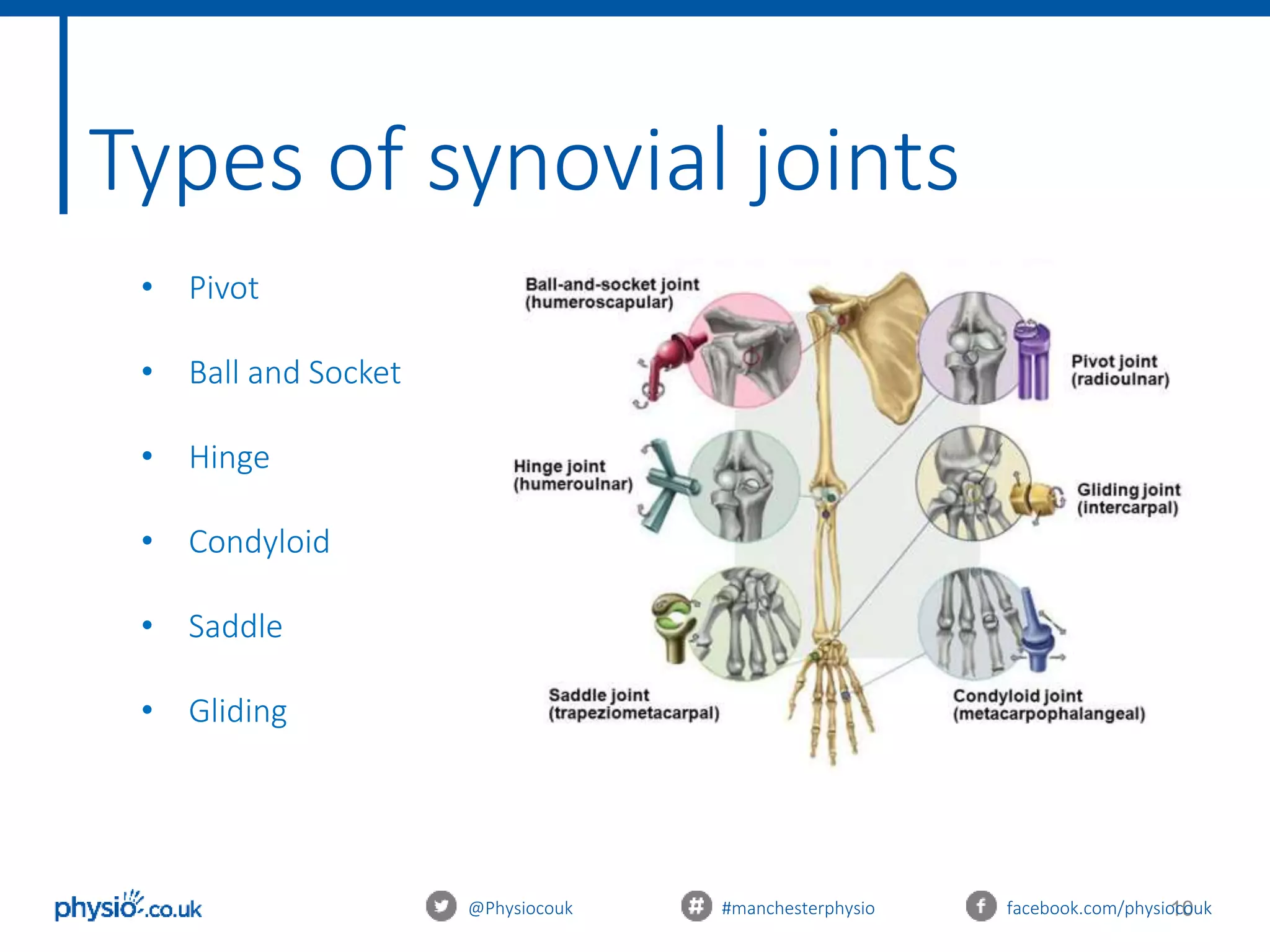
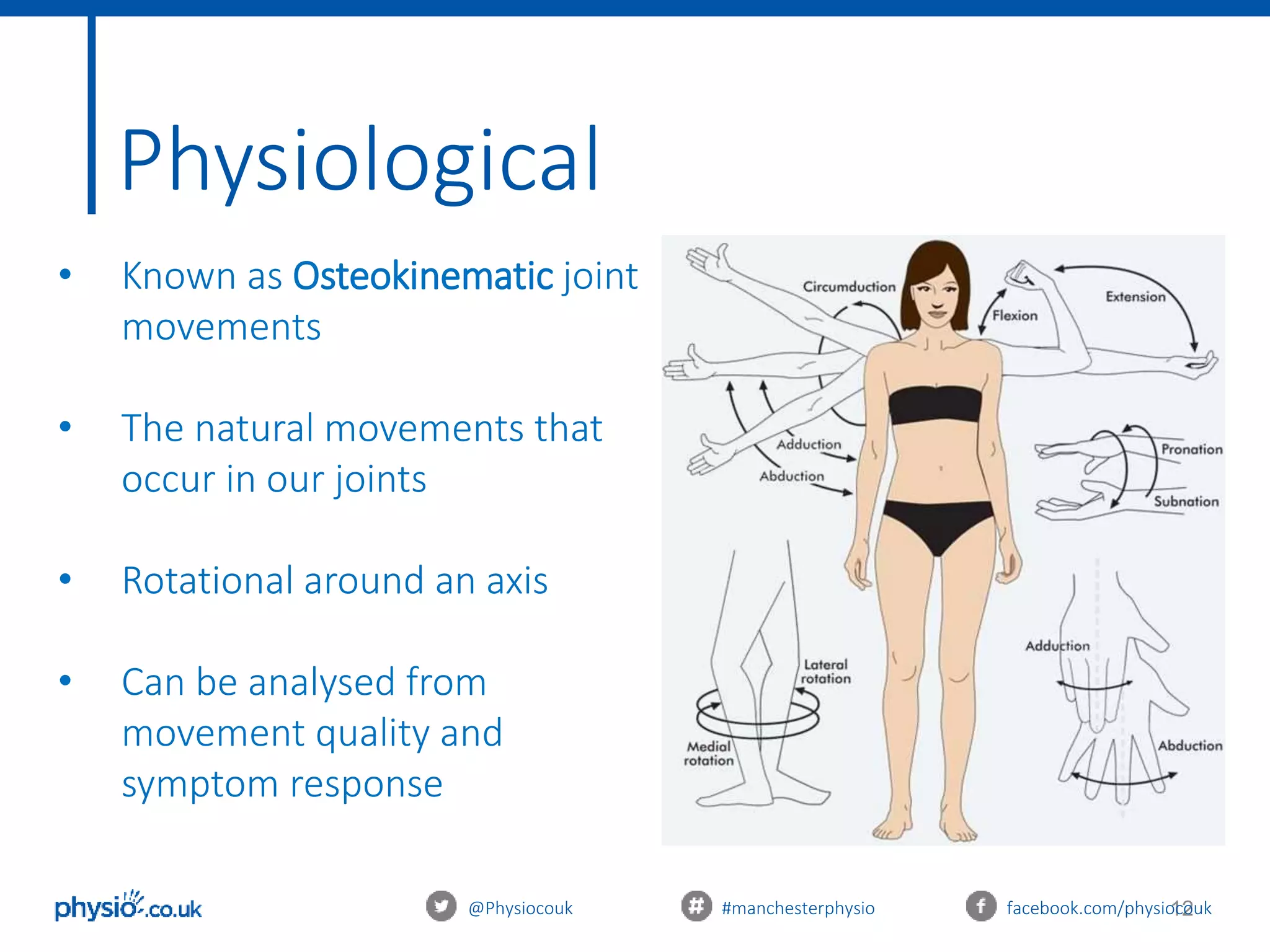
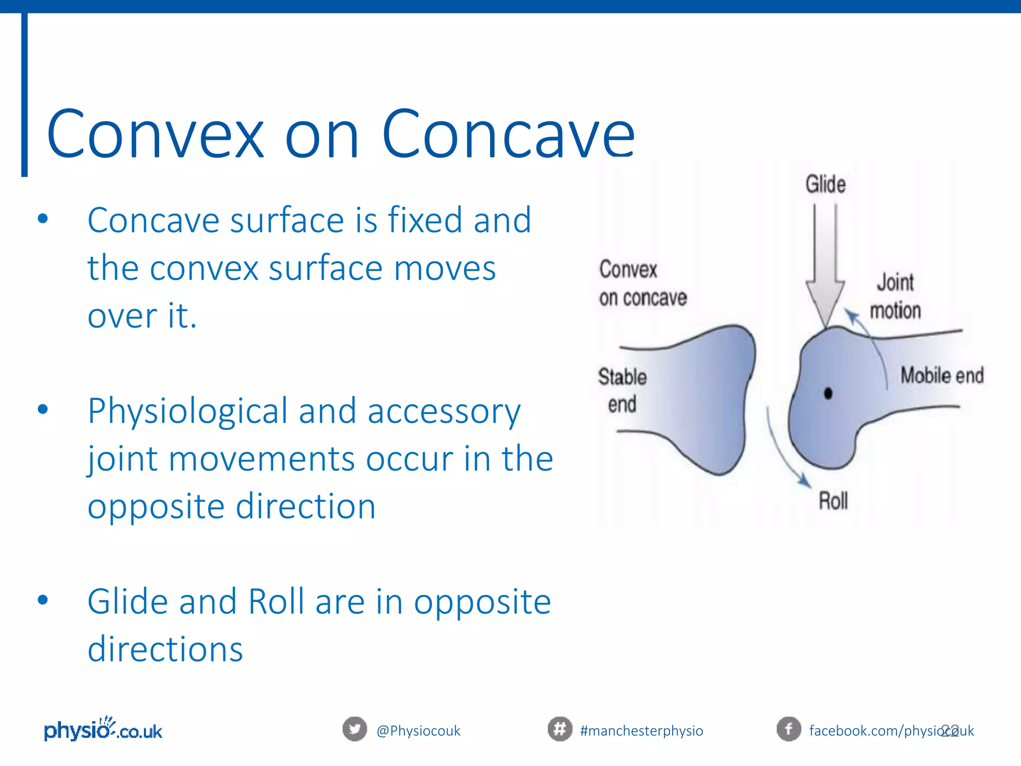
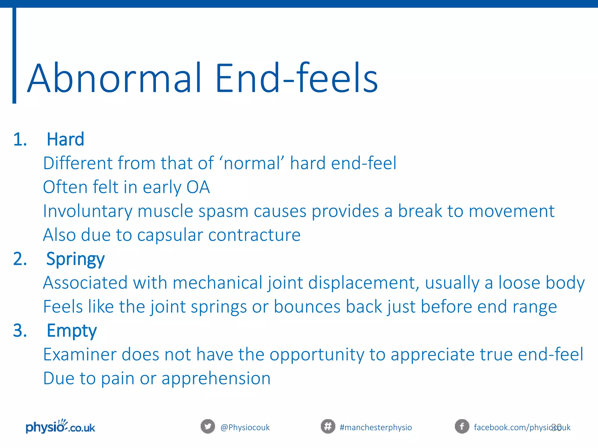
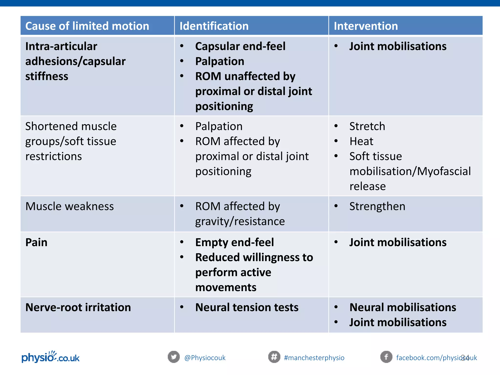
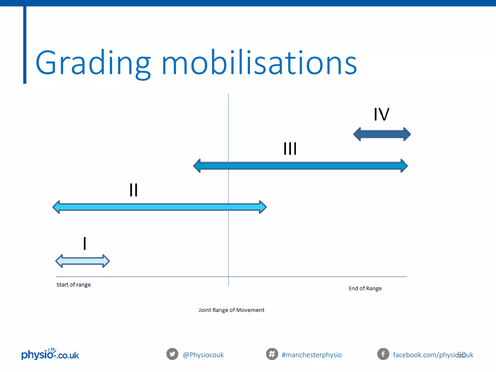
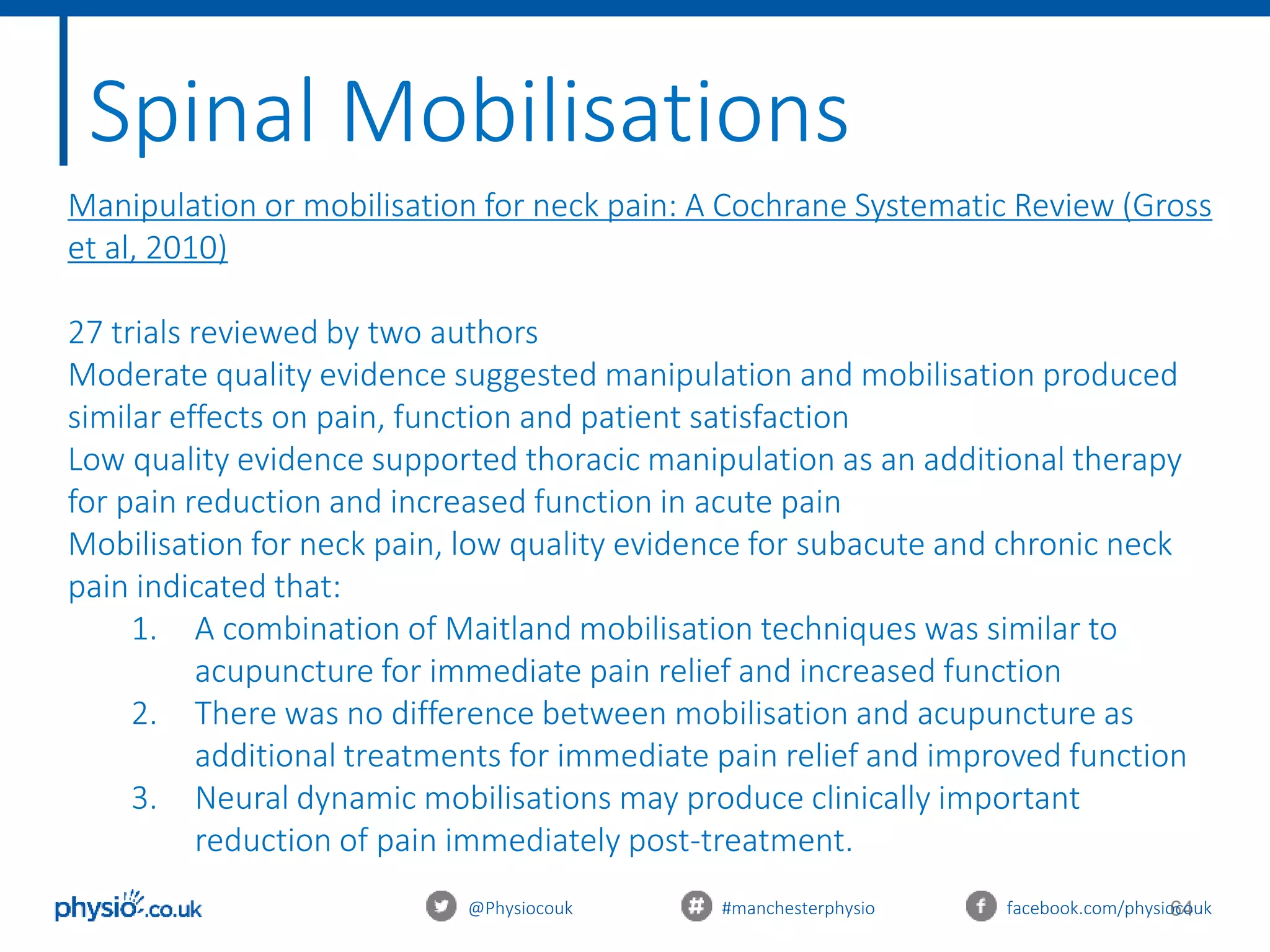
This document provides an introduction to joint mobilization techniques for sports and massage therapists. It begins with an overview of joint anatomy and kinematics, including physiological and accessory movements. Assessment techniques like evaluating range of motion, capsular patterns, and end-feels are described. The aims of mobilizations like restoring range of motion, reducing pain via the pain gate theory, and increasing local blood flow are explained. Precautions and contraindications are listed. Grading of mobilization techniques from grade 1 to 4 are defined based on amplitude and patient pain level. The document concludes with treatment principles to consider for safe and effective joint mobilizations.
2. Welcome
An introduction to mobilisation and manual therapy for
sports and massage therapists
With Katie Emmett & Richard Gregory
@Physiocouk #manchesterphysio facebook.com/physiocouk
4. 4
Aims of today
@Physiocouk #manchesterphysio facebook.com/physiocouk
Learn the theory of joint mobilisations
Learn how to assess a joint before mobilising
Practice different joint mobilisations and manual therapy
treatments
Learn the evidence and research behind joint mobilisations
5. 5
Itinerary
10.00 - 10.30: Induction/arrival
10.30 – 11.30: Theory: Mobilisations and Manual therapy
11.30 – 12.00: Assessment Practical
12.00 – 12.30: Lunch
12.30 - 13.30 : Practical: Mobilisations and Manual therapy
13.30 - 14.00: Evidence and recent research
14.00 – 15.00: Case studies and Practical
@Physiocouk #manchesterphysio facebook.com/physiocouk
7. 7
Definition of a joint
mobilisation
@Physiocouk #manchesterphysio facebook.com/physiocouk
A skilled passive movement of the articular surfaces
performed by a physical therapist to decrease pain
or increase joint mobility.
Edward P. Mulligan, 2001
8. 8@Physiocouk #manchesterphysio facebook.com/physiocouk
Anatomy of a synovial joint
• The synovial joint is the most common type of joint found in the
body
• Most evolved and therefore most mobile type of joints
• Articular surfaces are covered with hyaline cartilage
9. 9@Physiocouk #manchesterphysio facebook.com/physiocouk
Anatomy of a synovial joint
• Between the articular surfaces there is a joint cavity filled with
synovial fluid
• The joint is surrounded by an articular capsule which is fibrous
in nature and is lined by synovial membrane
• The synovial membrane lines the entire joint except the
articular surfaces covered by hyaline cartilage
15. 15@Physiocouk #manchesterphysio facebook.com/physiocouk
Rotation around an axis
• Imaginary line that is the pivotal/ rotational point at a joint
• Movement in the planes occurs around this point and it is
perpendicular to the planes
• Three axes of rotation:
Anterior-posterior axis
Mediolateral axis
Longitudinal axis
17. 17
Accessory
@Physiocouk #manchesterphysio facebook.com/physiocouk
• Known as Arthrokinematic joint movements
• Articular movements between two joint surfaces:
• Roll
• Glide
• Spin
• Occur with all active/passive physiological joint movement
• Necessary for full, pain-free range of movement
• Movements that we FEEL
18. 18
Arthrokinematic Roll
@Physiocouk #manchesterphysio facebook.com/physiocouk
• New points of one surface come into contact with
the other surface
• This can only occur when the two joint surfaces are
incongruent
• Analogy: wheel
19. 19
Arthrokinematic Glide
@Physiocouk #manchesterphysio facebook.com/physiocouk
• One joint surfaces slides or translates over the other
• Occurs when two surfaces are congruent and flat, or
congruent and curved
• Analogy: An ice-skater’s blade (one point) sliding
across the ice surface (many points)
20. 20
Arthrokinematic Spin
@Physiocouk #manchesterphysio facebook.com/physiocouk
• Rotation around a longitudinal axis
• One joint surface rotates around another
• Analogy: a top spinning on the table (if it
were to remain upright and in one place)
21. 21
Joint Morphology
@Physiocouk #manchesterphysio facebook.com/physiocouk
Joint surfaces can be described as
either:
1. Convex: Male, Arched, Rounded
2. Concave: Female, Shallow, Hollowed
Knowing that a joint surface is concave
or convex is important because shape
determines motion
22. 22
Convex on Concave
@Physiocouk #manchesterphysio facebook.com/physiocouk
• Concave surface is fixed and
the convex surface moves
over it.
• Physiological and accessory
joint movements occur in the
opposite direction
• Glide and Roll are in opposite
directions
23. 23
Concave on Convex
@Physiocouk #manchesterphysio facebook.com/physiocouk
• Convex segment is static
with the concave surface
moving over it
• Physiological and accessory
joint motions are in the
same direction
• Roll and glide are in the
same direction
25. 25
Assessing physiological joint
movements
@Physiocouk #manchesterphysio facebook.com/physiocouk
The therapist passively takes joints through their
available range.
Used to assess:
1. Available range of movement at a joint
2. Presence/absence of a capsular pattern
3. End-feel
4. Pain
26. 26
Capsular Patterns – Cyriax (1982)
@Physiocouk #manchesterphysio facebook.com/physiocouk
• A series of limitations of joint movement when the
joint capsule is a limiting structure.
• Usually represents pathology/restriction from within
the joint or capsule itself.
• Unique pattern to each synovial joint
• Assessed by evaluating the available ROM and ‘end-
feel’ in joints passively
27. 27@Physiocouk #manchesterphysio facebook.com/physiocouk
Joint Capsular Pattern (in order of most
limited)
Cervical Spine Side flexion & rotations equally limited,
extension
Thoracic Spine Side flexion & rotation equally limited,
extension
Lumbar Spine Extension, Side flexion & rotation equally
limited.
Shoulder (Glenohumeral) Lateral rotation, abduction, medial
rotation
Elbow (Humeroulnar) Flexion, extension
Wrist Flexion & extension equally limited
Hip Medial rotation, flexion, abduction
Knee Flexion, extension
Ankle (Talocrural) Plantar flexion, dorsi flexion
29. 29
Normal End-feels
@Physiocouk #manchesterphysio facebook.com/physiocouk
1. Hard
Bone-to-bone approximation
E.g. extension of the elbow
2. Soft
Characteristic of a stop to the movement due to
approximation of tissue
E.g. Knee flexion
3. Elastic
Felt when tissues are placed on a passive stretch causing an
elastic resistance
E.g. Lateral rotation of the hip or shoulder
30. 30
Abnormal End-feels
@Physiocouk #manchesterphysio facebook.com/physiocouk
1. Hard
Different from that of ‘normal’ hard end-feel
Often felt in early OA
Involuntary muscle spasm causes provides a break to movement
Also due to capsular contracture
2. Springy
Associated with mechanical joint displacement, usually a loose body
Feels like the joint springs or bounces back just before end range
3. Empty
Examiner does not have the opportunity to appreciate true end-feel
Due to pain or apprehension
37. @Physiocouk #manchesterphysio facebook.com/physiocouk
Restoring normal range of
movement
• Reduces pain (PGT)
• Enables normal biomechanics
• Functional movement
• Indication of proper muscle tone and balance around a joint
• Abnormal joint function are secondary to abnormal postures,
injury and stress
38. @Physiocouk #manchesterphysio facebook.com/physiocouk
Pain gate theory (PGT)
• Proposed in 1965 by Melzack and Wall
• Commonly used explanation of pain transmission
• Mobilisations increase excitation of a-Delta fibres
• 3 types of sensory nerves involved in the transmission
- a-beta fibres
Responsible for “sharp” pain, large diameter and
myelinated, fast transmission fibre
39. 39
Pain gate theory (PGT)
@Physiocouk #manchesterphysio facebook.com/physiocouk
- a-Delta fibres
Small diameter and myelinated, responsive to vibration
and light touch – fast reactive
- C – fibres
Small diameter and un-myelinated, throbbing or burning,
slow
• Size = bigger a nerve, the quicker its conduction
• Speed = increased with myelin sheath
• a – Delta purely sensory nerve
40. @Physiocouk #manchesterphysio facebook.com/physiocouk
Pain gate theory (PGT)
• All nerves synapse onto projection cells and travel up the CNS
to the brain
• Spinal cord has inhibitory interneurons acts as “gate keeper”
• When there is no sensation from the nerves the inhibitory
interneurons stop signals – no need for brain response (“gate
closed”)
• When smaller fibres are stimulated the inhibitory interneurons
do not act – “gate open”
• Pain is sensed
42. @Physiocouk #manchesterphysio facebook.com/physiocouk
Descending inhibition
• Mobilisations have shown to stimulate areas if the brain,
instrumental in experience of pain
• These areas include:
- Anterior cingulate cortex (ACC)
- Amygdyla
- Periaqueductal Gray (PAG)
- Rostral Ventromedial Medualla (RVM)
• The doral area of PAG and RVM, have been shown to selective
produce analgesia to cause sympatho-excitation and the
release of endorphins
43. @Physiocouk #manchesterphysio facebook.com/physiocouk
Increased local blood flow
Increased nutrition supply
Remove inflammatory exudate
Produces movement so that blood/fluid can move in and out of
articular cartilage within joints
Maintenance of healthy articular cartilage and proper joint
function.
Stimulates repair of cellular damage
Enhances the healing process
45. @Physiocouk #manchesterphysio facebook.com/physiocouk
Synovial sweep
• An oscillation/movement increases lubrication of
cartilage
• Provides nutrients to maintain healthy joints
• Elasticity increases range of movement
• Synovial fluid is found in the cavities of synovial
joints
• Egg white–like consistency, with the principal role of
reducing friction between the articulating surfaces
during movement.
• Lack of lubricated synovial fluid causes poor joint
dysfunction and secondary injuries
48. 48@Physiocouk #manchesterphysio facebook.com/physiocouk
Treatment Principles
Need to consider the following:
1. The Desired Effect - what effect of the mobilisation is the
therapist wanting? Relieve pain or stretch tissues?
2. The Starting Position - of patient and therapist to make the
treatment effective and comfortable.
3. The Direction - AP/PA; Cephalad/Caudad
49. 49@Physiocouk #manchesterphysio facebook.com/physiocouk
4. The Method of Application - The
position, grade, amplitude, rhythm and
duration of the technique.
5. The Expected Response - Should the
patient be pain-free, have an increased
range or have reduced soreness?
(Hengeveld and Banks, 2005)
Treatment Principles
1 oscillation per second = 30 oscillations if high SIN factor /
60 if low SIN factor (Donatelli, 2001)
51. 51@Physiocouk #manchesterphysio facebook.com/physiocouk
Small amplitude movement at the beginning of the
available ROM
Clinical Reasoning: Donatelli (2001)
• 7-10/10 VAS pain rating
• Pain before resistance upon palpation
• Acute phase of injury
• Inflammatory phase of healing
• Aim to reduce pain and neutralise joint pressures
Grade 1
52. 52@Physiocouk #manchesterphysio facebook.com/physiocouk
Grade 2
Large amplitude movement at within the available ROM
Clinical Reasoning: Donatelli (2001)
• 5-7/10 VAS pain rating
• Pain and resistance occur simultaneously upon
palpation
• Proliferation stage of recovery
• Aim to reduce pain and neutralise joint pressures
53. 53@Physiocouk #manchesterphysio facebook.com/physiocouk
Grade 3
Large amplitude movement that reaches the end ROM
Clinical Reasoning: Donatelli (2001)
• 3-5/10 VAS pain rating
• Resistance before pain
• Scar maturation/remodelling phase of healing
• Aim to treat stiffness/hypomobility
54. 54@Physiocouk #manchesterphysio facebook.com/physiocouk
Grade 4
Small amplitude movement at the very end range of motion
Clinical Reasoning: Donatelli (2001)
• 1-3/10 VAS pain rating
• Increase ROM through promotion of capsular mobility
and plastic deformation
62. 62@Physiocouk #manchesterphysio facebook.com/physiocouk
Upper Extremity
Comparison of Supervised Exercise With and Without Manual Physical
Therapy for Patients With Shoulder Impingement Syndrome (Bang et al,
2000):
Manual therapy combined with supervised clinical exercise resulted in
superior outcomes to exercise alone in patients with shoulder
impingement syndrome
The effect of joint mobilization as a component of comprehensive
treatment for primary shoulder impingement syndrome (Conroy et al,
1998):
Mobilisation decreased 24-hour pain and pain associated with
subacromial compression test in patients with shoulder impingement
syndrome
63. 63@Physiocouk #manchesterphysio facebook.com/physiocouk
Lower Extremity
A randomised controlled trial of a passive accessory joint mobilization on
acute ankle inversion sprains (Green et al, 2001)
Addition of talocrural mobilizations to the RICE protocol in the
management of inversion ankle injuries necessitated fewer treatments to
achieve pain-free dorsiflexion and to improve stride speed more than RICE
alone.
Effect of physical therapy on limited joint mobility in the diabetic foot. A
pilot study (Dijs et al, 2001)
Joint mobilization and physical therapy resulted in a significant, although
temporary, improvement in the mobility of the ankle and foot in diabetic
patients with limited joint mobility and neuropathy
64. 64@Physiocouk #manchesterphysio facebook.com/physiocouk
Spinal Mobilisations
Manipulation or mobilisation for neck pain: A Cochrane Systematic Review (Gross
et al, 2010)
27 trials reviewed by two authors
Moderate quality evidence suggested manipulation and mobilisation produced
similar effects on pain, function and patient satisfaction
Low quality evidence supported thoracic manipulation as an additional therapy
for pain reduction and increased function in acute pain
Mobilisation for neck pain, low quality evidence for subacute and chronic neck
pain indicated that:
1. A combination of Maitland mobilisation techniques was similar to
acupuncture for immediate pain relief and increased function
2. There was no difference between mobilisation and acupuncture as
additional treatments for immediate pain relief and improved function
3. Neural dynamic mobilisations may produce clinically important
reduction of pain immediately post-treatment.
65. 65@Physiocouk #manchesterphysio facebook.com/physiocouk
References
• Cyriax, J. (1982). Textbook of Orthopaedic Medicine, 8th edn. Bailliere Tindell,
London.
• Hengeveld, E. & Banks, K. (2005). Maitland's Peripheral Manipulation. 4th ed.
Elsevier: London.
• Donatelli 2001
• Bang, M. D., & Deyle, G. D. (2000). Comparison of supervised exercise with and
without manual physical therapy for patients with shoulder impingement
syndrome. Journal of Orthopaedic & Sports Physical Therapy, 30(3), 126-137.
• Conroy, D. E., & Hayes, K. W. (1998). The effect of joint mobilization as a
component of comprehensive treatment for primary shoulder impingement
syndrome. Journal of Orthopaedic & Sports Physical Therapy, 28(1), 3-14.
• Green, T., Refshauge, K., Crosbie, J., & Adams, R. (2001). A randomized controlled
trial of a passive accessory joint mobilization on acute ankle inversion
sprains. Physical therapy, 81(4), 984-994.
67. Lumbar spine:
Case study 1
@Physiocouk #manchesterphysio facebook.com/physiocouk
33 year old women who works as a social worker. Reports a
lifting and twisting injury 2 days ago. Immediate pain into lumbar
spine and referred unilateral leg sensations.
Aggravating factors are bending forwards and prolonged sitting.
Finds walking and bending backwards easing. She rates her pain
score 8/10 on the VAS scale.
• Diagnosis?
• What mobilisations would you perform to relieve symptoms?
• How many oscillations would you perform?
68. @Physiocouk #manchesterphysio facebook.com/physiocouk
55 year old taxi driver involved in a RTC 2 weeks ago. Reports
instant pain and reduced range of movement and now struggles
to check blind spot during driving. His current VAS score is 7/10.
Objective findings of limitation in bilateral side flexion and
rotation.
• Diagnosis?
• What mobilisations would you perform to relieve symptoms?
• How many oscillations would you perform?
Neck:
Case study 2
69. @Physiocouk #manchesterphysio facebook.com/physiocouk
SIJ:
Case study 3
23 year old female, sports therapist working full time.
Reports localised pain around lumbar spine/pelvis area. Left side
only. Gradual onset over the last 6 months and now reports
intermittent “clunks”. Vas scale: 4/10. Aggravating factors are
prolonged sitting and standing/ getting in and out of the car and
eases with heat.
• Diagnosis?
• What mobilisations would you perform to relieve symptoms?
• How many oscillations would you perform?
70. 70
Thanks for coming!
Don’t forget to follow us on Twitter: @physiocouk
@Physiocouk #manchesterphysio facebook.com/physiocouk
Editor's Notes
Introduction of company:
Manchester Physio has been established for quite a few years now
We offer a wide range of services along side physiotherapy
neurological physiotherapy
podiatry,
pilate classes
sports massage
And a specialised CIMT service.
We currently have 12 clinic locations across Manchester and Liverpool
Our locations in Liverpool come under Liverpool physio
The company is continuing to expand intp other others under the name physio.co.uk
Filming
Practical
Katie:
I’m Katie I graduated in sport and exercise therapy at Leeds Metropolitan University in 2013.
I started working for this company in the beginning of 2014.
My current role is to oversee our massage service
On a daily basis I treat a large massage and injury caseload
One of the most common problems I see is postural issues
So on a regular basis I assess and treat these and have found using kineso tape and some tapin techniques were going through today as an effective tool.
10.00 - 10.30 - Induction / arrival
10.30 – 12.00: Theory: Mobilisations and Manual Therapy
12.00 - 12.30 – Lunch
12.30 - 13.30 – Practical: Mobilisations and Manual Therapy
13.30 – 14.00 – Evidence and recent research
14.00 – 15.00 - Case Studies and Practica
The synovial joint is the most common type of joint found in the body
Most evolved and therefore most mobile type of joints
The characteristics include articular surfaces are covered with hyaline cartilage. This articular cartilage is avascular, non nervous and elastic. And Lubricated with synovial fluid
the cartilage forms slippery surfaces for free movements.
Between the articular surfaces there is a joint cavity filled with synovial fluid. The cavity may be partially or completely subdivided by an articular disc known as meniscus.
The joint is surrounded by an articular capsule which is fibrous in nature and is lined by synovial membrane. Because of its rich nerve supply the fibrous capsule is sensitive to stretches imposed by movements.
The synovial membrane lines the entire joint except the articular surfaces covered by hyaline cartilage.
It is this membrane that secretes synovial fluid which lubricates the joint and nourishes the articular cartilage
Pivot joint:
Movement – rotation of one bone around another. Example: atlas and axis bones
Ball and Socket
Movements: flexion/extension/abduction/adduction/internal and external rotation. Example: hip and shoulder
Hinge
Movements: flexion/extension. Example elbow and knee
Condyloid
Movements: flexion/extension/adduction/abduction/circumduction. example.: wrist, metacarpophalangel / metatarsophalangeal joint
Saddle
Movements: flexion, extension, adduction, abduction, circumduction. Example: carpometacarpal joint (thumb)
Gliding
Movements: intercarpal joints. example. Intercarpel joints ( articulations between the individual carpal bones)
observable movements of bones around joints are called Osteokinematic
natural movements that occur and Rotate around an axis
Movements described from the anatomical position
Can be analysed from quality and symptom response
Can be described from anatomical reference points…
Anatomical Neutral
This is the starting position for describing any movement. It is important that you know this to be able to understand what is meant by certain movement patterns. It is sometimes also called the anatomical starting position or fundamental starting position.
Anatomical neutral is:
Standing upright
Legs together and knee straight
Toes pointing straight forwards
Arms by the side
Palms facing forwards
Movement occurs in different planes0
Transverse Plane
This plane divides the body into top and bottom. Movements in this plane are rotational in nature, such as internal and external rotation, pronation and supination
Frontal Plane
The frontal plane divides the body into front and back. Movements in this plane are sideways movements, called abduction and adduction
Sagittal Plane
The Sagittal plane passes through the body front to back, so dividing it into left and right. Movements in this plane are the up and down movements of flexion and extension
Rotation around an axis
It is an imaginary line that is the pivotal/rotational point at a joint.
Movement in the planes occurs around this point and it is perpendicular to the planes, meaning that both lines—the plane and the axis—create a right (90°) angle.
Just as there are three planes of motion, there are three axes of rotation: the anterior-posterior axis, the mediolateral axis and the longitudinal axis.
Joints rotate in these axes, allowing movement to occur in the planes.
Known as Arthrokinematic joint movements
Articular movements between two joint surfaces:
Roll
Glide
Spin
Occur with all active/passive physiological joint movement
Necessary for full, pain-free range of movement
Movements that we FEEL
New points of one surface come into contact with the other surface
This can only occur when the two joint surfaces are incongruent
Analogy: wheel
Rotation around a longitudinal axis
One joint surface rotates around another
Analogy: a top spinning on the table (if it were to remain upright and in one place)
Joint surfaces can be described as either:
Convex: Male, Arched, Rounded
Concave: Female, Shallow, Hollowed
Knowing that a joint surface is concave or convex is important because shape determines motion
Concave surface is fixed and the convex surface moves over it.
Physiological and accessory joint movements occur in the opposite direction
Glide and Roll are in opposite directions
Convex segment is static with the concave surface moving over it
Physiological and accessory joint motions are in the same direction
Roll and glide are in the same direction
The therapist passively takes joints through their available range.
Used to assess:
Available range of movement at a joint
Presence/absence of a capsular pattern
End-feel
Pain
A series of limitations of joint movement when the joint capsule is a limiting structure.
Usually represents pathology/restriction from within the joint or capsule itself.
Unique pattern to each synovial joint
Assessed by evaluating the available ROM and ‘end-feel’ in joints passively
‘The specific sensation imparted through the examiner’s hands at the extreme of passive movement’
(Cyriax, 1982)
Can be categorised as either:
Normal end-feel
Abnormal end-feel
Hard
Bone-to-bone approximation
E.g. extension of the elbow
Soft
Characteristic of a stop to the movement due to approximation of tissue
E.g. Knee flexion
Elastic
Felt when tissues are placed on a passive stretch causing an elastic resistance
E.g. Lateral rotation of the hip or shoulder
Hard
Different from that of ‘normal’ hard end-feel
Often felt in early OA
Involuntary muscle spasm causes provides a break to movement
Also due to capsular contracture
Springy
Associated with mechanical joint displacement, usually a loose body
Feels like the joint springs or bounces back just before end range
Empty
Examiner does not have the opportunity to appreciate true end-feel
Due to pain or apprehension
Reduces pain (PGT)
Enables normal biomechanics
Functional movement
Indication of proper muscle tone and balance around a joint
Abnormal joint function are secondary to abnormal postures, injury and stress
Proposed in 1965 by Melzack and Wall
Commonly used explanation of pain transmission
Mobilisations increase excitation of a-Delta fibres
3 types of sensory nerves involved in the transmission
a-beta fibres
Responsible for “sharp” pain, large diameter and myelinated (nerve sheath- layer of myelin), fast transmission fibre
a-Delta fibres
Small diameter and myelinated, responsive to vibration and light touch – fast reactive
C – fibres
Small diameter and un-myelinated, throbbing or burning, slow
*If the nerve is covered with myelin sheath, the nerve impulse is faster. If we talk about unmyelinated neuron, this means the axon is not covered by this myelin sheath
Size = bigger a nerve, the quicker its conduction
Speed = increased with myelin sheath
a – Delta purely sensory nerve
All nerves synapse onto projection cells and travel up the CNS to the brain
Spinal cord has inhibitory interneurons acts as “gate keeper”
When there is no sensation from the nerves the inhibitory interneurons stop signals – no need for brain response (“gate closed”)
When smaller fibres are stimulated the inhibitory interneurons do not act – “gate open”
Pain is sensed
When the larger α-Delta fibres are stimulated they reach the inhibitory interneurons faster and, as larger fibres inhibit the interneuron from working, 'close' the gate. This is why after you have stubbed your toe, or bumped your head, rubbing it helps as you are stimulating the α-Delta fibres which close the gate
Mobilisations have shown to stimulate areas if the brain, instrumental in experience of pain
These areas include:
Anterior cingulate cortex (ACC)
Amygdyla
Periaqueductal Gray (PAG)
Rostral Ventromedial Medualla (RVM)
The doral area of PAG and RVM, have been shown to selective produce analgesia to cause sympatho-excitation and the release of endorphins
Increased nutrition supply
Remove inflammatory exudate
Produces movement so that blood/fluid can move in and out of articular cartilage within joints
Maintenance of healthy articular cartilage and proper joint function.
Stimulates repair of cellular damage
Enhances the healing process
An oscillation/movement increases lubrication of cartilage
Provides nutrients to maintain healthy joints
Elasticity increases increasing range of movement
Synovial fluid is found in the cavities of synovial joints
egg white–like consistency, with the principal role of reducing friction between the articulating surfaces during movement.
Lack of lubricated synovial fluid causes poor joint dysfunction and secondary injuries
contraindications
Need to consider the following:
The Desired Effect - what effect of the mobilisation is the therapist wanting? Relieve pain or stretch tissues?
The Starting Position - of patient and therapist to make the treatment effective and comfortable.
The Direction - AP/PA; Cephalad/Caudad
Small amplitude movement at the beginning of the available ROM
Clinical Reasoning: Donatelli (2001)
7-10/10 VAS pain rating
Pain before resistance upon palpation
Acute phase of injury
Inflammatory phase of healing
Aim to reduce pain and neutralise joint pressures
Large amplitude movement at within the available ROM
Clinical Reasoning: Donatelli (2001)
5-7/10 VAS pain rating
Pain and resistance occur simultaneously upon palpation
Proliferation stage of recovery
Aim to reduce pain and neutralise joint pressures
Large amplitude movement that reaches the end ROM
Clinical Reasoning: Donatelli (2001)
3-5/10 VAS pain rating
Resistance before pain
Scar maturation/remodelling phase of healing
Aim to treat stiffness/hypomobility
Small amplitude movement at the very end range of motion
Clinical Reasoning: Donatelli (2001)
1-3/10 VAS pain rating
Increase ROM through promotion of capsular mobility and plastic deformation
BANG 2000:
STUDY DESIGN:
A prospective randomized clinical trial.
OBJECTIVE:
To compare the effectiveness of 2 physical therapy treatment approaches for impingement syndrome of the shoulder.
BACKGROUND:
Manual physical therapy combined with exercise is a commonly applied but currently unproven clinical treatment for impingement syndrome of the shoulder.
METHODS AND MEASURES:
Thirty men and 22 women (age 43 years +/- 9.1) diagnosed with shoulder impingement syndrome were randomly assigned to 1 of 2 treatment groups. The exercise group performed supervised flexibility and strengthening exercises. The manual therapy group performed the same program and received manual physical therapy treatment. Both groups received the selected intervention 6 times over a 3-week period. The testers, who were blinded to group assignment, measured strength, pain, and function before treatment and after 6 physical therapy visits. Strength was a composite score of isometric strength tests for internal rotation, external rotation, and abduction. Pain was a composite score of visual analog scale measures during resisted break tests, active abduction, and functional activities. Function was measured with a functional assessment questionnaire. The visual analog scale used to measure pain with functional activities and the functional assessment questionnaire were also measured 2 months after the initiation of treatment.
RESULTS:
Subjects in both groups experienced significant decreases in pain and increases in function, but there was significantly more improvement in the manual therapy group compared to the exercise group. For example, pain in the manual therapy group was reduced from a pretreatment mean (+/- SD) of 575.8 (+/- 220.0) to a posttreatment mean of 174.4 (+/- 183.1). In contrast, pain in the exercise group was reduced from a pretreatment mean of 557.1 (+/- 237.2) to a posttreatment mean of 360.6 (+/- 272.3). Strength in the manual therapy group improved significantly while strength in the exercise group did not.
CONCLUSION:
Manual physical therapy applied by experienced physical therapists combined with supervised exercise in a brief clinical trial is better than exercise alone for increasing strength, decreasing pain, and improving function in patients with shoulder impingement syndrome.
CONROY 1998
examined whether subjects receiving joint mobilization and comprehensive treatment (hot packs, active range of motion, physiologic stretching, muscle strengthening, soft tissue mobilization, and patient education) would have improved pain, mobility, and function compared with similar patients receiving comprehensive treatment alone. Subjects were eight men and six women (mean age = 52.9 years)
One-tailed analyses of covariance (baseline values as covariates) showed that the experimental group had less 24-hour pain and pain with subacromial compression test but no differences in range of motion and function (Mann-Whitney U) compared with controls. The experimental group improved on all variables, while the control group improved only on mobility and function (one-tailed, paired t tests; Wilcoxon matched pairs).
Lumbar spine Case study 1:
Lumbar disc:
Presentation:
33 year old women – social worker
Reports Lifting and twisting injury 2 days ago. Immediate pain into lx sp and referred leg sensations
Agg factors: bending forwards, prolonged sitting
Eases: walking, bending backwards
VAS 8/10
Diagnosis?
What mobilisations would you perform to relieve symptoms?
What grade of mobilisation are you going to use?
How many oscillations would you perform?
Neck case study:
Whiplash injury
Presentation:
55 year old taxi driver
Involved in a RTC, instant pain and reduced range of movement
Struggling to check blind spot during driving
VAS score: 7/10
Objectively limited in bilateral side flexion and rotation
Diagnosis?
What mobilisation would you sue to increase neck range of movement?
What grade of mobilisation are you going to use?
How many oscillations would you perform?
SIJ case study
Unilateral Hypomobility
23 year old female, sports massage therapist – working full time
Localised pain around lx sp/pelvis . Left side only. Gradual onset over the last 6 months
Reports intermittent “clunks”
VAS 4/10
Agg: prolonged sitting and standing, getting in and out of car
eases: heat