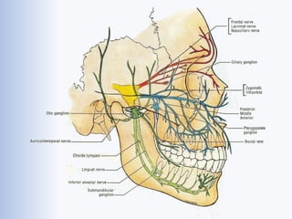
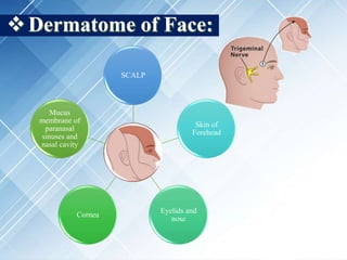
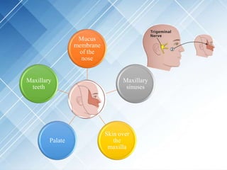
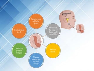
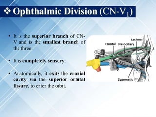
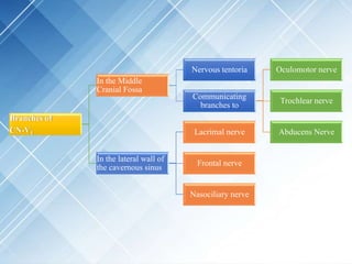
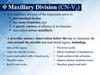
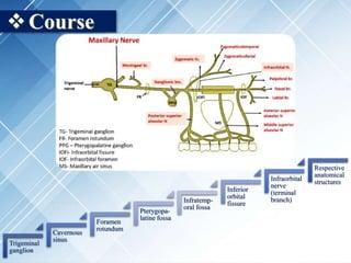
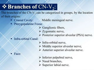
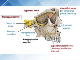
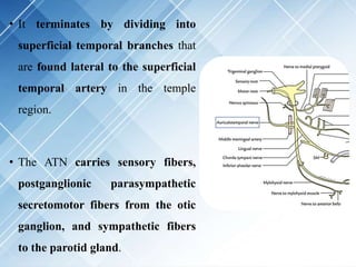
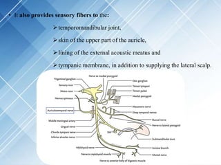
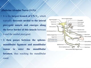
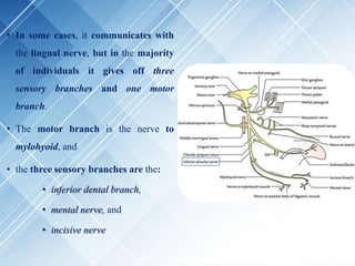
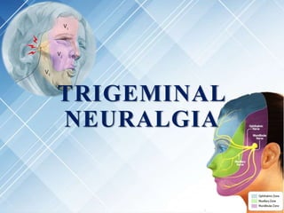
TRIGEMINAL NERVE
& its Applied Anatomy
by:
Dr. Mukesh Kumar Dey
MDS 1st Year,
Department of Pedodontics and
Preventive Dentistry, SPPGIDMS.
Contents
• Brief on Nervous System.
• Introduction to Trigeminal Nerve
• Trigeminal Nuclei
• Trigeminal Ganglia
• Divisions of the Trigeminal Nerve
• Dermatome of Face
• Ophthalmic Division of Trigeminal Nerve (CN-V1)
• Clinical Aspects of CN-V1
• Maxillary Division of Trigeminal Nerve (CN-V2)
• Course of CN-V2.
• Branches of CN-V2.
• Pterygopalatine ganglion.
• Clinical Significance of CN-V2.
• Mandibular Division of Trigeminal Nerve (CN-V3)
• Course of CN-V3.
• Branches of CN-V3.
• Associated ganglia.
• Clinical Significance of CN-V3.
• Summary.
• Trigeminal Neuralgia
• Introduction.
• Epidemiology.
• Classification.
• Etiology.
• Pathogenesis.
• Signs and Symptoms.
• Diagnosis.
• Management.
• References.
The human nervous system is
constituted by the:
o Central nervous system (CNS),
which is further constituted by
the:
 brain and
 spinal cord
o Peripheral nervous system, is a
vast network of spinal and cranial
nerves that are linked to the CNS.
peripheral
• The nerve trunks originating from the brain
and brainstem, are called cranial nerves.
They emerge out of the cranium via the
various foramina (21 in total).
• There are a total of 12 pairs of cranial nerves.
• Nerve trunks originating from the spinal cord
are called spinal nerves. The neural
foramen is the opening between every two
vertebrae where these nerve roots exit the
spine. There are two neural foramina between
each pair of vertebrae - one on each side.
• There are a total of 31 pairs of spinal nerves.
Foramen
Magnum
Names of the different cranial nerves (CN) are enlisted as under:
CN No. CN Name CN No. CN Name
0. Terminal Nerve I. Olfactory Nerve
II. Optic Nerve III. Oculomotor Nerve
IV. Trochlear Nerve V. Trigeminal Nerve
VI. Abducens Nerve VII. Facial Nerve
VIII. Vestibulocochlear Nerve IX. Glossopharyngeal Nerve
X. Vagus Nerve XI. Accessory Spinal Nerve
XII. Hypoglossal Nerve XIII. Terminal Nerve
The numbering of the cranial nerves is based on
the order in which they emerge from the brain and brainstem,
(anterior to posterior).
 Introduction to the Trigeminal Nerve
• It is the fifth pair of cranial nerves (CN-V).
• It is the largest cranial nerve and is also called:
oTrifacial Nerve or
oNerves Trigeminus.
• It is associated with the derivations of the first
pharyngeal arch.
• CN-V is a mixed nerve (i.e., serves both sensory as well as
motor functions).
• It is a mixed nerve for it serves.
oSensory functions to:
 Facial skin,
 Mucosa of cranial viscera,
except for base of tongue and pharynx.
oMotor functions to:
 Mylohyoid.
 Tensor tympani,
 Tensor villi palatini,
 Muscles of mastication,
 Anterior belly of digastric,
&
Trigeminal Nuclei
A cranial nerve nucleus is a collection of neurons in the
brainstem that is associated with one or more cranial nerves.
Trigeminal Nuclei
Sensory Nuclei
Mesencephalic
Nucleus
Principal Sensory
Nucleus
Spinal Nucleus
Motor Nucleus
 Sensory Nuclei:
i. Mesencephalic Nucleus:
• Cell body of pseudo-unipolar neuron.
• Situated in midbrain just lateral to the aqueduct.
• Relay proprioception from:
oMuscles of mastication,
oExtra ocular Muscles,
oFacial muscles.
ii. Principal Sensory Nucleus (PSN):
• Lies in pons, lateral to the motor nucleus.
• Relays touch sensation.
iii. Spinal Nucleus:
• Extends from caudal end of PSN in pons to 2nd or 3rd spinal
segment.
• It relays sensations of pain and temperature.
 Motor Nucleus:
• Derived from first branchial arch.
• Located in the pons.
• Innervates:
o Tensor palatini,
o Tensor tympani and
o Muscles of mastication.
Trigeminal Ganglia
• Also called:
oSemilunar ganglia
oGasserian ganglia
• Crescentic in shape, containing cell bodies of pseudo-unipolar
neurons.
• Lies in a bony fossa:
oat apex of the petrous part of temporal bone,
oon the floor of middle cranial fossa,
ojust lateral to posterior part of lateral wall of the
cavernous sinus.
• Covered in dural pouch, called:
oMeckel’s Cave or
oCavum Trigeminale.
• This region is lined by pia-mater and arachnoid thus the
ganglion is bathed in cerebro-spinal fluid.
• The trigeminal ganglion is about 2 mm in size.
• It is the largest of the cranial nerve ganglia and it is the widest
part of the trigeminal nerve.
• Anatomical relations of the CN-V ganglion:
SUPERIORLY LATERALLY INFERIORLY MEDIALLY
• Superior petrosal
sinus.
• Free margin of
tentorium
cerebelli.
• Uncus of
temporal lobe.
• Middle meningeal
artery and vein.
• Nervous
spinosum.
• Motor root of CN-
V.
• Greater petrosal
nerve.
• Petrous apex.
• Foramen lacerum.
• Posterior part of
lateral wall of
cavernous sinus.
• Internal carotid
artery with its
sympathetic
plexus.
Divisions of the Trigeminal Nerve
Three divisions of CN-V emerge
from the trigeminal ganglion,
viz.:
• Ophthalmic branch (CN-V1),
• Maxillary branch (CN-V2)
and
• Mandibular branch (CN-V3).
Dermatome of Face:
SCALP
Skin of
Forehead
Eyelids and
nose
Cornea
Mucus
membrane of
paranasal
sinuses and
nasal cavity
Mucus
membrane
of the
nose
Maxillary
sinuses
Skin over
the
maxilla
Palate
Maxillary
teeth
Temporoma
ndibular
joint
Skin over
the side of
head and
mandible
Skin of
cheek
Mucus
membrane
of oral
cavity
Anterior
part of
tongue
Mandibular
teeth
Ophthalmic Division (CN-V1)
• It is the superior branch of CN-
V and is the smallest branch of
the three.
• It is completely sensory.
• Anatomically, it exits the cranial
cavity via the superior orbital
fissure, to enter the orbit.
Branches of
CN-V1
In the Middle
Cranial Fossa
Nervous tentoria
Communicating
branches to
Oculomotor nerve
Trochlear nerve
Abducens Nerve
In the lateral wall of
the cavernous sinus
Lacrimal nerve
Frontal nerve
Nasociliary nerve
 Lacrimal Nerve:
• It is the smallest branch of the ophthalmic division.
• Innervates the lacrimal gland and the conjunctiva.
• It pierces through the orbital septum and ends in the
skin of the upper eyelid.
 Frontal Nerve:
• It is the largest branch and appears to be the direct
continuation of the ophthalmic division.
• It enters the orbit through the superior orbital fissure
and divides into two branches, viz.:
 Nasociliary Nerve:
• It is intermediate in size and runs more deeply.
• Its branches can be classified as follows:
Supraorbital Nerve Supratrochlear Nerve
• Larger of the two. • Relatively smaller of the two.
• Lateral to supratrochlear nerve. • Medial to supraorbital nerve.
• Innervates conjunctiva, SCALP (upto
vertex) and mucous membrane of
frontal sinus.
• Innervates the skin of the upper eyelid
and lower part of the forehead.
Branches of Nasociliary
Nerve
To the Orbit
Short Ciliary Nerves
Long Ciliary Nerves
Posterior Ethmoidal Nerve
Anterior Ethmoidal Nerve
Infratrochlear Nerve
To the Nasal Cavity
Innervates the mucous
membrane of the Nasal
Cavity
To the Face
Innervates the skin of the
bridge of the nose.
Innervates the skin of the
midle parts of eyelids and
the lacrimal sac.
Clinical Aspects of CN-V1:
 Corneal Reflex:
• When the ophthalmic nerve is injured, there is loss of
Corneal Blink Reflex.
• This reflex is mediated by:
 the CN-V1 which is the afferent pathway and
 The CN-VII, which sub-serves as the efferent pathway.
 Supraorbital Injuries:
• Trauma to the supraorbital margin may damage the
supraorbital and/or supratrochlear nerves, causing Sensory
Loss in the SCALP.
 Ethmoid Tumours:
• Malignant tumours of the mucous lining of the
ethmoidal air cells may expand into the orbits, damaging
the branches of V1, particularly the Ethmoidal Nerves.
• This may lead to displacement of the trigeminal, facial
and hypoglossal nerves.
Maxillary Division (CN-V2)
• The maxillary division of the trigeminal nerve is:
 intermediate in size,
 has many branches, and
 is purely sensory or afferent in its function.
 also called nervus maxillaris.
• It provides sensory innervation below the eye to structures in
and around the maxilla and mid-facial region, including:
• Skin of this region, • The lower eyelid,
• Upper lip and side of nose, • Mucus membrane of nasopharynx,
• Lateral and medial walls of nasal cavity, • Under-surface of the soft palate,
• Maxillary sinus, • Superior portion of palatine tonsil,
• Roof of oral cavity, • Maxillary gingiva and teeth.
Course
Trigeminal
ganglion
Cavernous
sinus
Foramen
rotundum
Pterygopa-
latine fossa
Infratemp-
oral fossa
Inferior
orbital
fissure
Infraorbital
nerve
(terminal
branch)
Respective
anatomical
structures
Branches of CN-V2:
The branches of the CN-V2 can be categorized in groups, by the location
of their origins:
 Cranial Cavity: Middle meningeal nerve.
 Pterygopalatine Fossa:
 Ganglionic fibers,
 Zygomatic nerve,
 Posterior superior alveolar (PSA) nerve.
 Infra-orbital Canal:
 Infra-orbital nerve,
 Middle superior alveolar nerve,
 Anterior superior alveolar nerve.
 Face:
 Inferior palpebral nerve,
 Nasal branches,
 Superior labial nerve.
 Middle Meningeal Nerve:
• Arises in the cranial cavity, just before
the maxillary nerve trunk enters the
cavernous sinus.
• It innervates the dura-mater in the
anterior part of the middle cranial fossa
with sensory fibers. Middle Meningeal Nerve
 Ganglionic Branches:
• The pterygopalatine nerves (also known as sphenopalatine
branches) are branches of the maxillary nerve within the
pterygopalatine fossa, which descend to the pterygopalatine
ganglion.
• However, they do not synapse (relay) at the ganglion.
Sphenopalatine
ganglion
 Zygomatic Nerve:
 Arises in the pterygopalatine fossa,
 Enters the infratemporal fossa and
 Passes through the inferior orbital
fissure,
 Thus entering the orbit.
• Its terminal branches contain
sensory axons, which provide
innervation to the skin
overlying the temporal and
zygomatic bones.
• The zygomatic nerve also carries postganglionic parasympathetic
axons, which have their cell bodies in the pterygopalatine ganglion.
• They travel from the ganglion to the zygomatic nerve and then to the
lacrimal nerve through a communicating branch.
• From the lacrimal nerve they enter the lacrimal gland and provide it
with secretomotor innervation.
Arises from the
trunk of the
maxillary nerve,
just before it
enters the
infraorbital
groove.
It descends to the
maxillary
tuberosity, giving
off several
branches to the
gingiva and
neigbouring parts
of the mucus
membrane of the
cheek.
 Posterior Superior Alveolar Nerve:
 Course:
It then enters the
alveolar canal on
the infra-temporal
surface of the
maxilla and
communicates with
the middle superior
alveolar nerve.
It gives off
branches to the
lining of maxillary
sinus, gingiva and
maxillary molar
teeth, from the
superior dental
plexus.
 Sites of Innervation:
• The PSA nerves innervates all roots of the maxillary second and third molars.
• Two out of the three roots (exception being the mesiobuccal root) of the maxillary
first molar.
 Infraorbital Nerve:
• It travels through the orbit and
enters the infraorbital canal, to exit
onto the face through
the infraorbital foramen.
• It provides sensory innervation to
the skin and mucous membranes
around the middle of the face.
 Middle Superior Alveolar Nerve:
• It leaves the infraorbital nerve in
the posterior portion of the
infraorbital canal and descends
anteriorly in the lateral wall of
the maxillary sinus to innervate
the maxillary premolars.
• This nerve has also been shown
to have anastomoses with the
PSA nerves.
 Anterior Superior Alveolar Nerve:
• The anterior superior alveolar nerve
leaves the infraorbital nerve in the
anterior aspect of the infraorbital
canal.
• It descends along the anterior wall of
the maxillary sinus, dividing into
branches that innervate the central
and lateral incisor and canine
teeth.
• This nerve communicates with the middle superior alveolar nerve to form a
dental plexus that gives rise to a nasal branch.
• This nasal branch passes through a small canal in the lateral wall of the
inferior meatus to innervate the mucus membrane of the inferior meatus
and nasal cavity floor.
 Terminal Branches on the Face:
• Inferior Palpebral branch
innervate the skin of the lower
eyelid, with sensory fibers.
• External Nasal branch
innervates the ala of nose with
sensory fibers.
• Superior Labial branch
provides sensory nerve fibers to the
skin of the upper lip.
Pterygopalatine Ganglion:
• It is the largest parasympathetic peripheral ganglion.
• It acts as a relay station for the secretomotor fibers to the:
 lacrimal gland and
 mucus glands of the:
 nose,
 palate,
 pharynx and
 paranasal sinuses.
• Topographically, it’s related to the CN-V2, but functionally it’s
associated to the facial nerve via greater petrosal nerve.
• It is situated deeply in the upper part of the pterygopalatine fossa,
suspended from CN-V2 by 2 short roots.
• Also called:
 Nasal ganglion,
 Meckel's ganglion,
 Ganglion of Hay Fever,
 Sphenopalatine ganglion,
 Anatomic Relations:
Superiorly
by the
maxillary
nerve.
Anteriorly
by
perpendicular
plate of
palatine
Medially by
sphenopalatine
foramen.
Posteriorly by
the pterygoid
canal.
Pterygopalatine Ganglion
 Branches of Pterygopalatine Ganglion:
• Orbital branch passes through the inferior orbital fissure, to
innervate the periosteum of the orbit and orbitalis muscle.
Orbital Lacrimal Nasal Palatine Pharyngeal
• The lacrimal branch as post ganglionic fibers, pass back in the
maxillary nerve to leave it through the zygomatic nerve and
innervate the lacrimal glands.
• Nasal branch enters the nasal cavity through the nasopalatine
foramen, and descends to the anterior part of hard palate
through the incisive foramen.
• These innervate the palatal
structures around maxillary
anterior teeth.
• The largest of these branches is
the nasopalatine nerve.
• Palatine branches descend through the greater palatine canal,
giving off two palatine branches, viz.:
1. The greater palatine nerve:
o Emerges upon the hard palate through the greater
palatine foramen.
o It innervates:
 the gingiva,
 the oral mucous membrane,
 glands of the hard palate, and
 communicates with the terminal filaments
of the nasopalatine nerve.
2. The lesser palatine nerve:
o Emerges upon the hard palate through the lesser
palatine foramen.
o It innervates:
 Uvula,
 Tonsils and
 Soft palate.
• Pharyngeal branches arises from the posterior part of the
pterygopalatine ganglion.
• It innervates the mucous membrane of the nasal part of the
pharynx, behind the auditory tube.
Clinical Significance:
• Sensitive circumstances, viz. hay fever or cold, cause aggravation of
nerve of the pterygoid canal / pterygopalatine ganglion, which causes
blockage of glands of the nose and palate and lacrimal glands.
• The effected individual suffers from running nose and eyes. Thus,
the nerve of pterygoid canal is termed as the nerve of Hay Fever and
the pterygopalatine ganglion is called Ganglion of Hay Fever.
• Maxillary nerve carries the afferent limb fibers of the Sneeze Reflex
as it carries general sensation from the nasal mucous membrane.
Mandibular Division (CN-V3)
• The mandibular division of the trigeminal
nerve is:
 largest in size,
 is functionally mixed (i.e., consists
of both sensory as well as motor
fibres).
 is the nerve of the first pharyngeal
arch, and innervates all the
structures originating from it.
 associated to the otic and
submandibular ganglia.
Course The origin point is in
the middle cranial
fossa, as 2 roots:
Large sensory
root
Smaller motor
root
leaves the cranial cavity
through foramen Ovale
to go into the Temporal
Fossa
originates from
Lateral Convex Part
of the Trigeminal
Ganglion
appears from the Pons
is located deep to the
trigeminal ganglion and
the sensory root.
Passes through foramen Ovale
to join the sensory root just
below the foramen ovale in the
infratemporal fossa,
thus creating the
key trunk.
The mandibular nerve trunk is short and after a brief course,
breaks up into:
 Smaller anterior and
 Larger posterior branch.
Branches:
All branches of the mandibular division of trigeminal nerve, can
be summarized as:
oof the Undivided Trunk:
Meningeal nerve
Tensor tympani nerve
Tensor veli palatini nerve
Medial pterygoid nerve
oof the Anterior Trunk:
Buccal nerve
Masseteric nerve
Deep temporal nerves
Nerve to the lateral pterygoid
oof the Posterior Trunk:
Auriculotemporal nerve
Inferior alveolar nerve
Nerve to the mylohyoid
Inferior dental branch and incisive branch
Mental nerve
Lingual nerve
Branches of the Undivided Trunk:
Meningeal Nerve:
• When the mandibular nerve emerges from the foramen ovale, it
is located between the:
tensor veli palatini muscle medially, and
lateral pterygoid muscle laterally.
• The otic ganglion is located on its medial surface.
• Here the nerve gives off a meningeal branch (also called, nervus
spinosus) that re-enters the cranium through the foramen
spinosum with the middle meningeal artery to innervate dura of
the middle cranial fossa.
Tensor Tympani Nerve contains proprioceptive and motor
fibers that provide innervation to the tensor tympani muscle.
This nerve passes through the otic ganglion to pierce the cartilage of
the auditory canal and enter the tensor tympani muscle.
Tensor Veli Palatini Nerve contains proprioceptive and motor fibers
that provide innervation to the tensor veli palatini muscle.
This nerve also passes through the otic ganglion to enter the tensor veli
palatini near its origin.
Medial Pterygoid Nerve contains proprioceptive and motor fibers that
provide innervation to the medial pterygoid muscle.
This nerve arises from the medial aspect of the undivided trunk and
passes through the otic ganglion en route to this muscle.
The Anterior Trunk
After entering the infratemporal fossa and giving off the afore-mentioned nerve
branches, the mandibular nerve divides into a smaller anterior and a larger
posterior trunk.
The anterior trunk consists mainly of motor fibers, with some sensory fibers,
and gives rise to the:
• Buccal nerve,
• Masseteric nerve,
• Posterior deep temporal nerves and
• Nerve to the lateral pterygoid muscle.
Branches of the Anterior Trunk:
Buccal Nerve:
• Commonly referred to as the long
buccal nerve is a sensory nerve
that emerges between the two
heads of the lateral pterygoid
muscle.
• Then descends toward the cheek
along the tendon of the temporalis
muscle.
• It provides sensory innervation to the skin over the anterior part
of the cheek.
• Some fibers pierce the buccinator to innervate the buccal mucosa
and buccal gingivae, adjacent to the second and third molars.
Masseteric Nerve:
• It is the most posterior branch of
the anterior trunk.
• It passes laterally between the
lateral pterygoid muscle and
infratemporal surface of the skull,
o anterior to the
temporomandibular joint
(TMJ) and
o posterior to the tendon of the
temporalis.
• The nerve crosses the posterior part of the mandibular notch with the
masseteric vessels to enter the deep surface of the masseter, to which
it provides motor fibers.
• It also provides a sensory branch to the TMJ.
Deep Temporal Nerves (DTN):
• DTN typically consists of anterior
and posterior branches that pass
superior to the lateral pterygoid
muscle, to enter the deep surface
of the temporalis muscle.
o The posterior branch arises with
the masseteric nerve, while
o the anterior branch arises with
the buccal nerve and ascends
superficial to the superior head of
the lateral pterygoid muscle.
Nerve to Lateral Pterygoid Muscle:
• The nerve to the lateral pterygoid has
the most variable origin of all the
branches of the trigeminal nerve.
• This nerve typically arises from the
anterior trunk of the mandibular
nerve,
• to enter the deep surface of the
lateral pterygoid muscle.
• However, it is common for this nerve
to arise from the buccal nerve.
The Posterior Trunk
The larger posterior trunk of the mandibular nerve consists mainly of sensory
fibers and descends medial to the lateral pterygoid muscle where it divides into
three main branches, viz.:
• Auriculotemporal nerve,
• Inferior alveolar nerve and
• Lingual nerve
Branches of the Posterior Trunk:
Auriculotemporal Nerve:
• It arises from the posterior trunk as two roots that characteristically
encircle the middle meningeal artery.
• These roots eventually join and the nerve courses posteriorly, to pass
between the spheno-mandibular ligament and the neck of the
mandible.
• The nerve then runs in a lateral direction behind the neck of the
mandible and the TMJ capsule and through the upper portion of the
parotid gland.
• Here, it gives off parotid branches into the gland, before ascending
to the lateral scalp.
• It terminates by dividing into
superficial temporal branches that
are found lateral to the superficial
temporal artery in the temple
region.
• The ATN carries sensory fibers,
postganglionic parasympathetic
secretomotor fibers from the otic
ganglion, and sympathetic fibers
to the parotid gland.
• It also provides sensory fibers to the:
temporomandibular joint,
skin of the upper part of the auricle,
lining of the external acoustic meatus and
tympanic membrane, in addition to supplying the lateral scalp.
Inferior Alveolar Nerve (IAN):
• It is the largest branch of CN-V3, which
typically descends medial to the lateral
pterygoid muscle and emerges along
the lower border of this muscle between
it and the medial pterygoid.
• It then passes between the spheno-
mandibular ligament and mandibular
ramus to enter the mandibular
foramen, thus reaching the mandibular
canal.
• In some cases, it communicates with
the lingual nerve, but in the majority
of individuals it gives off three
sensory branches and one motor
branch.
• The motor branch is the nerve to
mylohyoid, and
• the three sensory branches are the:
• inferior dental branch,
• mental nerve, and
• incisive nerve
Nerve to the Mylohyoid Muscle:
• It is given off just prior to the IAN
entering the mandibular foramen.
• This nerve proceeds anteriorly and
inferiorly within a short groove on the
mandibular ramus to course beneath
the mylohyoid line where it then
innervates both the:
 mylohyoid and
 anterior belly of the digastric.
Inferior Dental Branch:
• In the canal, the IAN courses anteriorly
and inferiorly, generally below the
apices of the mandibular teeth until it
reaches the first and second premolars.
• At this site, it divides into the terminal
incisive and mental branches.
• The inferior dental branches and the
incisive branch form an inferior dental
plexus located between the mandibular
canal and the roots of the mandibular
teeth, innervating the mandibular
teeth.
• The mental nerve is the terminal
branch of the IAN and emerges from
the mental foramen. It is pure sensory
to the:
• skin of the chin and lower lip,
• mucosa of the lip and
• the adjacent gum.
• The mental nerve, once it emerges from
the mental foramen, divides beneath the
depressor anguli oris into three
branches that communicate with the
marginal mandibular branch of the CN-
VII.
Lingual Nerve:
• It arises within the infratemporal fossa.
• The nerve emerges along the lower
border of the lateral pterygoid muscle.
• And descends along the superficial
surface of the medial pterygoid, deep to
the ramus of the mandible, before
entering the floor of the oral cavity just
medial to the third molar tooth.
• The LN supplies general sensation to
the:
anterior two-thirds of the
tongue,
floor of the mouth, and
associated gingiva along the
lingual side of the
mandibular teeth.
• Within the oral cavity, it courses along
the deep surface of the mylohyoid
muscle, lateral to the submandibular
duct, and then loops beneath it to enter
the ventral surface of the tongue.
• The submandibular ganglion is
connected to the lingual nerve near the
posterior edge of the mylohyoid muscle
as it enters the oral cavity.
Submandibular
Ganglion
Associated Ganglia:
The mandibular division of the trigeminal nerve is associated to two
ganglia, viz:
 The Otic Ganglion and
 The Submandibular Ganglion.
 The Otic Ganglion:
• It is a small parasympathetic ganglion.
• It joins the CN-V3 and provides a relay station to the secretomotor fibres,
traversing to the parotid gland.
• Topographically, it is closely associated with the mandibular nerve but
functionally it’s related to Glossopharyngeal Nerve.
 Anatomic Relations:
Posteriorly
by Middle
meningeal
artery.
Laterally
by
Mandibular
nerve.
Anteriorly
by Medial
pterygoid
muscle
Medially
by Tensor
palatini
muscle.
 Branches:
• It consists of three types of fibres, viz.:
 Sensory,
 Post ganglionic sympathetic and
 Post ganglionic parasympathetic.
• All these branches innervate the parotid gland, through the
auriculotemporal nerve.
 The Submandibular Ganglion:
• It’s a parasympathetic ganglion.
• Acts as a relay station for secretomotor fibres innervating the
submandibular (SM) and sublingual salivary glands.
• Topographically linked to the trigeminal nerve (lingual nerve more
specifically) but functionally it’s related to the facial nerve (via chorda
tympani).
 Anatomic Relations:
Superiorly
by lingual
nerve
Laterally by
the
superficial
portion of the
SM gland
Inferiorly
by the deep
part of the
SM gland
•Medially by
the
hyoglossus
muscle
 Branches:
• It consists of 5 to 6 branches, which innervate the
submandibular gland.
• Other fibres join the lingual nerve to innervate the sublingual
and anterior lingual glands.
Clinical Significance:
 Inferior Alveolar Nerve Block (IANB):
• Also referred to as 1A block.
• It’s the most frequently employed nerve block in dentistry to perform dental
procedures on mandibular teeth.
• The anesthetic agent is injected marginally above the entrance of IAN into
the mandibular foramen being overhung by the lingula.
• However, while performing inferior alveolar nerve block, if needle is inserted
too far posteriorly, it might go into the parotid gland and damage the facial
nerve resulting in transient facial palsy.
 Referred Pain is the pain often sent from one branch of the mandibular
nerve to the other.
• Consequently in patients suffering from tongue cancer, the pain radiates to
the ear and to the temporal fossa, in the area of distribution of auriculo-
temporal nerve.
• The pain is alleviated by splitting the lingual nerve below and behind the
last molar tooth.
• Likewise, pain from teeth is also referred to the ear and temporal region.
 The Lingual Nerve is at great risk during surgical removal of the impacted
third molar teeth.
The nerve is also at risk during surgical removal of the submandibular
salivary gland, during which the submandibular duct must be dissected out
thoroughly from the nerve.
Summary:
TRIGEMINAL
NEURALGIA
Introduction:
• Trigeminal neuralgia (TN) is a pain syndrome with a peripheral
cause but central pathogenesis.
• It is defined as,
“Sudden, usually unilateral, severe, brief, stabbing,
lancinating, recurring pain in the distribution of one or
more branches of trigeminal nerve.”
-International Association For Study Of Pain
(Clinical Journal Of Pain, 2002)
• It has been labeled as suicide disease due to insignificant number
of people taking their own lives because they are unable to have
their pain controlled by medication or surgery.
• Also called:
 Tic Douloureux,
 Fothergill’s Disease,
 Trigeminal Nerve Pain.
Epidemiology:
• The lifetime prevalence of TN is estimated to be 0.16% to 0.3%,
while the annual incidence is 4 to 29 per 1,00,000 person years.
• It is more prevalent in women (F) than in men (M), with the
F:M ≈ 3:2.
• The incidence increases with age, with a mean age of onset being the fifth
decade.
• The nerve of the right side is more affected than that of the left side.
• Furthermore, a recent pediatric headache clinic of 1040 identified five children
in the age range 9.5–16.5 years with TN.
Brameli A, Kachko L, Eidlitz-Markus T. Trigeminal neuralgia in children and adolescents:
experience of a tertiary pediatric headache clinic. Headache 2021;61:137-42.
Classification:
Eller et al. proposed a classification scheme in an attempt to
rationalize the language of facial pain, in 2005. They introduced a
new classification scheme that divides facial pain into several
distinct categories, viz:
 TN Type 1 is the classic form in which episodic lancinating pain predominates.
 TN Type 2 is the atypical form in which more constant pains (aching,
throbbing, burning) predominate.
 Trigeminal Neuropathic Pain results from incidental or accidental injury to
the trigeminal nerve or associated brain pathways.
 Trigeminal Deafferentation Pain that results from intentional injury to the
system in an attempt to treat trigeminal neuralgia (Numbness of the face is a
constant part of this syndrome, which has also been referred to as anesthesia
dolorosa or one of its variants.)
 Symptomatic Trigeminal Neuralgia which is associated with multiple
sclerosis.
 Postherpetic Neuralgia characterized with chronic facial pain that results from
an outbreak of herpes zoster (Shingles), usually in the ophthalmic division of
the trigeminal nerve on the face and that occurs usually in elderly patients.
 Geniculate Neuralgia characterized by pain typified as episodic and
lancinating, felt deep in the ear.
 Glossopharyngeal Neuralgia characterized by pain typified in the tonsillar
area or throat, usually triggered by talking or swallowing.
Headache Classification Subcommittee of The International Headache Society. The International
classification of headache disorders. 3rd edn. Cephalalgia, 2018:38.001–211.
Strict criteria for TN, as defined by the International Headache Society (IHS)
are as follows:
• A: Paroxysmal attacks of pain lasting from a fraction of a second to 2 minutes,
affecting 1 or more divisions of the CN-V and fulfilling criteria B and C.
• B: Pain has at least one of the following characteristics:
1. Intense, sharp, superficial or stabbing, or
2. Precipitated from trigger areas or by trigger factors.
• C: Attacks stereotyped in the individual patient.
• D: No clinically evident neurologic deficit.
• E: Not attributed to another disorder.
Etiology:
• Occurs as a result of multiple factors.
• Most cases are idiopathic, but compression of the trigeminal roots by
tumors or vascular anomalies may cause similar pain.
• Classical TN is considered idiopathic and includes the cases that are due to
arteries (such as the superior cerebellar artery or even a primitive trigeminal
artery) present in normal contact with the nerve.
• Secondary TN can have multiple origins, including:
• Tumors,
• Aneurysms,
• Chronic meningeal inflammation,
or other lesions may irritate trigeminal nerve roots along the pons causing
symptomatic trigeminal neuralgia.
Pathogenesis:
• The most widely accepted theory is that a majority of
cases of Classical TN are caused by an atherosclerotic
blood vessel pressing on and grooving the root of the
trigeminal nerve.
• This pressure results in focal demyelinization and
hyperexcitability of nerve fibers, which will then fire in
response to light touch, resulting in brief episodes of
intense pain.
• However, other investigators believe that a major factor
in the etiology of TN is a degeneration of the ganglion
rather than the nerve root.
CN-V Neuralgia
Signs & Symptoms:
• It presents as attacks of stabbing unilateral facial pain.
• The number of attacks may vary from
 less than 1 per day to
 12 or more per hour and
 up to hundreds per day.
• Triggers of pain attacks include:
 Drinking cold or hot fluids
 Chewing, talking or smiling,
 Touching, shaving, brushing teeth, blowing the nose,
 Encountering cold air from an open automobile window.
Pain has the following qualities:
 Characteristically severe, paroxysmal, and lancinating.
 Commences with a sensation of electrical shock in the affected area.
 Crescendos in less than 20 seconds to an excruciating discomfort felt
deep in the face.
 Begins to fade within seconds, only to give way to a burning ache
lasting seconds to minutes.
 Pain fully abates between attacks, even when they are severe and
frequent.
 Attacks may provoke patients to grimace, wince, or make an aversive
head movement, as if trying to escape the pain, thus producing an
obvious movement, or tic. Hence the term “tic douloureux”.
Diagnosis:
Diagnose is mainly based on description of the pain, including:
 Type: Pain related to trigeminal neuralgia is sudden, shock-like and
brief.
 Location: The pain commonly runs along the line dividing either:
 the mandibular and maxillary nerves or
 the maxillary and ophthalmic portions of the nerve.
 Triggers: Usually is brought on by light stimulation of cheeks,
such as from eating, talking or even encountering a cool breeze.
• Neurological Examination by touching and examining parts of face can help
to determine exactly where the pain is occurring and reflex tests also can helps
to determine if symptoms are caused by a compressed nerve or another
condition.
• Magnetic resonance imaging (MRI) scan of head to determine if multiple
sclerosis or a tumor is causing trigeminal neuralgia.
In some cases, a dye is injected into a blood vessel to view the arteries and
veins and highlight blood flow (Magnetic Resonance Angiogram).
Management:
Treatment can broadly be subdivided into:
 Pharmacologic therapy,
 Percutaneous procedures,
 Surgery and
 Radiation therapy.
However, adequate pharmacologic trials
should always precede the contemplation of a
more invasive approach.
 PHARMACOLOGIC APPROACH:
• Anticonvulsant drugs such as:
• Carbamazepine 100mg OD, BD or TID should be prescribed,
depending on the severity or frequency of pain.
 Maintenance dose: 400mg to 800mg per day.
 Maximum dose: 1,200mg per day.
• Phenytoin 300mg to 400mg daily.
• Skeletal muscle relaxants such as:
• Baclofen 8mg daily.
 SURGICAL METHODS:
• Microvascular Decompression.
• Peripheral Neurectomy (Nerve Avulsion).
• Brain Stereotactic Radiosurgery (Gamma Knife).
• Cryotherapy (for peripheral nerves).
• Alcoholic Injections.
 Microvascular Decompression:
• Involves relocating or removing blood
vessels that are in contact with the
trigeminal root to stop the nerve from
malfunctioning.
• It can successfully eliminate or reduce pain
most of the times, but pain can recur in
some people.
• Risks include:
 decreased hearing, facial weakness,
facial numbness, a stroke or other
complications.
 Most people who have this
procedure have no facial numbness
afterwards.
 Peripheral Neurectomy:
• It is the oldest and the most effective procedure.
• Simple to perform and relatively reliable.
• Indicated in patients in whom craniotomy is contraindicated due to age,
debility, limited life expectancy.
• Acts by interrupting the flow of a significant number of afferent impulses to
central trigeminal apparatus.
• Performed mostly on infraorbital, inferior alveolar, mental nerves and rarely
on lingual nerve.
 Brain Stereotactic Radiosurgery:
• In this procedure, a focused dose of radiation is directed to the root of
trigeminal nerve.
• This procedure uses radiation to damage the trigeminal nerve and reduce or
eliminate pain.
• Relief occurs gradually and may take up to a month.
• If pain recurs, the procedure can be repeated.
• Facial numbness can be a side effect.
 Cryotherapy:
• Used for peripheral nerves.
• Involves direct application of Cryotherapy Probe (Nitrous Oxide Probe).
• Temperature < -60ºC, for 2-3 minutes.
• This is repeated three times.
 Alcoholic Injections:
• Involves the injection of 95% absolute alcohol in small quantities (0.5 to 2 ml)
in peripheral branches of trigeminal nerve.
• However, repeated injections can cause:
 Fibrosis,
 Inflammation,
 Local tissue toxicity,
 Burning alcohol neuritis etc.
• Other surgical procedures include those such as rhizotomy, wherein the
surgeon destroys nerve fibers to reduce pain, and this causes some facial
numbness.
• Types of rhizotomy include:
• Glycerol injection.
• Balloon compression.
• Radiofrequency Thermal Lesioning or Peripheral Radiofrequency Neurolysis
Thermocoagulation.
 OTHER TREATMENT OPTIONS:
Transcutaneous Electrical Nerve Stimulation
• This relief tool is increasingly being used.
• Patients are treated with continuous transcutaneous electrical nerve stimulation
(TENS), at:
 a frequency of 250 Hz,
 for 20 minutes,
over the path of affected nerve for 5 days a week for 4 weeks.
• It can be used along side existing treatments or by itself.
• It is non-invasive, drug-free and fast acting.
• Pads are placed on the back, near the area of pain.
• TENS uses soothing pulses that are sent via these pads through the skin and
along the nerve fibers.
• The pulses suppress pain signals to the brain and encourage the body to
produce higher levels of its own natural pain killing chemicals, viz.:
• Endorphins and
• Encephalins.
References
• Gray, Henry. Gray’s Anatomy: With original illustrations by Henry Carter.
London, England: Arcturus Publishing Ltd, 2015;2.
• Chaurasia BD. Human Anatomy, CBS Publishers & distributers 2020;3,4.
• Romanes GJ. Cunningham’s Manual of Practical Medicine Volume three
Head Neck & Brain. Oxford Medical Publications;15.
• Fillmore EP, Seifert MF. Anatomy of the Trigeminal Nerve. Anatomy of
the Cranial Nerves 2015;1:319-350.
• Gibbins I. The Human Nervous System 2012;3.
• Lambru G, Zakrzewska J, Matharu M. Trigeminal neuralgia: a practical
guide. Pract Neurol 2021;21:392–402.
• Ovallath S. Textbook of Trigeminal Neuralgia. Nova Medicine & Health
2020;1:001-136.
• Brameli A, Kachko L, Eidlitz-Markus T. Trigeminal neuralgia in children
and adolescents: experience of a tertiary pediatric headache clinic. Headache
2021;61:137-42.
• Headache Classification Subcommittee of The International Headache
Society. The International classification of headache disorders. 3rd edn.
Cephalalgia, 2018:38.001–211.
to be
continued…

Trigeminal Nerve & its Applied Anatomy

  • 1.
    TRIGEMINAL NERVE & itsApplied Anatomy by: Dr. Mukesh Kumar Dey MDS 1st Year, Department of Pedodontics and Preventive Dentistry, SPPGIDMS.
  • 2.
    Contents • Brief onNervous System. • Introduction to Trigeminal Nerve • Trigeminal Nuclei • Trigeminal Ganglia • Divisions of the Trigeminal Nerve • Dermatome of Face • Ophthalmic Division of Trigeminal Nerve (CN-V1) • Clinical Aspects of CN-V1 • Maxillary Division of Trigeminal Nerve (CN-V2) • Course of CN-V2.
  • 3.
    • Branches ofCN-V2. • Pterygopalatine ganglion. • Clinical Significance of CN-V2. • Mandibular Division of Trigeminal Nerve (CN-V3) • Course of CN-V3. • Branches of CN-V3. • Associated ganglia. • Clinical Significance of CN-V3. • Summary.
  • 4.
    • Trigeminal Neuralgia •Introduction. • Epidemiology. • Classification. • Etiology. • Pathogenesis. • Signs and Symptoms. • Diagnosis. • Management. • References.
  • 5.
    The human nervoussystem is constituted by the: o Central nervous system (CNS), which is further constituted by the:  brain and  spinal cord o Peripheral nervous system, is a vast network of spinal and cranial nerves that are linked to the CNS. peripheral
  • 6.
    • The nervetrunks originating from the brain and brainstem, are called cranial nerves. They emerge out of the cranium via the various foramina (21 in total). • There are a total of 12 pairs of cranial nerves. • Nerve trunks originating from the spinal cord are called spinal nerves. The neural foramen is the opening between every two vertebrae where these nerve roots exit the spine. There are two neural foramina between each pair of vertebrae - one on each side. • There are a total of 31 pairs of spinal nerves. Foramen Magnum
  • 7.
    Names of thedifferent cranial nerves (CN) are enlisted as under: CN No. CN Name CN No. CN Name 0. Terminal Nerve I. Olfactory Nerve II. Optic Nerve III. Oculomotor Nerve IV. Trochlear Nerve V. Trigeminal Nerve VI. Abducens Nerve VII. Facial Nerve VIII. Vestibulocochlear Nerve IX. Glossopharyngeal Nerve X. Vagus Nerve XI. Accessory Spinal Nerve XII. Hypoglossal Nerve XIII. Terminal Nerve The numbering of the cranial nerves is based on the order in which they emerge from the brain and brainstem, (anterior to posterior).
  • 8.
     Introduction tothe Trigeminal Nerve • It is the fifth pair of cranial nerves (CN-V). • It is the largest cranial nerve and is also called: oTrifacial Nerve or oNerves Trigeminus. • It is associated with the derivations of the first pharyngeal arch. • CN-V is a mixed nerve (i.e., serves both sensory as well as motor functions).
  • 10.
    • It isa mixed nerve for it serves. oSensory functions to:  Facial skin,  Mucosa of cranial viscera, except for base of tongue and pharynx. oMotor functions to:  Mylohyoid.  Tensor tympani,  Tensor villi palatini,  Muscles of mastication,  Anterior belly of digastric, &
  • 11.
    Trigeminal Nuclei A cranialnerve nucleus is a collection of neurons in the brainstem that is associated with one or more cranial nerves. Trigeminal Nuclei Sensory Nuclei Mesencephalic Nucleus Principal Sensory Nucleus Spinal Nucleus Motor Nucleus
  • 12.
     Sensory Nuclei: i.Mesencephalic Nucleus: • Cell body of pseudo-unipolar neuron. • Situated in midbrain just lateral to the aqueduct. • Relay proprioception from: oMuscles of mastication, oExtra ocular Muscles, oFacial muscles.
  • 13.
    ii. Principal SensoryNucleus (PSN): • Lies in pons, lateral to the motor nucleus. • Relays touch sensation. iii. Spinal Nucleus: • Extends from caudal end of PSN in pons to 2nd or 3rd spinal segment. • It relays sensations of pain and temperature.
  • 14.
     Motor Nucleus: •Derived from first branchial arch. • Located in the pons. • Innervates: o Tensor palatini, o Tensor tympani and o Muscles of mastication.
  • 16.
    Trigeminal Ganglia • Alsocalled: oSemilunar ganglia oGasserian ganglia • Crescentic in shape, containing cell bodies of pseudo-unipolar neurons. • Lies in a bony fossa: oat apex of the petrous part of temporal bone, oon the floor of middle cranial fossa, ojust lateral to posterior part of lateral wall of the cavernous sinus.
  • 17.
    • Covered indural pouch, called: oMeckel’s Cave or oCavum Trigeminale. • This region is lined by pia-mater and arachnoid thus the ganglion is bathed in cerebro-spinal fluid. • The trigeminal ganglion is about 2 mm in size. • It is the largest of the cranial nerve ganglia and it is the widest part of the trigeminal nerve.
  • 18.
    • Anatomical relationsof the CN-V ganglion: SUPERIORLY LATERALLY INFERIORLY MEDIALLY • Superior petrosal sinus. • Free margin of tentorium cerebelli. • Uncus of temporal lobe. • Middle meningeal artery and vein. • Nervous spinosum. • Motor root of CN- V. • Greater petrosal nerve. • Petrous apex. • Foramen lacerum. • Posterior part of lateral wall of cavernous sinus. • Internal carotid artery with its sympathetic plexus.
  • 20.
    Divisions of theTrigeminal Nerve Three divisions of CN-V emerge from the trigeminal ganglion, viz.: • Ophthalmic branch (CN-V1), • Maxillary branch (CN-V2) and • Mandibular branch (CN-V3).
  • 21.
    Dermatome of Face: SCALP Skinof Forehead Eyelids and nose Cornea Mucus membrane of paranasal sinuses and nasal cavity
  • 22.
  • 23.
    Temporoma ndibular joint Skin over the sideof head and mandible Skin of cheek Mucus membrane of oral cavity Anterior part of tongue Mandibular teeth
  • 24.
    Ophthalmic Division (CN-V1) •It is the superior branch of CN- V and is the smallest branch of the three. • It is completely sensory. • Anatomically, it exits the cranial cavity via the superior orbital fissure, to enter the orbit.
  • 26.
    Branches of CN-V1 In theMiddle Cranial Fossa Nervous tentoria Communicating branches to Oculomotor nerve Trochlear nerve Abducens Nerve In the lateral wall of the cavernous sinus Lacrimal nerve Frontal nerve Nasociliary nerve
  • 27.
     Lacrimal Nerve: •It is the smallest branch of the ophthalmic division. • Innervates the lacrimal gland and the conjunctiva. • It pierces through the orbital septum and ends in the skin of the upper eyelid.  Frontal Nerve: • It is the largest branch and appears to be the direct continuation of the ophthalmic division. • It enters the orbit through the superior orbital fissure and divides into two branches, viz.:
  • 29.
     Nasociliary Nerve: •It is intermediate in size and runs more deeply. • Its branches can be classified as follows: Supraorbital Nerve Supratrochlear Nerve • Larger of the two. • Relatively smaller of the two. • Lateral to supratrochlear nerve. • Medial to supraorbital nerve. • Innervates conjunctiva, SCALP (upto vertex) and mucous membrane of frontal sinus. • Innervates the skin of the upper eyelid and lower part of the forehead.
  • 30.
    Branches of Nasociliary Nerve Tothe Orbit Short Ciliary Nerves Long Ciliary Nerves Posterior Ethmoidal Nerve Anterior Ethmoidal Nerve Infratrochlear Nerve To the Nasal Cavity Innervates the mucous membrane of the Nasal Cavity To the Face Innervates the skin of the bridge of the nose. Innervates the skin of the midle parts of eyelids and the lacrimal sac.
  • 31.
    Clinical Aspects ofCN-V1:  Corneal Reflex: • When the ophthalmic nerve is injured, there is loss of Corneal Blink Reflex. • This reflex is mediated by:  the CN-V1 which is the afferent pathway and  The CN-VII, which sub-serves as the efferent pathway.  Supraorbital Injuries: • Trauma to the supraorbital margin may damage the supraorbital and/or supratrochlear nerves, causing Sensory Loss in the SCALP.
  • 32.
     Ethmoid Tumours: •Malignant tumours of the mucous lining of the ethmoidal air cells may expand into the orbits, damaging the branches of V1, particularly the Ethmoidal Nerves. • This may lead to displacement of the trigeminal, facial and hypoglossal nerves.
  • 33.
    Maxillary Division (CN-V2) •The maxillary division of the trigeminal nerve is:  intermediate in size,  has many branches, and  is purely sensory or afferent in its function.  also called nervus maxillaris. • It provides sensory innervation below the eye to structures in and around the maxilla and mid-facial region, including: • Skin of this region, • The lower eyelid, • Upper lip and side of nose, • Mucus membrane of nasopharynx, • Lateral and medial walls of nasal cavity, • Under-surface of the soft palate, • Maxillary sinus, • Superior portion of palatine tonsil, • Roof of oral cavity, • Maxillary gingiva and teeth.
  • 34.
  • 35.
    Branches of CN-V2: Thebranches of the CN-V2 can be categorized in groups, by the location of their origins:  Cranial Cavity: Middle meningeal nerve.  Pterygopalatine Fossa:  Ganglionic fibers,  Zygomatic nerve,  Posterior superior alveolar (PSA) nerve.  Infra-orbital Canal:  Infra-orbital nerve,  Middle superior alveolar nerve,  Anterior superior alveolar nerve.  Face:  Inferior palpebral nerve,  Nasal branches,  Superior labial nerve.
  • 37.
     Middle MeningealNerve: • Arises in the cranial cavity, just before the maxillary nerve trunk enters the cavernous sinus. • It innervates the dura-mater in the anterior part of the middle cranial fossa with sensory fibers. Middle Meningeal Nerve
  • 38.
     Ganglionic Branches: •The pterygopalatine nerves (also known as sphenopalatine branches) are branches of the maxillary nerve within the pterygopalatine fossa, which descend to the pterygopalatine ganglion. • However, they do not synapse (relay) at the ganglion. Sphenopalatine ganglion
  • 39.
     Zygomatic Nerve: Arises in the pterygopalatine fossa,  Enters the infratemporal fossa and  Passes through the inferior orbital fissure,  Thus entering the orbit. • Its terminal branches contain sensory axons, which provide innervation to the skin overlying the temporal and zygomatic bones.
  • 40.
    • The zygomaticnerve also carries postganglionic parasympathetic axons, which have their cell bodies in the pterygopalatine ganglion. • They travel from the ganglion to the zygomatic nerve and then to the lacrimal nerve through a communicating branch. • From the lacrimal nerve they enter the lacrimal gland and provide it with secretomotor innervation.
  • 41.
    Arises from the trunkof the maxillary nerve, just before it enters the infraorbital groove. It descends to the maxillary tuberosity, giving off several branches to the gingiva and neigbouring parts of the mucus membrane of the cheek.  Posterior Superior Alveolar Nerve:  Course:
  • 42.
    It then entersthe alveolar canal on the infra-temporal surface of the maxilla and communicates with the middle superior alveolar nerve. It gives off branches to the lining of maxillary sinus, gingiva and maxillary molar teeth, from the superior dental plexus.  Sites of Innervation: • The PSA nerves innervates all roots of the maxillary second and third molars. • Two out of the three roots (exception being the mesiobuccal root) of the maxillary first molar.
  • 43.
     Infraorbital Nerve: •It travels through the orbit and enters the infraorbital canal, to exit onto the face through the infraorbital foramen. • It provides sensory innervation to the skin and mucous membranes around the middle of the face.
  • 44.
     Middle SuperiorAlveolar Nerve: • It leaves the infraorbital nerve in the posterior portion of the infraorbital canal and descends anteriorly in the lateral wall of the maxillary sinus to innervate the maxillary premolars. • This nerve has also been shown to have anastomoses with the PSA nerves.
  • 45.
     Anterior SuperiorAlveolar Nerve: • The anterior superior alveolar nerve leaves the infraorbital nerve in the anterior aspect of the infraorbital canal. • It descends along the anterior wall of the maxillary sinus, dividing into branches that innervate the central and lateral incisor and canine teeth.
  • 46.
    • This nervecommunicates with the middle superior alveolar nerve to form a dental plexus that gives rise to a nasal branch. • This nasal branch passes through a small canal in the lateral wall of the inferior meatus to innervate the mucus membrane of the inferior meatus and nasal cavity floor.
  • 47.
     Terminal Brancheson the Face: • Inferior Palpebral branch innervate the skin of the lower eyelid, with sensory fibers. • External Nasal branch innervates the ala of nose with sensory fibers. • Superior Labial branch provides sensory nerve fibers to the skin of the upper lip.
  • 48.
    Pterygopalatine Ganglion: • Itis the largest parasympathetic peripheral ganglion. • It acts as a relay station for the secretomotor fibers to the:  lacrimal gland and  mucus glands of the:  nose,  palate,  pharynx and  paranasal sinuses. • Topographically, it’s related to the CN-V2, but functionally it’s associated to the facial nerve via greater petrosal nerve.
  • 49.
    • It issituated deeply in the upper part of the pterygopalatine fossa, suspended from CN-V2 by 2 short roots. • Also called:  Nasal ganglion,  Meckel's ganglion,  Ganglion of Hay Fever,  Sphenopalatine ganglion,
  • 50.
     Anatomic Relations: Superiorly bythe maxillary nerve. Anteriorly by perpendicular plate of palatine Medially by sphenopalatine foramen. Posteriorly by the pterygoid canal. Pterygopalatine Ganglion
  • 51.
     Branches ofPterygopalatine Ganglion: • Orbital branch passes through the inferior orbital fissure, to innervate the periosteum of the orbit and orbitalis muscle. Orbital Lacrimal Nasal Palatine Pharyngeal
  • 52.
    • The lacrimalbranch as post ganglionic fibers, pass back in the maxillary nerve to leave it through the zygomatic nerve and innervate the lacrimal glands.
  • 53.
    • Nasal branchenters the nasal cavity through the nasopalatine foramen, and descends to the anterior part of hard palate through the incisive foramen. • These innervate the palatal structures around maxillary anterior teeth. • The largest of these branches is the nasopalatine nerve.
  • 55.
    • Palatine branchesdescend through the greater palatine canal, giving off two palatine branches, viz.: 1. The greater palatine nerve: o Emerges upon the hard palate through the greater palatine foramen. o It innervates:  the gingiva,  the oral mucous membrane,  glands of the hard palate, and  communicates with the terminal filaments of the nasopalatine nerve.
  • 57.
    2. The lesserpalatine nerve: o Emerges upon the hard palate through the lesser palatine foramen. o It innervates:  Uvula,  Tonsils and  Soft palate.
  • 58.
    • Pharyngeal branchesarises from the posterior part of the pterygopalatine ganglion. • It innervates the mucous membrane of the nasal part of the pharynx, behind the auditory tube.
  • 59.
    Clinical Significance: • Sensitivecircumstances, viz. hay fever or cold, cause aggravation of nerve of the pterygoid canal / pterygopalatine ganglion, which causes blockage of glands of the nose and palate and lacrimal glands. • The effected individual suffers from running nose and eyes. Thus, the nerve of pterygoid canal is termed as the nerve of Hay Fever and the pterygopalatine ganglion is called Ganglion of Hay Fever. • Maxillary nerve carries the afferent limb fibers of the Sneeze Reflex as it carries general sensation from the nasal mucous membrane.
  • 60.
    Mandibular Division (CN-V3) •The mandibular division of the trigeminal nerve is:  largest in size,  is functionally mixed (i.e., consists of both sensory as well as motor fibres).  is the nerve of the first pharyngeal arch, and innervates all the structures originating from it.  associated to the otic and submandibular ganglia.
  • 61.
    Course The originpoint is in the middle cranial fossa, as 2 roots: Large sensory root Smaller motor root leaves the cranial cavity through foramen Ovale to go into the Temporal Fossa originates from Lateral Convex Part of the Trigeminal Ganglion appears from the Pons is located deep to the trigeminal ganglion and the sensory root. Passes through foramen Ovale to join the sensory root just below the foramen ovale in the infratemporal fossa, thus creating the key trunk.
  • 63.
    The mandibular nervetrunk is short and after a brief course, breaks up into:  Smaller anterior and  Larger posterior branch.
  • 64.
    Branches: All branches ofthe mandibular division of trigeminal nerve, can be summarized as: oof the Undivided Trunk: Meningeal nerve Tensor tympani nerve Tensor veli palatini nerve Medial pterygoid nerve oof the Anterior Trunk: Buccal nerve Masseteric nerve
  • 65.
    Deep temporal nerves Nerveto the lateral pterygoid oof the Posterior Trunk: Auriculotemporal nerve Inferior alveolar nerve Nerve to the mylohyoid Inferior dental branch and incisive branch Mental nerve Lingual nerve
  • 66.
    Branches of theUndivided Trunk: Meningeal Nerve: • When the mandibular nerve emerges from the foramen ovale, it is located between the: tensor veli palatini muscle medially, and lateral pterygoid muscle laterally.
  • 67.
    • The oticganglion is located on its medial surface. • Here the nerve gives off a meningeal branch (also called, nervus spinosus) that re-enters the cranium through the foramen spinosum with the middle meningeal artery to innervate dura of the middle cranial fossa.
  • 68.
    Tensor Tympani Nervecontains proprioceptive and motor fibers that provide innervation to the tensor tympani muscle. This nerve passes through the otic ganglion to pierce the cartilage of the auditory canal and enter the tensor tympani muscle.
  • 69.
    Tensor Veli PalatiniNerve contains proprioceptive and motor fibers that provide innervation to the tensor veli palatini muscle. This nerve also passes through the otic ganglion to enter the tensor veli palatini near its origin.
  • 70.
    Medial Pterygoid Nervecontains proprioceptive and motor fibers that provide innervation to the medial pterygoid muscle. This nerve arises from the medial aspect of the undivided trunk and passes through the otic ganglion en route to this muscle.
  • 71.
    The Anterior Trunk Afterentering the infratemporal fossa and giving off the afore-mentioned nerve branches, the mandibular nerve divides into a smaller anterior and a larger posterior trunk. The anterior trunk consists mainly of motor fibers, with some sensory fibers, and gives rise to the: • Buccal nerve, • Masseteric nerve, • Posterior deep temporal nerves and • Nerve to the lateral pterygoid muscle.
  • 73.
    Branches of theAnterior Trunk: Buccal Nerve: • Commonly referred to as the long buccal nerve is a sensory nerve that emerges between the two heads of the lateral pterygoid muscle. • Then descends toward the cheek along the tendon of the temporalis muscle.
  • 75.
    • It providessensory innervation to the skin over the anterior part of the cheek. • Some fibers pierce the buccinator to innervate the buccal mucosa and buccal gingivae, adjacent to the second and third molars.
  • 76.
    Masseteric Nerve: • Itis the most posterior branch of the anterior trunk. • It passes laterally between the lateral pterygoid muscle and infratemporal surface of the skull, o anterior to the temporomandibular joint (TMJ) and o posterior to the tendon of the temporalis.
  • 77.
    • The nervecrosses the posterior part of the mandibular notch with the masseteric vessels to enter the deep surface of the masseter, to which it provides motor fibers. • It also provides a sensory branch to the TMJ.
  • 78.
    Deep Temporal Nerves(DTN): • DTN typically consists of anterior and posterior branches that pass superior to the lateral pterygoid muscle, to enter the deep surface of the temporalis muscle. o The posterior branch arises with the masseteric nerve, while o the anterior branch arises with the buccal nerve and ascends superficial to the superior head of the lateral pterygoid muscle.
  • 79.
    Nerve to LateralPterygoid Muscle: • The nerve to the lateral pterygoid has the most variable origin of all the branches of the trigeminal nerve. • This nerve typically arises from the anterior trunk of the mandibular nerve, • to enter the deep surface of the lateral pterygoid muscle. • However, it is common for this nerve to arise from the buccal nerve.
  • 80.
    The Posterior Trunk Thelarger posterior trunk of the mandibular nerve consists mainly of sensory fibers and descends medial to the lateral pterygoid muscle where it divides into three main branches, viz.: • Auriculotemporal nerve, • Inferior alveolar nerve and • Lingual nerve
  • 82.
    Branches of thePosterior Trunk: Auriculotemporal Nerve: • It arises from the posterior trunk as two roots that characteristically encircle the middle meningeal artery. • These roots eventually join and the nerve courses posteriorly, to pass between the spheno-mandibular ligament and the neck of the mandible. • The nerve then runs in a lateral direction behind the neck of the mandible and the TMJ capsule and through the upper portion of the parotid gland. • Here, it gives off parotid branches into the gland, before ascending to the lateral scalp.
  • 83.
    • It terminatesby dividing into superficial temporal branches that are found lateral to the superficial temporal artery in the temple region. • The ATN carries sensory fibers, postganglionic parasympathetic secretomotor fibers from the otic ganglion, and sympathetic fibers to the parotid gland.
  • 84.
    • It alsoprovides sensory fibers to the: temporomandibular joint, skin of the upper part of the auricle, lining of the external acoustic meatus and tympanic membrane, in addition to supplying the lateral scalp.
  • 85.
    Inferior Alveolar Nerve(IAN): • It is the largest branch of CN-V3, which typically descends medial to the lateral pterygoid muscle and emerges along the lower border of this muscle between it and the medial pterygoid. • It then passes between the spheno- mandibular ligament and mandibular ramus to enter the mandibular foramen, thus reaching the mandibular canal.
  • 86.
    • In somecases, it communicates with the lingual nerve, but in the majority of individuals it gives off three sensory branches and one motor branch. • The motor branch is the nerve to mylohyoid, and • the three sensory branches are the: • inferior dental branch, • mental nerve, and • incisive nerve
  • 88.
    Nerve to theMylohyoid Muscle: • It is given off just prior to the IAN entering the mandibular foramen. • This nerve proceeds anteriorly and inferiorly within a short groove on the mandibular ramus to course beneath the mylohyoid line where it then innervates both the:  mylohyoid and  anterior belly of the digastric.
  • 89.
    Inferior Dental Branch: •In the canal, the IAN courses anteriorly and inferiorly, generally below the apices of the mandibular teeth until it reaches the first and second premolars. • At this site, it divides into the terminal incisive and mental branches. • The inferior dental branches and the incisive branch form an inferior dental plexus located between the mandibular canal and the roots of the mandibular teeth, innervating the mandibular teeth.
  • 91.
    • The mentalnerve is the terminal branch of the IAN and emerges from the mental foramen. It is pure sensory to the: • skin of the chin and lower lip, • mucosa of the lip and • the adjacent gum. • The mental nerve, once it emerges from the mental foramen, divides beneath the depressor anguli oris into three branches that communicate with the marginal mandibular branch of the CN- VII.
  • 92.
    Lingual Nerve: • Itarises within the infratemporal fossa. • The nerve emerges along the lower border of the lateral pterygoid muscle. • And descends along the superficial surface of the medial pterygoid, deep to the ramus of the mandible, before entering the floor of the oral cavity just medial to the third molar tooth.
  • 93.
    • The LNsupplies general sensation to the: anterior two-thirds of the tongue, floor of the mouth, and associated gingiva along the lingual side of the mandibular teeth. • Within the oral cavity, it courses along the deep surface of the mylohyoid muscle, lateral to the submandibular duct, and then loops beneath it to enter the ventral surface of the tongue. • The submandibular ganglion is connected to the lingual nerve near the posterior edge of the mylohyoid muscle as it enters the oral cavity.
  • 94.
  • 95.
    Associated Ganglia: The mandibulardivision of the trigeminal nerve is associated to two ganglia, viz:  The Otic Ganglion and  The Submandibular Ganglion.
  • 96.
     The OticGanglion: • It is a small parasympathetic ganglion. • It joins the CN-V3 and provides a relay station to the secretomotor fibres, traversing to the parotid gland. • Topographically, it is closely associated with the mandibular nerve but functionally it’s related to Glossopharyngeal Nerve.
  • 97.
     Anatomic Relations: Posteriorly byMiddle meningeal artery. Laterally by Mandibular nerve. Anteriorly by Medial pterygoid muscle Medially by Tensor palatini muscle.
  • 99.
     Branches: • Itconsists of three types of fibres, viz.:  Sensory,  Post ganglionic sympathetic and  Post ganglionic parasympathetic. • All these branches innervate the parotid gland, through the auriculotemporal nerve.
  • 100.
     The SubmandibularGanglion: • It’s a parasympathetic ganglion. • Acts as a relay station for secretomotor fibres innervating the submandibular (SM) and sublingual salivary glands. • Topographically linked to the trigeminal nerve (lingual nerve more specifically) but functionally it’s related to the facial nerve (via chorda tympani).
  • 101.
     Anatomic Relations: Superiorly bylingual nerve Laterally by the superficial portion of the SM gland Inferiorly by the deep part of the SM gland •Medially by the hyoglossus muscle
  • 103.
     Branches: • Itconsists of 5 to 6 branches, which innervate the submandibular gland. • Other fibres join the lingual nerve to innervate the sublingual and anterior lingual glands.
  • 104.
    Clinical Significance:  InferiorAlveolar Nerve Block (IANB): • Also referred to as 1A block. • It’s the most frequently employed nerve block in dentistry to perform dental procedures on mandibular teeth. • The anesthetic agent is injected marginally above the entrance of IAN into the mandibular foramen being overhung by the lingula. • However, while performing inferior alveolar nerve block, if needle is inserted too far posteriorly, it might go into the parotid gland and damage the facial nerve resulting in transient facial palsy.
  • 105.
     Referred Painis the pain often sent from one branch of the mandibular nerve to the other. • Consequently in patients suffering from tongue cancer, the pain radiates to the ear and to the temporal fossa, in the area of distribution of auriculo- temporal nerve. • The pain is alleviated by splitting the lingual nerve below and behind the last molar tooth. • Likewise, pain from teeth is also referred to the ear and temporal region.
  • 106.
     The LingualNerve is at great risk during surgical removal of the impacted third molar teeth. The nerve is also at risk during surgical removal of the submandibular salivary gland, during which the submandibular duct must be dissected out thoroughly from the nerve.
  • 107.
  • 108.
  • 109.
    Introduction: • Trigeminal neuralgia(TN) is a pain syndrome with a peripheral cause but central pathogenesis. • It is defined as, “Sudden, usually unilateral, severe, brief, stabbing, lancinating, recurring pain in the distribution of one or more branches of trigeminal nerve.” -International Association For Study Of Pain (Clinical Journal Of Pain, 2002)
  • 110.
    • It hasbeen labeled as suicide disease due to insignificant number of people taking their own lives because they are unable to have their pain controlled by medication or surgery. • Also called:  Tic Douloureux,  Fothergill’s Disease,  Trigeminal Nerve Pain.
  • 111.
    Epidemiology: • The lifetimeprevalence of TN is estimated to be 0.16% to 0.3%, while the annual incidence is 4 to 29 per 1,00,000 person years. • It is more prevalent in women (F) than in men (M), with the F:M ≈ 3:2. • The incidence increases with age, with a mean age of onset being the fifth decade. • The nerve of the right side is more affected than that of the left side. • Furthermore, a recent pediatric headache clinic of 1040 identified five children in the age range 9.5–16.5 years with TN. Brameli A, Kachko L, Eidlitz-Markus T. Trigeminal neuralgia in children and adolescents: experience of a tertiary pediatric headache clinic. Headache 2021;61:137-42.
  • 112.
    Classification: Eller et al.proposed a classification scheme in an attempt to rationalize the language of facial pain, in 2005. They introduced a new classification scheme that divides facial pain into several distinct categories, viz:  TN Type 1 is the classic form in which episodic lancinating pain predominates.  TN Type 2 is the atypical form in which more constant pains (aching, throbbing, burning) predominate.
  • 113.
     Trigeminal NeuropathicPain results from incidental or accidental injury to the trigeminal nerve or associated brain pathways.  Trigeminal Deafferentation Pain that results from intentional injury to the system in an attempt to treat trigeminal neuralgia (Numbness of the face is a constant part of this syndrome, which has also been referred to as anesthesia dolorosa or one of its variants.)  Symptomatic Trigeminal Neuralgia which is associated with multiple sclerosis.
  • 114.
     Postherpetic Neuralgiacharacterized with chronic facial pain that results from an outbreak of herpes zoster (Shingles), usually in the ophthalmic division of the trigeminal nerve on the face and that occurs usually in elderly patients.  Geniculate Neuralgia characterized by pain typified as episodic and lancinating, felt deep in the ear.  Glossopharyngeal Neuralgia characterized by pain typified in the tonsillar area or throat, usually triggered by talking or swallowing.
  • 115.
    Headache Classification Subcommitteeof The International Headache Society. The International classification of headache disorders. 3rd edn. Cephalalgia, 2018:38.001–211.
  • 116.
    Strict criteria forTN, as defined by the International Headache Society (IHS) are as follows: • A: Paroxysmal attacks of pain lasting from a fraction of a second to 2 minutes, affecting 1 or more divisions of the CN-V and fulfilling criteria B and C. • B: Pain has at least one of the following characteristics: 1. Intense, sharp, superficial or stabbing, or 2. Precipitated from trigger areas or by trigger factors. • C: Attacks stereotyped in the individual patient. • D: No clinically evident neurologic deficit. • E: Not attributed to another disorder.
  • 117.
    Etiology: • Occurs asa result of multiple factors. • Most cases are idiopathic, but compression of the trigeminal roots by tumors or vascular anomalies may cause similar pain. • Classical TN is considered idiopathic and includes the cases that are due to arteries (such as the superior cerebellar artery or even a primitive trigeminal artery) present in normal contact with the nerve. • Secondary TN can have multiple origins, including: • Tumors, • Aneurysms, • Chronic meningeal inflammation, or other lesions may irritate trigeminal nerve roots along the pons causing symptomatic trigeminal neuralgia.
  • 118.
    Pathogenesis: • The mostwidely accepted theory is that a majority of cases of Classical TN are caused by an atherosclerotic blood vessel pressing on and grooving the root of the trigeminal nerve. • This pressure results in focal demyelinization and hyperexcitability of nerve fibers, which will then fire in response to light touch, resulting in brief episodes of intense pain. • However, other investigators believe that a major factor in the etiology of TN is a degeneration of the ganglion rather than the nerve root. CN-V Neuralgia
  • 119.
    Signs & Symptoms: •It presents as attacks of stabbing unilateral facial pain. • The number of attacks may vary from  less than 1 per day to  12 or more per hour and  up to hundreds per day. • Triggers of pain attacks include:  Drinking cold or hot fluids  Chewing, talking or smiling,  Touching, shaving, brushing teeth, blowing the nose,  Encountering cold air from an open automobile window.
  • 120.
    Pain has thefollowing qualities:  Characteristically severe, paroxysmal, and lancinating.  Commences with a sensation of electrical shock in the affected area.  Crescendos in less than 20 seconds to an excruciating discomfort felt deep in the face.  Begins to fade within seconds, only to give way to a burning ache lasting seconds to minutes.  Pain fully abates between attacks, even when they are severe and frequent.  Attacks may provoke patients to grimace, wince, or make an aversive head movement, as if trying to escape the pain, thus producing an obvious movement, or tic. Hence the term “tic douloureux”.
  • 122.
    Diagnosis: Diagnose is mainlybased on description of the pain, including:  Type: Pain related to trigeminal neuralgia is sudden, shock-like and brief.  Location: The pain commonly runs along the line dividing either:  the mandibular and maxillary nerves or  the maxillary and ophthalmic portions of the nerve.  Triggers: Usually is brought on by light stimulation of cheeks, such as from eating, talking or even encountering a cool breeze.
  • 123.
    • Neurological Examinationby touching and examining parts of face can help to determine exactly where the pain is occurring and reflex tests also can helps to determine if symptoms are caused by a compressed nerve or another condition. • Magnetic resonance imaging (MRI) scan of head to determine if multiple sclerosis or a tumor is causing trigeminal neuralgia. In some cases, a dye is injected into a blood vessel to view the arteries and veins and highlight blood flow (Magnetic Resonance Angiogram).
  • 125.
    Management: Treatment can broadlybe subdivided into:  Pharmacologic therapy,  Percutaneous procedures,  Surgery and  Radiation therapy. However, adequate pharmacologic trials should always precede the contemplation of a more invasive approach.
  • 126.
     PHARMACOLOGIC APPROACH: •Anticonvulsant drugs such as: • Carbamazepine 100mg OD, BD or TID should be prescribed, depending on the severity or frequency of pain.  Maintenance dose: 400mg to 800mg per day.  Maximum dose: 1,200mg per day. • Phenytoin 300mg to 400mg daily. • Skeletal muscle relaxants such as: • Baclofen 8mg daily.
  • 127.
     SURGICAL METHODS: •Microvascular Decompression. • Peripheral Neurectomy (Nerve Avulsion). • Brain Stereotactic Radiosurgery (Gamma Knife). • Cryotherapy (for peripheral nerves). • Alcoholic Injections.
  • 128.
     Microvascular Decompression: •Involves relocating or removing blood vessels that are in contact with the trigeminal root to stop the nerve from malfunctioning. • It can successfully eliminate or reduce pain most of the times, but pain can recur in some people. • Risks include:  decreased hearing, facial weakness, facial numbness, a stroke or other complications.  Most people who have this procedure have no facial numbness afterwards.
  • 129.
     Peripheral Neurectomy: •It is the oldest and the most effective procedure. • Simple to perform and relatively reliable. • Indicated in patients in whom craniotomy is contraindicated due to age, debility, limited life expectancy. • Acts by interrupting the flow of a significant number of afferent impulses to central trigeminal apparatus. • Performed mostly on infraorbital, inferior alveolar, mental nerves and rarely on lingual nerve.
  • 130.
     Brain StereotacticRadiosurgery: • In this procedure, a focused dose of radiation is directed to the root of trigeminal nerve. • This procedure uses radiation to damage the trigeminal nerve and reduce or eliminate pain. • Relief occurs gradually and may take up to a month. • If pain recurs, the procedure can be repeated. • Facial numbness can be a side effect.
  • 131.
     Cryotherapy: • Usedfor peripheral nerves. • Involves direct application of Cryotherapy Probe (Nitrous Oxide Probe). • Temperature < -60ºC, for 2-3 minutes. • This is repeated three times.
  • 132.
     Alcoholic Injections: •Involves the injection of 95% absolute alcohol in small quantities (0.5 to 2 ml) in peripheral branches of trigeminal nerve. • However, repeated injections can cause:  Fibrosis,  Inflammation,  Local tissue toxicity,  Burning alcohol neuritis etc.
  • 133.
    • Other surgicalprocedures include those such as rhizotomy, wherein the surgeon destroys nerve fibers to reduce pain, and this causes some facial numbness. • Types of rhizotomy include: • Glycerol injection. • Balloon compression. • Radiofrequency Thermal Lesioning or Peripheral Radiofrequency Neurolysis Thermocoagulation.
  • 134.
     OTHER TREATMENTOPTIONS: Transcutaneous Electrical Nerve Stimulation • This relief tool is increasingly being used. • Patients are treated with continuous transcutaneous electrical nerve stimulation (TENS), at:  a frequency of 250 Hz,  for 20 minutes, over the path of affected nerve for 5 days a week for 4 weeks.
  • 135.
    • It canbe used along side existing treatments or by itself. • It is non-invasive, drug-free and fast acting. • Pads are placed on the back, near the area of pain. • TENS uses soothing pulses that are sent via these pads through the skin and along the nerve fibers. • The pulses suppress pain signals to the brain and encourage the body to produce higher levels of its own natural pain killing chemicals, viz.: • Endorphins and • Encephalins.
  • 136.
    References • Gray, Henry.Gray’s Anatomy: With original illustrations by Henry Carter. London, England: Arcturus Publishing Ltd, 2015;2. • Chaurasia BD. Human Anatomy, CBS Publishers & distributers 2020;3,4. • Romanes GJ. Cunningham’s Manual of Practical Medicine Volume three Head Neck & Brain. Oxford Medical Publications;15. • Fillmore EP, Seifert MF. Anatomy of the Trigeminal Nerve. Anatomy of the Cranial Nerves 2015;1:319-350. • Gibbins I. The Human Nervous System 2012;3. • Lambru G, Zakrzewska J, Matharu M. Trigeminal neuralgia: a practical guide. Pract Neurol 2021;21:392–402. • Ovallath S. Textbook of Trigeminal Neuralgia. Nova Medicine & Health 2020;1:001-136. • Brameli A, Kachko L, Eidlitz-Markus T. Trigeminal neuralgia in children and adolescents: experience of a tertiary pediatric headache clinic. Headache 2021;61:137-42. • Headache Classification Subcommittee of The International Headache Society. The International classification of headache disorders. 3rd edn. Cephalalgia, 2018:38.001–211.
  • 137.