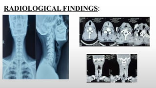
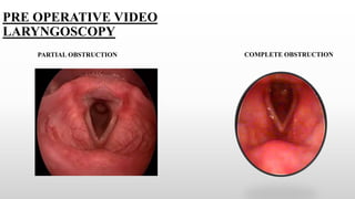
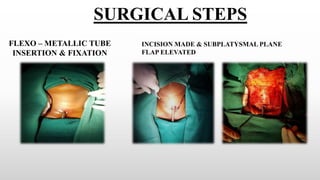
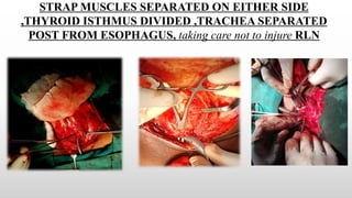
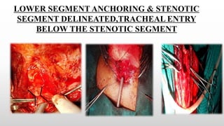
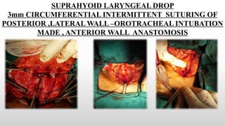
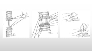
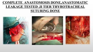
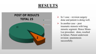
A LIFELINE TO THE LIFE’S
CONDUIT”
-A CASE SERIES ANALYSIS ON TRACHEAL
STENOSIS & ITS MANAGEMENT
AUTHORS :
PROF.DR.R.MUTHUKUMAR,MS,DLO,DNB
PROF.DR.K.SEMMANASELVAN,MS,DLO
INTRODUCTION
Management of tracheal stenosis is an enigma for the ENT surgeons
Until recently surgery for tracheal stenosis was under the realm of the
cardiothoracic surgeon.
Presently, cervical tracheal stenosis is increasingly managed by ENT
surgeons, avoiding unnecessary midline sternotomy.
MAIN GOAL –
To reduce the morbidity of the patient and to provide a
Normal Functioning Airway
STUDY METHODOLOGY:
STUDY DESIGN:
- A Case Series analysis from
2011-2021 ,10 year study period
STUDY SUBJECTS:
- 25 patients
AGE GROUP NUMBER PERCENTAG
E
%
<25 Years 18 72
26 – 45 Years 7 28
>45 Years - -
TOTAL 25
RANGE 19- 36 YEARS
AGE DISTRIBUTION:
MALE
FEMALE
GENDER
DISTRIBUTION:
25 Cases done in a period of 10 years
- 3 cases done along with CTS
- 22 cases done independently
In 4 cases cricotracheal anastamosis ,
rest were tracheotracheal anastomosis
Suprahyoid Laryngeal drop was done
in 21 cases
In 23 of our cases the cause was a
postintubation stenosis and 2 cases -
post traumatic
In 20 cases prior tracheostomy done-
hence anaesthetized via the
tracheostomy site
In 2 cases – no tracheostomy –ET
tube kept proximal to the stenotic site
In 3 cases – Intra Operative
tracheostomy done & proceeded
MATERIALS & METHODS
STENOTIC
SEGMENT
NUMBER OF
PATIENTS
1-2 tracheal rings 3
3-4 tracheal rings 8
4-5 tracheal rings 13
6-7 tracheal rings 1
1.Flexible bronchoscopy
- Flexible fibreoptic bronchoscopy
is the best investigation for exact
site & size.
2. CT scan with reconstruction
Helical CT with high resolution
and 3 dimensional reconstruction
- Site,length,surrounding anatomy
3.VLS & Direct laryngoscopy
4.Lung function tests
RADIOLOGICAL FINDINGS:
PRE OPERATIVE VIDEO
LARYNGOSCOPY
COMPLETE OBSTRUCTION
PARTIAL OBSTRUCTION
FLEXO – METALLIC TUBE
INSERTION & FIXATION
SURGICAL STEPS
INCISION MADE & SUBPLATYSMAL PLANE
FLAP ELEVATED
STRAP MUSCLES SEPARATED ON EITHER SIDE
,THYROID ISTHMUS DIVIDED ,TRACHEA SEPARATED
POST FROM ESOPHAGUS, taking care not to injure RLN
LOWER SEGMENT ANCHORING & STENOTIC
SEGMENT DELINEATED,TRACHEAL ENTRY
BELOW THE STENOTIC SEGMENT
SUPRAHYOID LARYNGEAL DROP
3mm CIRCUMFERENTIAL INTERMITTENT SUTURING OF
POSTERIOR ,LATERAL WALL –OROTRACHEAL INTUBATION
MADE , ANTERIOR WALL ANASTOMOSIS
COMPLETE ANASTOMOSIS DONE,ANASTOMATIC
LEAKAGE TESTED ,II TIER THYROTRACHEAL
SUTURING DONE
WOUND CLOSURE IN
LAYERS
THIRD TIER TENSION
RELIEVING SUTURE-CHIN TO
CHEST MADE
POST OP CARE
1. Patient is maintained in chin flexed position with adequate back
support for 7 days.
2. Patient allowed to converse but with minimal necktwisting
movements.
3. Sympathetic counselling to the patients.
4. Steam nebulisation
5. Adequate hydration
6. Antibiotics, Mucolytics
7. Supplementation steroids
8. RT feeding for 3 days, followed by straw feeding
9.Physiotherapy-not possible
GRILLO’S
STITCHES
/GUARDIAN
SUTURE
POST OPERATIVE POSITION
PRE OP-POST OP VDL
PATHOPHYSIOLOGY OF LTS:
TRAUMA
SUBMUCOSAL
HAEMATOMA
GRANULATION
TISSUE
COLLAGEN
FORMATION
FIBROSIS AND
LUMINAL STENOSIS
ULCERATION
SUPERFICIAL
NO STENOSIS
DEEP
CARTILAGE
NECROSIS AND
HOUR GLASS
STENOSIS
- LTS is the end result of inflammation/trauma to larynx
- This is further compounded by hypoxia which coexists with the condition
DISCUSSION
FACTORS FAVOURING DEVELOPMENT OF
LARYNGOTRACHEAL STENOSIS IN POST
INTUBATION PATIENTS
Pressure necrosis
Tracheal mucosal blood flow
Pressure and volume of cuff
Duration of intubation
Head and neck position
Systemic hypotension
Local infection
MEYER – COTTON
STAGING SYSTEMS
UPPER AIRWAY STENOSIS ( GRILLO
TYPES)
TYPE A – High tracheal stenosis (easily treated by segmental
resection and tracheotracheal anastomosis.)
TYPE B – Stenosis reaching lower border of cricoid- Cricotracheal
anastomosis
TYPE C – Stenosis of lower subglottic larynx and upper trachea
-involves anterior portion of cricoid cartilage(LTR/PCTR)
TYPE D – Stenosis that reaches to glottis
No subglottic space for an effective anastomosis.
- LTR/PCTR/Glottic reconstruction
All types require Cervical approach without sternotomy
CLINICAL FEATURES :
 Ineffective cough
 Stridor ( Insp / Exp)
 Dyspnoea
 - Clinical signs of stenosis appear when lumen
obliterated >50%
PEARLS & PITFALLS
To use LOW PRESSURE HIGH
VOLUME Endo tracheal &
Tracheostomy tubes
When a stenotic patient presents with
stridor –Tracheostomy should be done
at or just below the stenotic site, So
that cartilage is not wasted and trachea
can be saved for future anastomosis
Laryngeal drop-aids in
additional 2-3cm tracheal
mobilisation MONTGOMERY DROP
/SUPRAHYOID RELEASE
POST OP COMPLICATIONS
Granulation
Separation – excessive anastamotic tension
Air leakage
Cord dysfunction
Aspiration
Hypoxemia
Quadriplegia
RESULTS
23
POST OP RESULTS
TOTAL 25 UNEVENTFUL
RESTENOSIS
2
 In 1 case – revision surgery
done and patient is doing well.
 In another case – post
traumatic stenosis with long
stenotic segment. Hence Shian-
Lee procedure done, resulted
in failure. Patient underwent
revision anastomosis
successfully.
CONCLUSION
 Tracheal resection and reconstruction can be challenging and should be undertaken by
centres with experience.
 Standard treatment consists of resection of pathologic segment of trachea with end
to end anastomosis ( > 95% success rate).
 Careful attention to matching the geometry of the ends to be anastomosed is important to
avoid gaps or weak points and for air tight seal anastomosis. (However some amount
telescoping of the anastomosis ends is permitted )
 Adequate planning of ENT surgeon & anaesthesia support is essential for ventilation,
haemostasis and better visualisation.
 Second and third tier reinforcement suturing is essential to relieve the tension at the
anastomotic site for successful wound healing .
REFERENCES
Surgery of the trachea and bronchi,Hermes c.Grillo,MD,2004
Laryngeal and tracheobronchial stenosis,Guri s.sandhu
MD,FRCS,2016
D'Andrilli, Antonio et al. “Subglottic tracheal stenosis.” Journal of
thoracic disease vol. 8,Suppl 2 (2016): S140-7.
Melkane AE, Matar NE, Haddad AC, Nassar MN, Almoutran HG,
Management of postintubation tracheal stenosis: appropriate
indications make outcome differences. Respiration. 2010;79(5):395-
401. doi: 10.1159/000279225. Epub 2010 Jan 26. PMID: 20110646.

tracheal stenosis Dr.muthukumar dr.semmanaselvan

  • 1.
    A LIFELINE TOTHE LIFE’S CONDUIT” -A CASE SERIES ANALYSIS ON TRACHEAL STENOSIS & ITS MANAGEMENT AUTHORS : PROF.DR.R.MUTHUKUMAR,MS,DLO,DNB PROF.DR.K.SEMMANASELVAN,MS,DLO
  • 2.
    INTRODUCTION Management of trachealstenosis is an enigma for the ENT surgeons Until recently surgery for tracheal stenosis was under the realm of the cardiothoracic surgeon. Presently, cervical tracheal stenosis is increasingly managed by ENT surgeons, avoiding unnecessary midline sternotomy. MAIN GOAL – To reduce the morbidity of the patient and to provide a Normal Functioning Airway
  • 3.
    STUDY METHODOLOGY: STUDY DESIGN: -A Case Series analysis from 2011-2021 ,10 year study period STUDY SUBJECTS: - 25 patients AGE GROUP NUMBER PERCENTAG E % <25 Years 18 72 26 – 45 Years 7 28 >45 Years - - TOTAL 25 RANGE 19- 36 YEARS AGE DISTRIBUTION: MALE FEMALE GENDER DISTRIBUTION:
  • 4.
    25 Cases donein a period of 10 years - 3 cases done along with CTS - 22 cases done independently In 4 cases cricotracheal anastamosis , rest were tracheotracheal anastomosis Suprahyoid Laryngeal drop was done in 21 cases In 23 of our cases the cause was a postintubation stenosis and 2 cases - post traumatic In 20 cases prior tracheostomy done- hence anaesthetized via the tracheostomy site In 2 cases – no tracheostomy –ET tube kept proximal to the stenotic site In 3 cases – Intra Operative tracheostomy done & proceeded
  • 5.
    MATERIALS & METHODS STENOTIC SEGMENT NUMBEROF PATIENTS 1-2 tracheal rings 3 3-4 tracheal rings 8 4-5 tracheal rings 13 6-7 tracheal rings 1 1.Flexible bronchoscopy - Flexible fibreoptic bronchoscopy is the best investigation for exact site & size. 2. CT scan with reconstruction Helical CT with high resolution and 3 dimensional reconstruction - Site,length,surrounding anatomy 3.VLS & Direct laryngoscopy 4.Lung function tests
  • 6.
  • 7.
    PRE OPERATIVE VIDEO LARYNGOSCOPY COMPLETEOBSTRUCTION PARTIAL OBSTRUCTION
  • 8.
    FLEXO – METALLICTUBE INSERTION & FIXATION SURGICAL STEPS INCISION MADE & SUBPLATYSMAL PLANE FLAP ELEVATED
  • 9.
    STRAP MUSCLES SEPARATEDON EITHER SIDE ,THYROID ISTHMUS DIVIDED ,TRACHEA SEPARATED POST FROM ESOPHAGUS, taking care not to injure RLN
  • 10.
    LOWER SEGMENT ANCHORING& STENOTIC SEGMENT DELINEATED,TRACHEAL ENTRY BELOW THE STENOTIC SEGMENT
  • 11.
    SUPRAHYOID LARYNGEAL DROP 3mmCIRCUMFERENTIAL INTERMITTENT SUTURING OF POSTERIOR ,LATERAL WALL –OROTRACHEAL INTUBATION MADE , ANTERIOR WALL ANASTOMOSIS
  • 13.
    COMPLETE ANASTOMOSIS DONE,ANASTOMATIC LEAKAGETESTED ,II TIER THYROTRACHEAL SUTURING DONE
  • 14.
    WOUND CLOSURE IN LAYERS THIRDTIER TENSION RELIEVING SUTURE-CHIN TO CHEST MADE
  • 15.
    POST OP CARE 1.Patient is maintained in chin flexed position with adequate back support for 7 days. 2. Patient allowed to converse but with minimal necktwisting movements. 3. Sympathetic counselling to the patients. 4. Steam nebulisation 5. Adequate hydration 6. Antibiotics, Mucolytics 7. Supplementation steroids 8. RT feeding for 3 days, followed by straw feeding 9.Physiotherapy-not possible GRILLO’S STITCHES /GUARDIAN SUTURE
  • 16.
  • 17.
  • 18.
    PATHOPHYSIOLOGY OF LTS: TRAUMA SUBMUCOSAL HAEMATOMA GRANULATION TISSUE COLLAGEN FORMATION FIBROSISAND LUMINAL STENOSIS ULCERATION SUPERFICIAL NO STENOSIS DEEP CARTILAGE NECROSIS AND HOUR GLASS STENOSIS - LTS is the end result of inflammation/trauma to larynx - This is further compounded by hypoxia which coexists with the condition DISCUSSION
  • 19.
    FACTORS FAVOURING DEVELOPMENTOF LARYNGOTRACHEAL STENOSIS IN POST INTUBATION PATIENTS Pressure necrosis Tracheal mucosal blood flow Pressure and volume of cuff Duration of intubation Head and neck position Systemic hypotension Local infection
  • 20.
    MEYER – COTTON STAGINGSYSTEMS UPPER AIRWAY STENOSIS ( GRILLO TYPES) TYPE A – High tracheal stenosis (easily treated by segmental resection and tracheotracheal anastomosis.) TYPE B – Stenosis reaching lower border of cricoid- Cricotracheal anastomosis TYPE C – Stenosis of lower subglottic larynx and upper trachea -involves anterior portion of cricoid cartilage(LTR/PCTR) TYPE D – Stenosis that reaches to glottis No subglottic space for an effective anastomosis. - LTR/PCTR/Glottic reconstruction All types require Cervical approach without sternotomy CLINICAL FEATURES :  Ineffective cough  Stridor ( Insp / Exp)  Dyspnoea  - Clinical signs of stenosis appear when lumen obliterated >50%
  • 21.
    PEARLS & PITFALLS Touse LOW PRESSURE HIGH VOLUME Endo tracheal & Tracheostomy tubes When a stenotic patient presents with stridor –Tracheostomy should be done at or just below the stenotic site, So that cartilage is not wasted and trachea can be saved for future anastomosis Laryngeal drop-aids in additional 2-3cm tracheal mobilisation MONTGOMERY DROP /SUPRAHYOID RELEASE
  • 22.
    POST OP COMPLICATIONS Granulation Separation– excessive anastamotic tension Air leakage Cord dysfunction Aspiration Hypoxemia Quadriplegia
  • 23.
    RESULTS 23 POST OP RESULTS TOTAL25 UNEVENTFUL RESTENOSIS 2  In 1 case – revision surgery done and patient is doing well.  In another case – post traumatic stenosis with long stenotic segment. Hence Shian- Lee procedure done, resulted in failure. Patient underwent revision anastomosis successfully.
  • 24.
    CONCLUSION  Tracheal resectionand reconstruction can be challenging and should be undertaken by centres with experience.  Standard treatment consists of resection of pathologic segment of trachea with end to end anastomosis ( > 95% success rate).  Careful attention to matching the geometry of the ends to be anastomosed is important to avoid gaps or weak points and for air tight seal anastomosis. (However some amount telescoping of the anastomosis ends is permitted )  Adequate planning of ENT surgeon & anaesthesia support is essential for ventilation, haemostasis and better visualisation.  Second and third tier reinforcement suturing is essential to relieve the tension at the anastomotic site for successful wound healing .
  • 25.
    REFERENCES Surgery of thetrachea and bronchi,Hermes c.Grillo,MD,2004 Laryngeal and tracheobronchial stenosis,Guri s.sandhu MD,FRCS,2016 D'Andrilli, Antonio et al. “Subglottic tracheal stenosis.” Journal of thoracic disease vol. 8,Suppl 2 (2016): S140-7. Melkane AE, Matar NE, Haddad AC, Nassar MN, Almoutran HG, Management of postintubation tracheal stenosis: appropriate indications make outcome differences. Respiration. 2010;79(5):395- 401. doi: 10.1159/000279225. Epub 2010 Jan 26. PMID: 20110646.