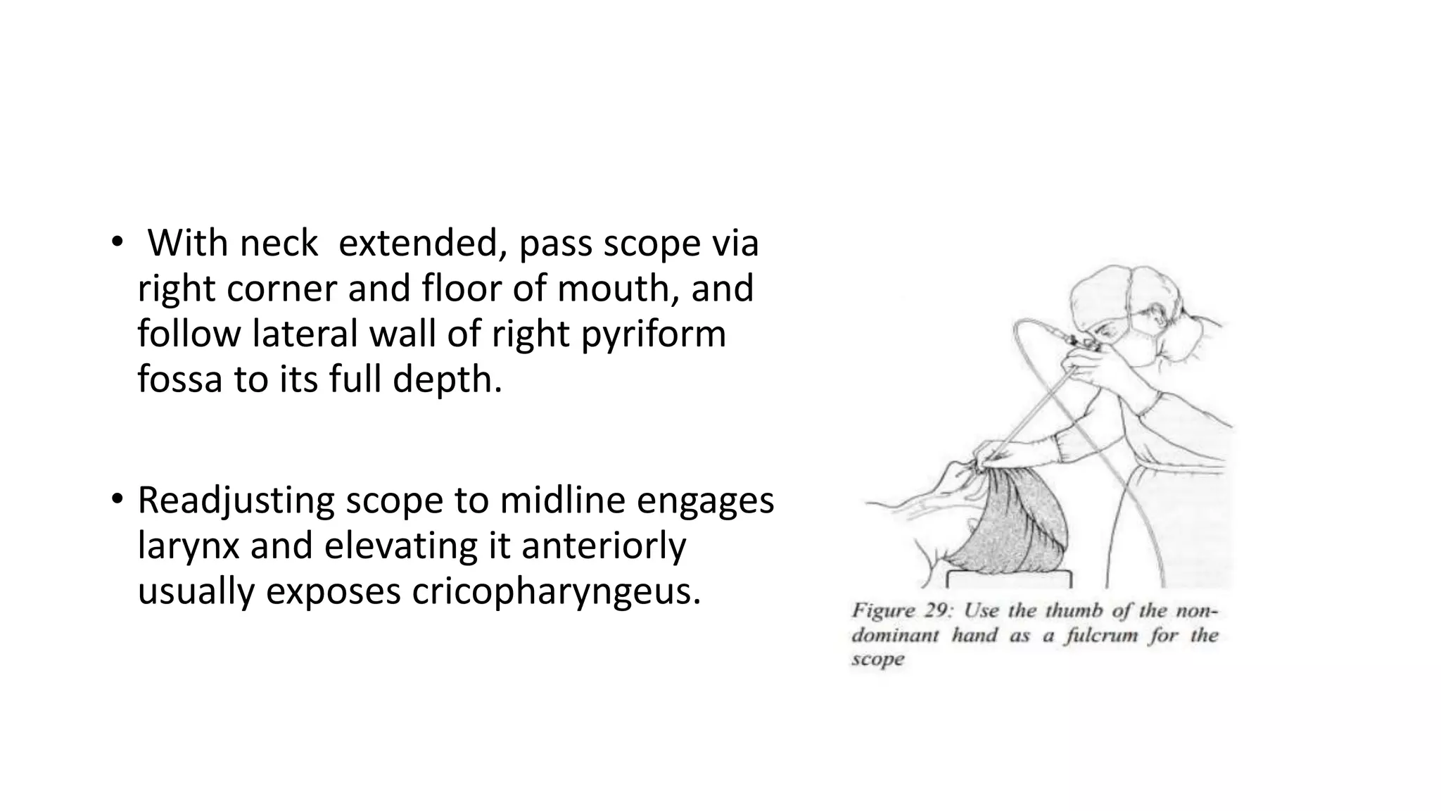
The document provides a history of the development of endoscopy of the esophagus and related structures. It describes innovations from the early 19th century using candles for illumination to the modern development of flexible fiberoptic endoscopy in the 1960s. It also summarizes the anatomy of the esophagus including its parts, walls, blood supply, lymphatics and innervation. Finally, it discusses techniques for rigid and flexible esophagoscopy along with potential complications.
Esophagoscopy continues to be a reliable diagnostic and therapeutic tool with a wide variety of applications, including biopsy, dilatation of strictures, repair of Zenker's diverticulum, placement of stents, and retrieval of foreign bodies.
Tracheostomy decannulation is always challenging and this presentation address the various issues, indications, contra-indications, problems and solutions.
Google drive:-https://www.youtube.com/redirect?q=https%3A%2F%2Fdrive.google.com%2Fopen%3Fid%3D1ZET4JzZalyUfM1KWXemKZsQQXMzrYpcJ&v=WHOggpW5Ee8&event=video_description&redir_token=77oOekaJs8_u0RLfrUH8z68tJFt8MTU2MDY1Njc4N0AxNTYwNTcwMzg3
Youtube:-https://www.youtube.com/watch?v=WHOggpW5Ee8
thyroidectomy-surgical seminare, prepared by Dr. Siddharth JINDAL, third year resident in dept. of general surgery at P.D.U. Government Medical College and Civil Hospital, Rajkot, Gujarat.
TRACHEOSTOMY is a surgical procedure to maintain a patent airway to the person who is in airway distress or electively in certain surgical procedures like oncological resections to maintain an adequate oxygenation to the patient by creating a stoma on the trachea
Adv. biopharm. APPLICATION OF PHARMACOKINETICS : TARGETED DRUG DELIVERY SYSTEMSAkankshaAshtankar
MIP 201T & MPH 202T
ADVANCED BIOPHARMACEUTICS & PHARMACOKINETICS : UNIT 5
APPLICATION OF PHARMACOKINETICS : TARGETED DRUG DELIVERY SYSTEMS By - AKANKSHA ASHTANKAR
Title: Sense of Smell
Presenter: Dr. Faiza, Assistant Professor of Physiology
Qualifications:
MBBS (Best Graduate, AIMC Lahore)
FCPS Physiology
ICMT, CHPE, DHPE (STMU)
MPH (GC University, Faisalabad)
MBA (Virtual University of Pakistan)
Learning Objectives:
Describe the primary categories of smells and the concept of odor blindness.
Explain the structure and location of the olfactory membrane and mucosa, including the types and roles of cells involved in olfaction.
Describe the pathway and mechanisms of olfactory signal transmission from the olfactory receptors to the brain.
Illustrate the biochemical cascade triggered by odorant binding to olfactory receptors, including the role of G-proteins and second messengers in generating an action potential.
Identify different types of olfactory disorders such as anosmia, hyposmia, hyperosmia, and dysosmia, including their potential causes.
Key Topics:
Olfactory Genes:
3% of the human genome accounts for olfactory genes.
400 genes for odorant receptors.
Olfactory Membrane:
Located in the superior part of the nasal cavity.
Medially: Folds downward along the superior septum.
Laterally: Folds over the superior turbinate and upper surface of the middle turbinate.
Total surface area: 5-10 square centimeters.
Olfactory Mucosa:
Olfactory Cells: Bipolar nerve cells derived from the CNS (100 million), with 4-25 olfactory cilia per cell.
Sustentacular Cells: Produce mucus and maintain ionic and molecular environment.
Basal Cells: Replace worn-out olfactory cells with an average lifespan of 1-2 months.
Bowman’s Gland: Secretes mucus.
Stimulation of Olfactory Cells:
Odorant dissolves in mucus and attaches to receptors on olfactory cilia.
Involves a cascade effect through G-proteins and second messengers, leading to depolarization and action potential generation in the olfactory nerve.
Quality of a Good Odorant:
Small (3-20 Carbon atoms), volatile, water-soluble, and lipid-soluble.
Facilitated by odorant-binding proteins in mucus.
Membrane Potential and Action Potential:
Resting membrane potential: -55mV.
Action potential frequency in the olfactory nerve increases with odorant strength.
Adaptation Towards the Sense of Smell:
Rapid adaptation within the first second, with further slow adaptation.
Psychological adaptation greater than receptor adaptation, involving feedback inhibition from the central nervous system.
Primary Sensations of Smell:
Camphoraceous, Musky, Floral, Pepperminty, Ethereal, Pungent, Putrid.
Odor Detection Threshold:
Examples: Hydrogen sulfide (0.0005 ppm), Methyl-mercaptan (0.002 ppm).
Some toxic substances are odorless at lethal concentrations.
Characteristics of Smell:
Odor blindness for single substances due to lack of appropriate receptor protein.
Behavioral and emotional influences of smell.
Transmission of Olfactory Signals:
From olfactory cells to glomeruli in the olfactory bulb, involving lateral inhibition.
Primitive, less old, and new olfactory systems with different path
These simplified slides by Dr. Sidra Arshad present an overview of the non-respiratory functions of the respiratory tract.
Learning objectives:
1. Enlist the non-respiratory functions of the respiratory tract
2. Briefly explain how these functions are carried out
3. Discuss the significance of dead space
4. Differentiate between minute ventilation and alveolar ventilation
5. Describe the cough and sneeze reflexes
Study Resources:
1. Chapter 39, Guyton and Hall Textbook of Medical Physiology, 14th edition
2. Chapter 34, Ganong’s Review of Medical Physiology, 26th edition
3. Chapter 17, Human Physiology by Lauralee Sherwood, 9th edition
4. Non-respiratory functions of the lungs https://academic.oup.com/bjaed/article/13/3/98/278874
Recomendações da OMS sobre cuidados maternos e neonatais para uma experiência pós-natal positiva.
Em consonância com os ODS – Objetivos do Desenvolvimento Sustentável e a Estratégia Global para a Saúde das Mulheres, Crianças e Adolescentes, e aplicando uma abordagem baseada nos direitos humanos, os esforços de cuidados pós-natais devem expandir-se para além da cobertura e da simples sobrevivência, de modo a incluir cuidados de qualidade.
Estas diretrizes visam melhorar a qualidade dos cuidados pós-natais essenciais e de rotina prestados às mulheres e aos recém-nascidos, com o objetivo final de melhorar a saúde e o bem-estar materno e neonatal.
Uma “experiência pós-natal positiva” é um resultado importante para todas as mulheres que dão à luz e para os seus recém-nascidos, estabelecendo as bases para a melhoria da saúde e do bem-estar a curto e longo prazo. Uma experiência pós-natal positiva é definida como aquela em que as mulheres, pessoas que gestam, os recém-nascidos, os casais, os pais, os cuidadores e as famílias recebem informação consistente, garantia e apoio de profissionais de saúde motivados; e onde um sistema de saúde flexível e com recursos reconheça as necessidades das mulheres e dos bebês e respeite o seu contexto cultural.
Estas diretrizes consolidadas apresentam algumas recomendações novas e já bem fundamentadas sobre cuidados pós-natais de rotina para mulheres e neonatos que recebem cuidados no pós-parto em unidades de saúde ou na comunidade, independentemente dos recursos disponíveis.
É fornecido um conjunto abrangente de recomendações para cuidados durante o período puerperal, com ênfase nos cuidados essenciais que todas as mulheres e recém-nascidos devem receber, e com a devida atenção à qualidade dos cuidados; isto é, a entrega e a experiência do cuidado recebido. Estas diretrizes atualizam e ampliam as recomendações da OMS de 2014 sobre cuidados pós-natais da mãe e do recém-nascido e complementam as atuais diretrizes da OMS sobre a gestão de complicações pós-natais.
O estabelecimento da amamentação e o manejo das principais intercorrências é contemplada.
Recomendamos muito.
Vamos discutir essas recomendações no nosso curso de pós-graduação em Aleitamento no Instituto Ciclos.
Esta publicação só está disponível em inglês até o momento.
Prof. Marcus Renato de Carvalho
www.agostodourado.com
Tom Selleck Health: A Comprehensive Look at the Iconic Actor’s Wellness Journeygreendigital
Tom Selleck, an enduring figure in Hollywood. has captivated audiences for decades with his rugged charm, iconic moustache. and memorable roles in television and film. From his breakout role as Thomas Magnum in Magnum P.I. to his current portrayal of Frank Reagan in Blue Bloods. Selleck's career has spanned over 50 years. But beyond his professional achievements. fans have often been curious about Tom Selleck Health. especially as he has aged in the public eye.
Follow us on: Pinterest
Introduction
Many have been interested in Tom Selleck health. not only because of his enduring presence on screen but also because of the challenges. and lifestyle choices he has faced and made over the years. This article delves into the various aspects of Tom Selleck health. exploring his fitness regimen, diet, mental health. and the challenges he has encountered as he ages. We'll look at how he maintains his well-being. the health issues he has faced, and his approach to ageing .
Early Life and Career
Childhood and Athletic Beginnings
Tom Selleck was born on January 29, 1945, in Detroit, Michigan, and grew up in Sherman Oaks, California. From an early age, he was involved in sports, particularly basketball. which played a significant role in his physical development. His athletic pursuits continued into college. where he attended the University of Southern California (USC) on a basketball scholarship. This early involvement in sports laid a strong foundation for his physical health and disciplined lifestyle.
Transition to Acting
Selleck's transition from an athlete to an actor came with its physical demands. His first significant role in "Magnum P.I." required him to perform various stunts and maintain a fit appearance. This role, which he played from 1980 to 1988. necessitated a rigorous fitness routine to meet the show's demands. setting the stage for his long-term commitment to health and wellness.
Fitness Regimen
Workout Routine
Tom Selleck health and fitness regimen has evolved. adapting to his changing roles and age. During his "Magnum, P.I." days. Selleck's workouts were intense and focused on building and maintaining muscle mass. His routine included weightlifting, cardiovascular exercises. and specific training for the stunts he performed on the show.
Selleck adjusted his fitness routine as he aged to suit his body's needs. Today, his workouts focus on maintaining flexibility, strength, and cardiovascular health. He incorporates low-impact exercises such as swimming, walking, and light weightlifting. This balanced approach helps him stay fit without putting undue strain on his joints and muscles.
Importance of Flexibility and Mobility
In recent years, Selleck has emphasized the importance of flexibility and mobility in his fitness regimen. Understanding the natural decline in muscle mass and joint flexibility with age. he includes stretching and yoga in his routine. These practices help prevent injuries, improve posture, and maintain mobilit
Ozempic: Preoperative Management of Patients on GLP-1 Receptor Agonists Saeid Safari
Preoperative Management of Patients on GLP-1 Receptor Agonists like Ozempic and Semiglutide
ASA GUIDELINE
NYSORA Guideline
2 Case Reports of Gastric Ultrasound
CDSCO and Phamacovigilance {Regulatory body in India}NEHA GUPTA
The Central Drugs Standard Control Organization (CDSCO) is India's national regulatory body for pharmaceuticals and medical devices. Operating under the Directorate General of Health Services, Ministry of Health & Family Welfare, Government of India, the CDSCO is responsible for approving new drugs, conducting clinical trials, setting standards for drugs, controlling the quality of imported drugs, and coordinating the activities of State Drug Control Organizations by providing expert advice.
Pharmacovigilance, on the other hand, is the science and activities related to the detection, assessment, understanding, and prevention of adverse effects or any other drug-related problems. The primary aim of pharmacovigilance is to ensure the safety and efficacy of medicines, thereby protecting public health.
In India, pharmacovigilance activities are monitored by the Pharmacovigilance Programme of India (PvPI), which works closely with CDSCO to collect, analyze, and act upon data regarding adverse drug reactions (ADRs). Together, they play a critical role in ensuring that the benefits of drugs outweigh their risks, maintaining high standards of patient safety, and promoting the rational use of medicines.
- Video recording of this lecture in English language: https://youtu.be/kqbnxVAZs-0
- Video recording of this lecture in Arabic language: https://youtu.be/SINlygW1Mpc
- Link to download the book free: https://nephrotube.blogspot.com/p/nephrotube-nephrology-books.html
- Link to NephroTube website: www.NephroTube.com
- Link to NephroTube social media accounts: https://nephrotube.blogspot.com/p/join-nephrotube-on-social-media.html
Flu Vaccine Alert in Bangalore Karnatakaaddon Scans
As flu season approaches, health officials in Bangalore, Karnataka, are urging residents to get their flu vaccinations. The seasonal flu, while common, can lead to severe health complications, particularly for vulnerable populations such as young children, the elderly, and those with underlying health conditions.
Dr. Vidisha Kumari, a leading epidemiologist in Bangalore, emphasizes the importance of getting vaccinated. "The flu vaccine is our best defense against the influenza virus. It not only protects individuals but also helps prevent the spread of the virus in our communities," he says.
This year, the flu season is expected to coincide with a potential increase in other respiratory illnesses. The Karnataka Health Department has launched an awareness campaign highlighting the significance of flu vaccinations. They have set up multiple vaccination centers across Bangalore, making it convenient for residents to receive their shots.
To encourage widespread vaccination, the government is also collaborating with local schools, workplaces, and community centers to facilitate vaccination drives. Special attention is being given to ensuring that the vaccine is accessible to all, including marginalized communities who may have limited access to healthcare.
Residents are reminded that the flu vaccine is safe and effective. Common side effects are mild and may include soreness at the injection site, mild fever, or muscle aches. These side effects are generally short-lived and far less severe than the flu itself.
Healthcare providers are also stressing the importance of continuing COVID-19 precautions. Wearing masks, practicing good hand hygiene, and maintaining social distancing are still crucial, especially in crowded places.
Protect yourself and your loved ones by getting vaccinated. Together, we can help keep Bangalore healthy and safe this flu season. For more information on vaccination centers and schedules, residents can visit the Karnataka Health Department’s official website or follow their social media pages.
Stay informed, stay safe, and get your flu shot today!
2. HISTORY OF AERODIGESTIVE
ENDOSCOPY
• In 1806,Bozzini fashioned an angled speculum with a mirror insert for
examination of the larynx, using a single wax candle for illumination.
• In 1829, Babbington described a glottiscope, a three-bladed device
including a stainless steel mirror and tongue retractors.
• Desmoreaux, the “father of endoscopy,” redesigned it in 1853 by
attaching a gaslight and condensers to project a beam of light down
the tube.
3. HISTORY OF AERODIGESTIVE
ENDOSCOPY
• Killian In 1897,developed a direct laryngoscope using a headlight for
illumination.
• He was the first to remove a foreign body in the bronchial tree using
direct upper bronchoscopy.
• In the early 1900s, Chevalier Jackson invented distal lighting for
endoscopic equipment, and designed a variety of endoscopic
instruments
4. HISTORY OF AERODIGESTIVE
ENDOSCOPY
• The use of magnification to enhance endoscopy was developed by
Brunings and Jackson.
• The addition of the Zeiss operating microscope for direct
pharyngolaryngoscopy was seen in the mid-1950s.
• Adolf Kussmaul was the first to use a straight tube after studying the
techniques of sword swallowers to visualize the osephagus.
5. HISTORY OF AERODIGESTIVE
ENDOSCOPY
• In the 1960s, flexible fiber-optic bronchoscopes were developed and
introduced into clinical use by Shigeto Ikeda.
• Fiber-optic esophagoscopes, shortly thereafter, came into use.
6. ANATOMY
• The esophagus is a vertical muscular tube that extends from the
hypopharynx to the stomach.
• 23 to 25 cm in length in the adult.2 cm Width.It’s Lumen is flattened
anteroposteriorly.Normally it’s kept closed (collapsed) and opens
(dilates) only during the passage of the food.
• The esophagus starts in the midline at the lower border of the cricoid
cartilage (C6), extending to the cardiac orifice of the stomach(T11).
7. • PARTS OF THE ESOPHAGUS
• The esophagus is split into the
following 3 parts
• Cervical part (4 cm in length).The
cervical part extends from the
lower border of cricoid cartilage
to the superior border of
manubrium sterni.
8. • Borders of cervical oseophagus
• Anteriorly by the trachea
• Posteriorly by the vertebral
column
• Laterally by the carotid sheaths
and the thyroid gland
9. • Thoracic part (20 cm in length)
• The thoracic part extends from superior border of manubrium sterni
to the esophageal opening in the diaphragm
• In the superior mediastinum, the esophagus is posterior to the
trachea and in contact with the common carotid arteries.
• The recurrent laryngeal nerves lie in the angle between the
esophagus and the trachea
• The thoracic duct lies on its left side
11. • The esophagus passes posterior and to the right of the aortic arch
and proceeds along the right side of the descending aorta.
• Inferior to the arch of the aorta, the left bronchus crosses and indents
the esophagus anteriorly.
• The thoracic duct is posterior to the lower portion of the esophagus,
and the azygos vein is to the right side of the esophagus in the thorax.
• The right vagus nerve descends posterior to and the left vagus
descends anterior to the esophagus
13. • Abdominal part (1-2 cm in
length).
• The abdominal part extends
create esophageal opening in
the diaphragm to the cardiac
end of the stomach.
• The short abdominal part of the
oesophagus (about 1 cm) forms
a groove in the left lobe in the
liver.
14. Anatomical Position
• The oesophagus begins in the
midline at the level of the lower
cricoid border (C6).
• It then deviates to the left at the
root of the neck.
• It returns to the midline in the
mediastinum at T5.
15. • When it reaches T7, it once
again deviates to the left to
reach the gastric cardia.
• It passes anteriorly into the
oesophageal hiatus of the
diaphragm at T10.
• It ends at the level of T11 in the
gastric cardiac orifice
16. Topography and Constrictions of Esophagus
• The distance of each constriction is measured from the upper incisor
teeth.
15 cm
17.
18. CLINICAL IMPORTANCE OF
ESOPHAGEAL CONSTRICTIONS
• The anatomical constrictions of esophagus are of considerable clinical
importance because of the following reasons.
• These are the sites where swallowed foreign bodies may stuck in the
esophagus.
• These are the sites where strictures develop after ingestion of caustic
substances.
• These are sites via which it might be difficult to pass
ophagoscope/gastric tube
19. The wall of the esophagus: four layers
• The esophagus is lined by nonkeratinizing stratified squamous
epithelium.It has a thin lamina propria layer.
• The muscularis mucosae are composed of smooth muscle fibers
• The submucosa consists primarily of thick collagenous and coarse
elastic fibers.It also contains mucous glands and Meissner’s plexus.
• The outer coat, or fibrosa, consists of loose fibroelastic tissue
20. Musculature of Esophagus
• The esophagus consists of
– Striated (voluntary) muscle in its upper third,
– Smooth (involuntary) muscle in its lower third,
– Mixture of striated and smooth muscle in between
• The muscle of the esophagus is composed of an inner circular layer
and an outer longitudinal layer.
• Auerbach’s myenteric plexus lies between the two muscle layers.
22. Oesophageal Sphincters
• There are two sphincters present in the oesophagus, known as
–The upper upper esophageal sphincter is a high-pressure zone in
esophageal manometry and corresponds with the cricopharyngeal
muscle.
–The lower oesophageal sphincter is approximately 3 cm long.
• A single distinct muscle responsible for the lower sphincter action has
not been identified.
23. ARTERIAL SUPPLY
• A.The cervical part is by
– Inferior thyroid arteries
B.The thoracic part is by
– Esophageal branches of
– Descending thoracic aorta, and
– Bronchial arteries.
24. ARTERIAL SUPPLY
• C.The abdominal part is by
– Esophageal branches of
--Left gastric artery, and
--Left inferior phrenic artery
25. VENOUS DRAINAGE
• A. Cervical part is drained by
inferior thyroid veins.
• B. Thoracic part is drained by
azygos and hemiazygos veins.
26. VENOUS DRAINAGE
• C. Abdominal part is drained by
2 venous channels
• Hemiazygos vein, a tributary of
inferior vena cava.
• Left gastric vein, a tributary of
portal vein.
• Thus abdominal part of
esophagus is the site of
portocaval( porto-systemic )
anastomosis.
27. Lymphatics
• The lymphatic drainage of the
oesophagus is divided into thirds
• Superior third :Deep cervical
lymph nodes
• Middle third:Superior and
posterior mediastinal nodes
29. NERVE SUPPLY
• The esophagus is supplied by both parasympathetic and sympathetic
fibres.
• The parasympathetic fibres are originated from recurrent laryngeal
nerves and esophageal plexuses created by vagus nerves.
• They supply sensory, motor, and secretomotor supply to the
esophagus.
30. • The sympathetic fibres are
originated from T5-T9 spinal
segments are sensory and
vasomotor
31. Rigid esophagoscopy
• Indications
1. Exclude 2nd primaries in SCC of upper aerodigestive tract
2. Remove foreign bodies
3. Biopsy, dilate or stent tumours
4. Determine distal extent of hypopharyngeal and oesophageal
carcinoma
5. Dilate strictures
6. Exclude traumatic perforations with Penetrating injury of neck
7. Inject oesophageal varices
32. • 8.Blunt trauma to the neck with associated subcutaneous air
• 9..Radiographic evidence of esophageal masses or strictures.
• 1o.Congenital anomalies.
• 11.Vocal fold palsies
34. Technique
• Proximal oesophagus follows lordosis of Cervical & thoracic spine;
bring both into straight line by elevating head.
• The patient is placed in supine position with a pillow placed
underneath the shoulders and neck to achieve minor extension of
neck and marked head extension at the atlanto-occipital joint.
• Thumb of non-dominant hand as a fulcrum to protect teeth
35. • With neck extended, pass scope via
right corner and floor of mouth, and
follow lateral wall of right pyriform
fossa to its full depth.
• Readjusting scope to midline engages
larynx and elevating it anteriorly
usually exposes cricopharyngeus.
36. • Scope comes to a dead-stop and pharyngeal lumen disappears as one
reaches cricopharyngeal sphincter
• Ensure that bevel of scope is pointing upward
• Elevate tip of scope against post surface of cricoid with non-dominant
thumb.
• Look for oesophageal lumen to appear while applying steady, firm
pressure against contracted cricopharyngeus.
37. • Slowly advance tip of scope always keeping lumen in view.
• Always consider possibility of pharyngeal pouch (zenker’s
diverticulum).Tightly inflated ET tube cuff may compress esophagus.
• Once esophagoscope has been passed all the way, carefully inspect
for pathology & mucosal trauma while slowly retracting scope.
• Biopsy lesions with long biopsy forceps.
38. • Pathology seen at rigid
oesophagoscopy recorded as its
distance from upper incisors
39. FIBER-OPTIC ESOPHAGOSCOPY
• Fiber-optic esophagoscopy can
be performed under general or
local anesthesia.
• Transnasal or oral
• Topical anesthetic agents are
sprayed into the oral cavity and
oropharynx, a bite guard is
placed, and intravenous
sedation.
40. Technique
• The esophagoscope can be inserted blindly, by feel, through the
esophageal inlet.
• The tip of the endoscope is gently advanced into the hypopharynx to
a distance approximately 18 cm from the incisors upto the level of the
upper esophageal sphincter or muscle.
• The cricopharyngeus is closed at rest and opens during swallowing
41.
42. • The esophageal inlet is visualized, and the endoscope is gently
advanced into the esophagus as the patient swallows.
• Examination is performed to a distance of 40 cm.This is also referred
to as the squamocolumnar mucosal junction or Z line.
• Z line, demarcating the junction between pearly, esophageal,
stratified squamous mucosa and the redder gastric columnar
epithelium.
43. Complications of esophagoscopy
• Injury to lip, teeth & tongue.
• Glottic trauma may involve vocal cord injury or dislocation of
arytenoid cartilages.
• Laryngeal edema.
• Aspiration of gastric contents.
• Cervical spinal cord injury.
• Tachycardia, arrhythmias, hypertension, and myocardial ischemia or
infarction due to sympathetic stimulation.
44. Complications of esophagoscopy
• Mucosal tears/lacerations
• Esophageal perforation
• Mediastinitis.
• Insufflation used during fiber-optic esophagoscopy can cause
abdominal distention, contributing to respiratory compromise