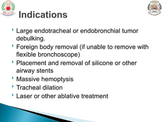
 Rigid bronchoscopy is a technique that visualizes the
trachea and proximal bronchi.
 It is most commonly used to manage patients who
have obstruction of either their trachea or a proximal
bronchus, since the rigid bronchoscope’s large lumen
facilitates suctioning and the removal of debris, or for
interventional procedures such as insertion of airway
stents.
 Large endotracheal or endobronchial tumor
debulking.
 Foreign body removal (if unable to remove with
flexible bronchoscope)
 Placement and removal of silicone or other
airway stents
 Massive hemoptysis
 Tracheal dilation
 Laser or other ablative treatment
 Current or recent myocardial ischemia.
 Poorly controlled heart failure.
 Significant hypotension, hypertension.
 Arrhythmia: bradycardia, or tachycardia.
 Exacerbation of asthma or chronic obstructive
pulmonary disease; severe hypoxemia.
 Additional contraindications exist when
brushing, biopsy, or needle aspiration is
planned, which are related to bleeding risk.
 They include recent anti-platelet agents
(eg, aspirin , clopidogrel ), anticoagulant
therapy, thrombocytopenia, coagulopathy,
elevated blood urea nitrogen (BUN), or
elevated serum creatinine.
 Other contraindications include:
 Inability to cooperate with the procedure.
 Unstable cervical spine, an immobile
cervical spine.
 Limited motion of the temporomandibular
joint
 Not suitable for patients with cervical spine
fracture or trauma.
 Done under general anaesthesia.
 Narrow field of vision.
 Trauma to teeth, pharynx, trachea.
 During rigid bronchoscopy, a rigid telescope and light
source are generally inserted through a rigid
bronchoscope to visualize the airways.
 However, a few systems allow direct visualization
through the rigid bronchoscope using light conducted
through a tube that extends the length of the rigid
bronchoscope or through a prism at the proximal end.
A flexible bronchoscope inserted through the rigid
bronchoscope is a reasonable alternative
Intubation with the rigid bronchoscope usually prompts
the following questions:
 What anesthesia and patient preparation techniques
are commonly used?
 How is direct intubation performed?
 Is laryngoscopically-guided intubation possible?
 Can intubation be performed through a
tracheostomy?
 What complications arise from faulty technique?
 Topical anesthesia is applied
using lidocaine or tetracaine .
 Additional topical anesthetic is usually administered
to the tracheobronchial tree after intubation with the
rigid bronchoscope.
 Patients are oxygenated by mask, and pharyngeal
secretions are aspirated.
 Dentures are removed, and the teeth and gums are
carefully inspected. The teeth may be protected with
gauze pads, foam rubber, or plastic mouth guards.
Patient preparation
 Combination of intravenous general anesthesia
with propofol and assisted spontaneous ventilation is
currently the most frequently used anesthesia technique
for rigid bronchoscopy .
 Complete muscle relaxation is occasionally desirable,
particularly if rigid bronchoscopy is stimulating and if
work is being performed within the lower airways.
 Ventilatory support can be provided using assisted
spontaneous ventilation or closed-circuit positive
pressure ventilation.
Intubation with the rigid bronchoscope usually prompts
the following questions:
 What anesthesia and patient preparation techniques
are commonly used?
 How is direct intubation performed?
 Is laryngoscopically-guided intubation possible?
 Can intubation be performed through a
tracheostomy?
 What complications arise from faulty technique?
 DIRECT INTUBATION: Direct intubation using a rigid telescope is
the method of choice for rigid bronchoscopic intubation. Steps for
direct intubation using a rigid telescope:
 The bronchoscopist stands directly behind the head of the supine
patient.
 The rigid bronchoscope is held in the right hand with its tip
uppermost. The middle finger of the left hand is used to protect the
upper teeth and gums and to control head movements. The
telescope should not extend beyond the edge of the rigid
bronchoscope.
 The bronchoscope is inserted with its tip facing forward. Looking
through the telescope, the bronchoscopist identifies the uvula
posteriorly, and the bronchoscope is advanced along the route of the
tongue.
 The rigid bronchoscope is gently lifted upwards and the epiglottis
is brought into view . The anterior aspect of the beveled tip of the
bronchoscope is then slid under the epiglottis. Gentle
advancement of the rigid tube provides further access to the
larynx.
 After both arytenoids are identified, the rigid tube is lifted more
anteriorly and the vocal cords are seen. As the vocal cords are
approached, the tip of the bronchoscope is rotated 90 degrees
laterally so that the beveled tip lies between them.
 The bronchoscope is advanced and rotated to enter the trachea
without traumatizing the larynx.
 Once beyond the level of the cricoid cartilage, the bronchoscope
may be rotated so that the beveled tip lies along the posterior wall
of the trachea
Intubation with the rigid bronchoscope usually prompts
the following questions:
 What anesthesia and patient preparation techniques
are commonly used?
 How is direct intubation performed?
 Is laryngoscopically-guided intubation possible?
 Can intubation be performed through a
tracheostomy?
 What complications arise from faulty technique?
 INTUBATION WITH LARYNGOSCOPY: For operators
who are uncomfortable with direct intubation but who are
adept at laryngoscopy, the vocal cords may be
visualized using the rigid laryngoscope alone. Either
straight or curved instruments may be used with
intubation during rigid laryngoscopy:
 With the laryngoscope in the midline and after the
epiglottis has been lifted, the rigid bronchoscope is
inserted into the lateral aspect of the mouth and directed
toward the larynx. Care must be taken to avoid
obstruction of the larynx by the rigid tube.
 As the bronchoscope is inserted between the vocal
cords, it is rotated slightly and kept in the midline to
avoid striking the beveled edge of the bronchoscope
against the lateral wall of the subglottis.
 Once the bronchoscope is inserted through the cords,
the laryngoscope is removed, and the rigid telescope
can be placed into the rigid bronchoscope. The
bronchoscope is then advanced further into the tracheal
lumen.
Intubation with the rigid bronchoscope usually prompts
the following questions:
 What anesthesia and patient preparation techniques
are commonly used?
 How is direct intubation performed?
 Is laryngoscopically-guided intubation possible?
 Can intubation be performed through a
tracheostomy?
 What complications arise from faulty technique?
 INTUBATION VIA TRACHEOSTOMY : is a relatively simple
technique. After topical anesthesia with viscous
lidocaine applied to the edges of the tracheostomy, the rigid
bronchoscope can be inserted directly through the stoma
from a lateral position.
 Tracheostomy intubation is easiest if the bronchoscopist
stands behind the patient and slightly to the patient's left or
right. Care is taken to insert a long, rigid tube obliquely in
order to avoid striking the beveled edge of the rigid
bronchoscope against the posterior wall of the trachea.
Intubation with the rigid bronchoscope usually prompts
the following questions:
 What anesthesia and patient preparation techniques
are commonly used?
 How is direct intubation performed?
 Is laryngoscopically-guided intubation possible?
 Can intubation be performed through a
tracheostomy?
 What complications arise from faulty technique?
1. Hypoxemia
2. Hypercapnia
3. Cardiac arrhythmias
4. Laryngospasm
5. Bronchospasm
6. Cross-infection.
7. Fever.
8. Transient bacteraemia and septicaemia.
9. Spill over into unaffected lung.
10. Pneumothorax
11. Epistaxis
12. Haemoptysis
13. Subglottic oedema
14. Mucosal oedema
 Damage to structures of the mouth and
oropharynx : Careful inspection of the mouth and
teeth is essential to avoid dislodging a loose tooth. In
addition, the gums should not be traumatized.
 Laryngeal swelling: Laryngeal swelling may occur
during or immediately following rigid bronchoscopy.
Inhalation of beta agonists prior to procedure or the
administration of intravenous corticosteroids before
and after rigid bronchoscopy may be beneficial in
preventing this problem.
 Spinal cord injury: Spinal cord injuries are possible
in patients with cervical spine disease and severe
osteoporosis.
 Airway perforation and injury to the vocal cords
and arytenoids: Two major complications that must
be avoided are airway wall perforation (particularly of
the posterior wall of the trachea and the subglottic
region) and laceration at the level of the vocal cords
and arytenoids.
Thank you

Rigid bronchoscopy

  • 2.
     Rigid bronchoscopyis a technique that visualizes the trachea and proximal bronchi.  It is most commonly used to manage patients who have obstruction of either their trachea or a proximal bronchus, since the rigid bronchoscope’s large lumen facilitates suctioning and the removal of debris, or for interventional procedures such as insertion of airway stents.
  • 3.
     Large endotrachealor endobronchial tumor debulking.  Foreign body removal (if unable to remove with flexible bronchoscope)  Placement and removal of silicone or other airway stents  Massive hemoptysis  Tracheal dilation  Laser or other ablative treatment
  • 4.
     Current orrecent myocardial ischemia.  Poorly controlled heart failure.  Significant hypotension, hypertension.  Arrhythmia: bradycardia, or tachycardia.  Exacerbation of asthma or chronic obstructive pulmonary disease; severe hypoxemia.
  • 5.
     Additional contraindicationsexist when brushing, biopsy, or needle aspiration is planned, which are related to bleeding risk.  They include recent anti-platelet agents (eg, aspirin , clopidogrel ), anticoagulant therapy, thrombocytopenia, coagulopathy, elevated blood urea nitrogen (BUN), or elevated serum creatinine.
  • 6.
     Other contraindicationsinclude:  Inability to cooperate with the procedure.  Unstable cervical spine, an immobile cervical spine.  Limited motion of the temporomandibular joint
  • 7.
     Not suitablefor patients with cervical spine fracture or trauma.  Done under general anaesthesia.  Narrow field of vision.  Trauma to teeth, pharynx, trachea.
  • 8.
     During rigidbronchoscopy, a rigid telescope and light source are generally inserted through a rigid bronchoscope to visualize the airways.  However, a few systems allow direct visualization through the rigid bronchoscope using light conducted through a tube that extends the length of the rigid bronchoscope or through a prism at the proximal end. A flexible bronchoscope inserted through the rigid bronchoscope is a reasonable alternative
  • 15.
    Intubation with therigid bronchoscope usually prompts the following questions:  What anesthesia and patient preparation techniques are commonly used?  How is direct intubation performed?  Is laryngoscopically-guided intubation possible?  Can intubation be performed through a tracheostomy?  What complications arise from faulty technique?
  • 16.
     Topical anesthesiais applied using lidocaine or tetracaine .  Additional topical anesthetic is usually administered to the tracheobronchial tree after intubation with the rigid bronchoscope.  Patients are oxygenated by mask, and pharyngeal secretions are aspirated.  Dentures are removed, and the teeth and gums are carefully inspected. The teeth may be protected with gauze pads, foam rubber, or plastic mouth guards. Patient preparation
  • 17.
     Combination ofintravenous general anesthesia with propofol and assisted spontaneous ventilation is currently the most frequently used anesthesia technique for rigid bronchoscopy .  Complete muscle relaxation is occasionally desirable, particularly if rigid bronchoscopy is stimulating and if work is being performed within the lower airways.  Ventilatory support can be provided using assisted spontaneous ventilation or closed-circuit positive pressure ventilation.
  • 18.
    Intubation with therigid bronchoscope usually prompts the following questions:  What anesthesia and patient preparation techniques are commonly used?  How is direct intubation performed?  Is laryngoscopically-guided intubation possible?  Can intubation be performed through a tracheostomy?  What complications arise from faulty technique?
  • 19.
     DIRECT INTUBATION:Direct intubation using a rigid telescope is the method of choice for rigid bronchoscopic intubation. Steps for direct intubation using a rigid telescope:  The bronchoscopist stands directly behind the head of the supine patient.  The rigid bronchoscope is held in the right hand with its tip uppermost. The middle finger of the left hand is used to protect the upper teeth and gums and to control head movements. The telescope should not extend beyond the edge of the rigid bronchoscope.  The bronchoscope is inserted with its tip facing forward. Looking through the telescope, the bronchoscopist identifies the uvula posteriorly, and the bronchoscope is advanced along the route of the tongue.
  • 20.
     The rigidbronchoscope is gently lifted upwards and the epiglottis is brought into view . The anterior aspect of the beveled tip of the bronchoscope is then slid under the epiglottis. Gentle advancement of the rigid tube provides further access to the larynx.  After both arytenoids are identified, the rigid tube is lifted more anteriorly and the vocal cords are seen. As the vocal cords are approached, the tip of the bronchoscope is rotated 90 degrees laterally so that the beveled tip lies between them.  The bronchoscope is advanced and rotated to enter the trachea without traumatizing the larynx.  Once beyond the level of the cricoid cartilage, the bronchoscope may be rotated so that the beveled tip lies along the posterior wall of the trachea
  • 21.
    Intubation with therigid bronchoscope usually prompts the following questions:  What anesthesia and patient preparation techniques are commonly used?  How is direct intubation performed?  Is laryngoscopically-guided intubation possible?  Can intubation be performed through a tracheostomy?  What complications arise from faulty technique?
  • 22.
     INTUBATION WITHLARYNGOSCOPY: For operators who are uncomfortable with direct intubation but who are adept at laryngoscopy, the vocal cords may be visualized using the rigid laryngoscope alone. Either straight or curved instruments may be used with intubation during rigid laryngoscopy:  With the laryngoscope in the midline and after the epiglottis has been lifted, the rigid bronchoscope is inserted into the lateral aspect of the mouth and directed toward the larynx. Care must be taken to avoid obstruction of the larynx by the rigid tube.
  • 23.
     As thebronchoscope is inserted between the vocal cords, it is rotated slightly and kept in the midline to avoid striking the beveled edge of the bronchoscope against the lateral wall of the subglottis.  Once the bronchoscope is inserted through the cords, the laryngoscope is removed, and the rigid telescope can be placed into the rigid bronchoscope. The bronchoscope is then advanced further into the tracheal lumen.
  • 24.
    Intubation with therigid bronchoscope usually prompts the following questions:  What anesthesia and patient preparation techniques are commonly used?  How is direct intubation performed?  Is laryngoscopically-guided intubation possible?  Can intubation be performed through a tracheostomy?  What complications arise from faulty technique?
  • 25.
     INTUBATION VIATRACHEOSTOMY : is a relatively simple technique. After topical anesthesia with viscous lidocaine applied to the edges of the tracheostomy, the rigid bronchoscope can be inserted directly through the stoma from a lateral position.  Tracheostomy intubation is easiest if the bronchoscopist stands behind the patient and slightly to the patient's left or right. Care is taken to insert a long, rigid tube obliquely in order to avoid striking the beveled edge of the rigid bronchoscope against the posterior wall of the trachea.
  • 26.
    Intubation with therigid bronchoscope usually prompts the following questions:  What anesthesia and patient preparation techniques are commonly used?  How is direct intubation performed?  Is laryngoscopically-guided intubation possible?  Can intubation be performed through a tracheostomy?  What complications arise from faulty technique?
  • 27.
    1. Hypoxemia 2. Hypercapnia 3.Cardiac arrhythmias 4. Laryngospasm 5. Bronchospasm 6. Cross-infection. 7. Fever. 8. Transient bacteraemia and septicaemia. 9. Spill over into unaffected lung. 10. Pneumothorax 11. Epistaxis 12. Haemoptysis 13. Subglottic oedema 14. Mucosal oedema
  • 28.
     Damage tostructures of the mouth and oropharynx : Careful inspection of the mouth and teeth is essential to avoid dislodging a loose tooth. In addition, the gums should not be traumatized.  Laryngeal swelling: Laryngeal swelling may occur during or immediately following rigid bronchoscopy. Inhalation of beta agonists prior to procedure or the administration of intravenous corticosteroids before and after rigid bronchoscopy may be beneficial in preventing this problem.
  • 29.
     Spinal cordinjury: Spinal cord injuries are possible in patients with cervical spine disease and severe osteoporosis.  Airway perforation and injury to the vocal cords and arytenoids: Two major complications that must be avoided are airway wall perforation (particularly of the posterior wall of the trachea and the subglottic region) and laceration at the level of the vocal cords and arytenoids.
  • 30.