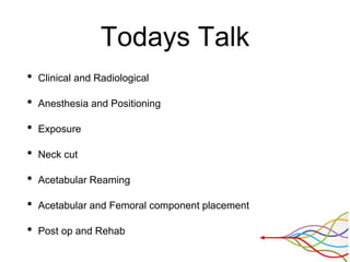
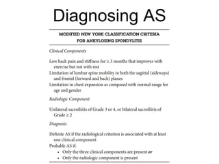
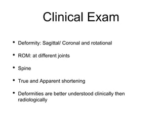
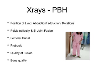
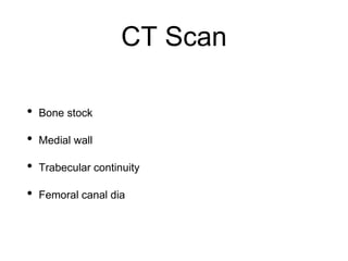
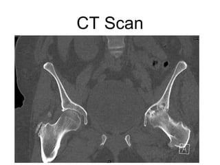
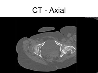
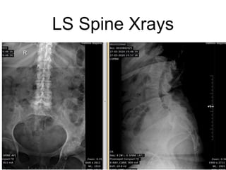
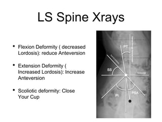
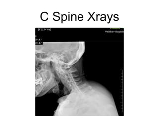
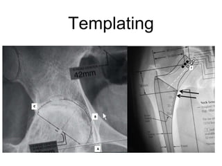
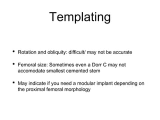
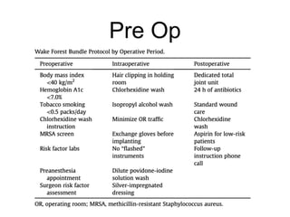
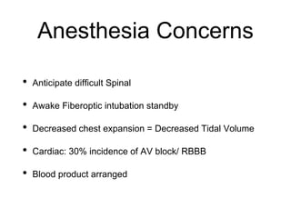
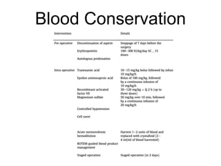
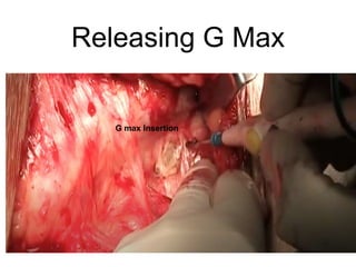
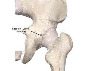
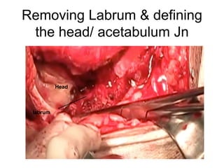
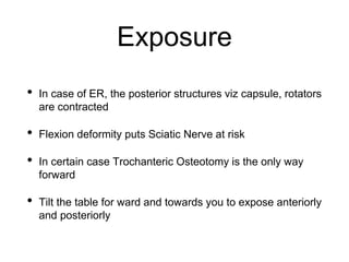
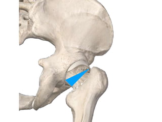
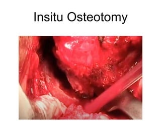
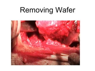
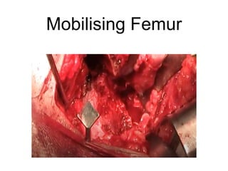
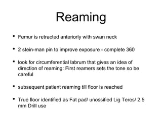
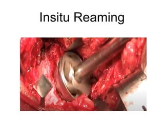
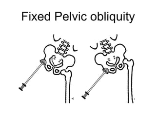
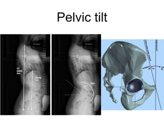
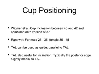
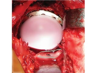
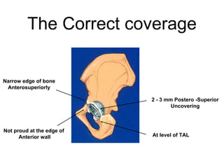
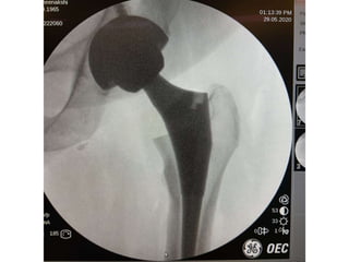
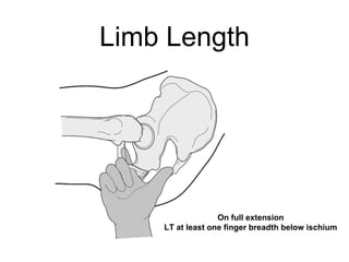
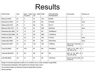
Tips for THR in
Ankylosing Spondylitis
Dr Vaibhav Bagaria
Director - Orthopaedics
Sir HN Reliance Foundation Hospital
President - SICOT India
Mumbai, India
It’s Elemental! Series
Todays Talk
• Clinical and Radiological
• Anesthesia and Positioning
• Exposure
• Neck cut
• Acetabular Reaming
• Acetabular and Femoral component placement
• Post op and Rehab
Diagnosing AS
Clinical Exam
• Deformity: Sagittal/ Coronal and rotational
• ROM: at different joints
• Spine
• True and Apparent shortening
• Deformities are better understood clinically then
radiologically
Xrays - PBH
• Position of Limb: Abduction/ adduction/ Rotations
• Pelvic obliquity & SI Joint Fusion
• Femoral Canal
• Protrusio
• Quality of Fusion
• Bone quality
Xrays
CT Scan
• Bone stock
• Medial wall
• Trabecular continuity
• Femoral canal dia
CT Scan
CT - Axial
LS Spine Xrays
LS Spine Xrays
• Flexion Deformity ( decreased
Lordosis): reduce Anteversion
• Extension Deformity (
Increased Lordosis): Increase
Anteversion
• Scoliotic deformity: Close
Your Cup
C Spine Xrays
Templating
Templating
• Rotation and obliquity: difficult/ may not be accurate
• Femoral size: Sometimes even a Dorr C may not
accomodate smallest cemented stem
• May indicate if you need a modular implant depending on
the proximal femoral morphology
Pre Op
Anesthesia Concerns
• Anticipate difficult Spinal
• Awake Fiberoptic intubation standby
• Decreased chest expansion = Decreased Tidal Volume
• Cardiac: 30% incidence of AV block/ RBBB
• Blood product arranged
Blood Conservation
Positioning
• Lateral Decubitus with Sacral and Pubic support
• Commonest deformities are: Flexion, Abduction and
external rotation
• Bilateral deformities are not uncommon
• Adequate padding
• Multiple pillow or head rings for C Spine stiffness.
Before start
• Table: Should be able to Tilted; Patient strapped
• Image Intensifier
• Tranexa/ Antibiotics/ Blood reconfirmation
Steps
• Exposure
• Removing Head
• Preparing Acetabulum
• Preparing Femur
Exposure
• Release G Max
• TFL : Fractional Lengthening / W shaped release
• Identify the head acetabulum junction
• Remove labrum
• Complete visualisation of neck and soft tissue release is
the key
• Dual Approach: If the Hip is in External Rotation
Releasing G Max
G max Insertion
Capsulo - Labral
Junction
Removing Labrum & defining
the head/ acetabulum Jn
Head
labrum
Exposure
• In case of ER, the posterior structures viz capsule, rotators
are contracted
• Flexion deformity puts Sciatic Nerve at risk
• In certain case Trochanteric Osteotomy is the only way
forward
• Tilt the table for ward and towards you to expose anteriorly
and posteriorly
Osteotomy
• Wafer Osteotomy
• Do not damage abductor
• Does not extend to GT or acetabular wall
• Combination of Saw/Osteotome
• No Bone splintering
Marking Osteotomy
Insitu Osteotomy
Removing Wafer
Mobilising Femur
Reaming
• Femur is retracted anteriorly with swan neck
• 2 stein-man pin to improve exposure - complete 360
• look for circumferential labrum that gives an idea of
direction of reaming: First reamers sets the tone so be
careful
• subsequent patient reaming till floor is reached
• True floor identified as Fat pad/ unossified Lig Teres/ 2.5
mm Drill use
Insitu Reaming
Fixed Pelvic obliquity
Pelvic tilt
Cup Positioning
• Widmer et al: Cup Inclination between 40 and 42 and
combined ante version of 37
• Ranawat: For male 25 - 35; female 35 - 45
• TAL can be used as guide: parallel to TAL
• TAL also useful for inclination: Typically the posterior edge
slightly medial to TAL
TAL
The Correct coverage
At level of TAL
2 - 3 mm Postero -Superior
Uncovering
Narrow edge of bone
Anterosuperiorly
Not proud at the edge of
Anterior wall
Femoral Component
• Anteversion ranges from 17 - 30 degree
• Concept of combined ante version important
• As stated earlier templating may not be accurate.
• Reaming before rasping may be required in certain case
• In younger patients - preserve the bone
Femoral Anteversion
• Think Posterolateral
• Calcar is good clue
• Palpate the epicondyles at Knee
• Judging - Easier with the neck on
Limb Length
On full extension
LT at least one finger breadth below ischium
Closure
• Posterior structure may not be available for a tight
posterior closure
• Quadratus is a saviour: translocate a sleeve anteriorly
• Adductor tenotomy may be required postoperatively
• Suction: only if oozing is significant intro
Preventing Heterotropic
Ossification
• Copious Wash: At least 3 litre to clear the soft tissue of
any bone debris
• Avoid overzealous retraction; Counsel the team about
need to respect soft tissues
• Use new fresh blade for good sharp bone cuts
• No periosteal stripping
• Indomethacin 25 -75 mg if creat OK for 3 weeks.
Results
Pearls
• Good prep work up and planning
• Adequate exposure and releases
• Neck cut without splintering and damage to walls/ abductors
• Restoring biomechanics of the hip
• Identifying true acetabulum
• Restoring Acetabular centre ( correcting Protrusio)
• Correct acetabular and femoral component placement
• THINK 3D…
It’s Elemental! Series
vaibhavbagaria.com

Total Hip replacement for Ankylosing Spondylitis: Planning & Execution

  • 1.
    Tips for THRin Ankylosing Spondylitis Dr Vaibhav Bagaria Director - Orthopaedics Sir HN Reliance Foundation Hospital President - SICOT India Mumbai, India It’s Elemental! Series
  • 2.
    Todays Talk • Clinicaland Radiological • Anesthesia and Positioning • Exposure • Neck cut • Acetabular Reaming • Acetabular and Femoral component placement • Post op and Rehab
  • 3.
  • 4.
    Clinical Exam • Deformity:Sagittal/ Coronal and rotational • ROM: at different joints • Spine • True and Apparent shortening • Deformities are better understood clinically then radiologically
  • 5.
    Xrays - PBH •Position of Limb: Abduction/ adduction/ Rotations • Pelvic obliquity & SI Joint Fusion • Femoral Canal • Protrusio • Quality of Fusion • Bone quality
  • 6.
  • 7.
    CT Scan • Bonestock • Medial wall • Trabecular continuity • Femoral canal dia
  • 8.
  • 9.
  • 10.
  • 11.
    LS Spine Xrays •Flexion Deformity ( decreased Lordosis): reduce Anteversion • Extension Deformity ( Increased Lordosis): Increase Anteversion • Scoliotic deformity: Close Your Cup
  • 12.
  • 13.
  • 14.
    Templating • Rotation andobliquity: difficult/ may not be accurate • Femoral size: Sometimes even a Dorr C may not accomodate smallest cemented stem • May indicate if you need a modular implant depending on the proximal femoral morphology
  • 15.
  • 16.
    Anesthesia Concerns • Anticipatedifficult Spinal • Awake Fiberoptic intubation standby • Decreased chest expansion = Decreased Tidal Volume • Cardiac: 30% incidence of AV block/ RBBB • Blood product arranged
  • 17.
  • 18.
    Positioning • Lateral Decubituswith Sacral and Pubic support • Commonest deformities are: Flexion, Abduction and external rotation • Bilateral deformities are not uncommon • Adequate padding • Multiple pillow or head rings for C Spine stiffness.
  • 19.
    Before start • Table:Should be able to Tilted; Patient strapped • Image Intensifier • Tranexa/ Antibiotics/ Blood reconfirmation
  • 20.
    Steps • Exposure • RemovingHead • Preparing Acetabulum • Preparing Femur
  • 21.
    Exposure • Release GMax • TFL : Fractional Lengthening / W shaped release • Identify the head acetabulum junction • Remove labrum • Complete visualisation of neck and soft tissue release is the key • Dual Approach: If the Hip is in External Rotation
  • 22.
    Releasing G Max Gmax Insertion
  • 23.
  • 24.
    Removing Labrum &defining the head/ acetabulum Jn Head labrum
  • 25.
    Exposure • In caseof ER, the posterior structures viz capsule, rotators are contracted • Flexion deformity puts Sciatic Nerve at risk • In certain case Trochanteric Osteotomy is the only way forward • Tilt the table for ward and towards you to expose anteriorly and posteriorly
  • 26.
    Osteotomy • Wafer Osteotomy •Do not damage abductor • Does not extend to GT or acetabular wall • Combination of Saw/Osteotome • No Bone splintering
  • 28.
  • 29.
  • 30.
  • 31.
  • 32.
    Reaming • Femur isretracted anteriorly with swan neck • 2 stein-man pin to improve exposure - complete 360 • look for circumferential labrum that gives an idea of direction of reaming: First reamers sets the tone so be careful • subsequent patient reaming till floor is reached • True floor identified as Fat pad/ unossified Lig Teres/ 2.5 mm Drill use
  • 33.
  • 34.
  • 35.
  • 40.
    Cup Positioning • Widmeret al: Cup Inclination between 40 and 42 and combined ante version of 37 • Ranawat: For male 25 - 35; female 35 - 45 • TAL can be used as guide: parallel to TAL • TAL also useful for inclination: Typically the posterior edge slightly medial to TAL
  • 41.
  • 42.
    The Correct coverage Atlevel of TAL 2 - 3 mm Postero -Superior Uncovering Narrow edge of bone Anterosuperiorly Not proud at the edge of Anterior wall
  • 43.
    Femoral Component • Anteversionranges from 17 - 30 degree • Concept of combined ante version important • As stated earlier templating may not be accurate. • Reaming before rasping may be required in certain case • In younger patients - preserve the bone
  • 44.
    Femoral Anteversion • ThinkPosterolateral • Calcar is good clue • Palpate the epicondyles at Knee • Judging - Easier with the neck on
  • 47.
    Limb Length On fullextension LT at least one finger breadth below ischium
  • 48.
    Closure • Posterior structuremay not be available for a tight posterior closure • Quadratus is a saviour: translocate a sleeve anteriorly • Adductor tenotomy may be required postoperatively • Suction: only if oozing is significant intro
  • 49.
    Preventing Heterotropic Ossification • CopiousWash: At least 3 litre to clear the soft tissue of any bone debris • Avoid overzealous retraction; Counsel the team about need to respect soft tissues • Use new fresh blade for good sharp bone cuts • No periosteal stripping • Indomethacin 25 -75 mg if creat OK for 3 weeks.
  • 50.
  • 51.
    Pearls • Good prepwork up and planning • Adequate exposure and releases • Neck cut without splintering and damage to walls/ abductors • Restoring biomechanics of the hip • Identifying true acetabulum • Restoring Acetabular centre ( correcting Protrusio) • Correct acetabular and femoral component placement • THINK 3D…
  • 52.