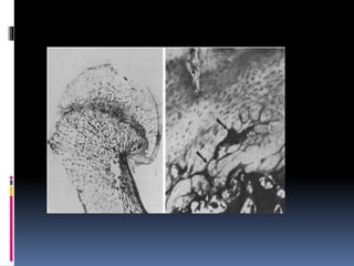
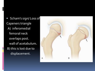
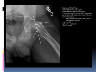
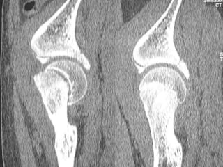
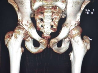
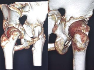
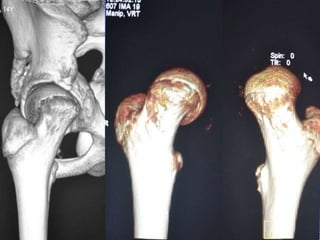
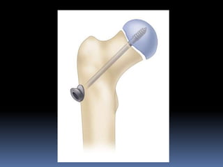
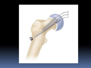
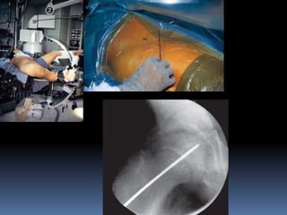
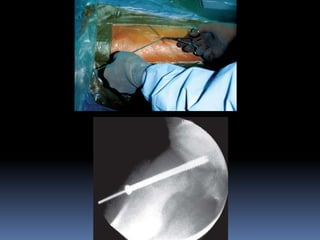
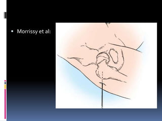
PRESENTED BY
DR. ANUBHAVVERMA on
7th JUNE 2016
CHAIRPERSON:
DR. ARUNODHAYA S
 History:
 First described by- Muller in 1889
 First series on osteotomy –Whitman in 1909
 First stabilisation with pin – Boyd in 1949
 Synonyms-
 Slipped upper femoral epiphysis
 Adolescent coxa vara
 Epiphyseal coxa vara
 Epiphysiolisthesis
 Epiphyseolysis
 Posterior & inferior displacement of proximal femoral
epiphysis in adolescents during rapid growth period.
 Physis layers:
1. Germinal layer – zone of resting cartilage cells.
2. Proliferative zone – actively growing cartilage cells.
3. Zone of hypertrophy – chondrocytes become swollen & vacuolated.
4. Zone of calcification – chondrocytes become calcified.
 Idiopathic
 Predisposing factors-
 Age- boys-12 to 16 yrs
Girls-10 to 14 yrs
 Sex- males >>females 5:2
 Blacks> whites
 Left side>right side
 Bilateral-25%
 Seasonal variations – june & july.
 HORMONALTHEORY-
 Growth hormone increase rate of proliferation of chondrocytes .
 Sex hormones especially estrogen inhibits GH.
 InAdiposogenital syndrome ,sex hormones decrease leading to increased
ratio of GH to sex hormones.
 In tall thin child GH is in excess.
 Hypothryoidism – loss of hydrostatic pressure in physeal matrix.
• Trauma-trivial injury.
• Periosteal thinning- in adolescent.
• Associated conditions – crf , irradiation, klinefelter’s syndrome.
 Epiphysis displaces posteriorly & inferiorly.
 Neck shifting upward & rotating anteriorly- anteverted position.
 Results in varus & adduction & E.R.
 In early stages-synovial membrane is swollen edematous,hyperemic &
villous.
 After several weeks- synovial membrane becomes less vascular & more
fibrotic & inelastic.
 After several months – epiphyseal junc heals & exposed portion of neck
covered –fibrocartilage.
 EXCESSIVE DISPLACEMENT-ABD & I.R LIMITED
MAINLY BY:
1. Large hump of fibrocartilage anteriorly.
2. Capital epiphysis is fixed posteriorly & inferiorly.
DEPENDING ON CLINICAL COURSE-
1. Acute slip- 11% ,sudden onset , <3 weeks.
2. Chronic slip- 60% , gradual onset , >3 weeks.
3. Acute on chronic- 23% , sudden exacerbation after trivial injury , >1
month .
4. Pre slip- 6% , x-ray finding ;irregularity ,widening & indistinctness of
epiphyseal plate.
 DEPENDING ON AMOUNT OF DISPLACEMENT:
1. Grade 1 – mild slip – neck displaced <1/3 diameter of head in lat view,
head shaft angle deviates by 30degree on both ap & lat.
2. Grade 2 –moderate slip – neck displaced between 1/3 to ½ , head shaft
angle between 30 to 60 degrees.
3. Grade 3 – severe slip – neck displaced >1/2 , head shaft angle >60
degrees.
 Head shaft angle.
 Angle b/w axis of
femur shaft &
perpendicular to
base of epiphysis.
 BASED ON STABILITY BY LODER:
1. Unstable – severe pain which prevents from walking even with crutches.
2. Stable – walking possible with or without crutches.
 PRE SLIP STAGE:
Slight discomfort in groin usually after work.
Discomfort radiates ant & med thigh to inner aspect of knee.
Slight stiffness & occasional limp.
No objective findings are apparent.
 CHRONIC SLIP STAGE:
Pain increases gradually in intensity.
Antalgic limp pronounced & persistent.
Objective finding – tenderness in hip , limitation of motion esp abduction &
I.R.
Limb develops – adduction & E.R deformity.
True shortening – upward displacement of femur
Apparent shortening – adduction deformity.
Hip flexion – E.R deformity accentuated.
Extreme slip – gluteus medius is inadequate & results in positive
trendelenburg test.
Bilateral severe slip – waddling gait.
 ACUTE ON CHRONIC:
Symptoms present for 1 month with sudden , acute & severe
exacerbation.
Usually there is history of trivial trauma- stumbling over a step or slipping
on stairs.
 STAGE OF FIXED DEFORMITY:
Pain & muscle spasm disappears.
Limp , E.R , adduction & shortening persists.
 RADIOLOGICAL APPEARANCES-
 Plain x-rays are taken in AP & lateral view (frog leg)
 Metaphyseal blanch sign
of steel- crescent shaped
Increased density lies over
Metaphysis of femur neck
Adjacent to physis
 Scham’s sign/ Loss of
Capeners triangle
A) inferomedial
femoral neck
overlaps post.
wall of acetabulum.
B) this is lost due to
displacement.
 PRESLIP STAGE:
 Minimal slip – absence of normal shoulder on upper aspect of neck & head-
trethowan sign ;line drawn along superior surface of neck will pass above
head.
 Head is sickle shaped.
 Epiphyseal plate widened with streaks of sclerosis.
 Lat view – slightest backward displacement
 EARLY STAGE:
 Head of femur rotated - displaced downwards & laterally.
 Lower margin of head projecting as beak like process below the lower
margin of neck.
 Upper margin of head thinned out.
 Femur neck – upper border is lengthened & convex upward , lower border
is concave & shortened.
 ADVANCED STAGE:
 Head is atrophic , grossly rotated & displaced.
 Projecting lower edge of head is curved laterally & upwards – is in contact
with lower border of neck.
 Neck is thick & short , sharply bowed upwards.
 Neck-shaft angle appears decreased to about 90 deg.
 Severe displacement – head is completely separated from neck & lies loose
in acetabulum.
 CT SCAN-
Accurately measures extent of epiphyseal displacement & angulation.
 TECHNETIUM-99 BONE SCAN-
Increased uptake in capital epiphysis of involved hip.
 USG-
Detection of early slip – joint effusion & step b/w femoral neck &
epiphysis , >6mm was diagnostic.
 MRI-
Early detection but expensive.
• TUBERCULOSISOF HIP –
Adducted & medially rotated , ROM restricted in all directions.
X-ray – extensive demineralisation of femoral head & acetabulum without
epiphyseal displacement.
• PERTHE’S DISEASE –
 Age – rarely begins after ten yrs.
 X-ray – head is not displaced but deformed.
 CONGENITAL DISLOCATIONOF HEAD:
 Long history of lameness.
 Head of femur palpated outside acetabulum & telescopy test positive .
 Analgesics
 Once diagnosed – absolute bed rest is essential.
 Longitudinal traction is applied – counteract muscle spasm.
 Traction along with medial rotation – reduce displacement in acute slip.
 Spica cast for 8- 16weeks.
 SURGICAL MANAGEMENT–
AIMS:
To reduce epiphyseal displacement.
To stabilise the capital epiphysis to femoral neck to prevent further
slipping.
To effect early closure of epiphyseal plate.
 METHODS OF OPERATIVETREATMENT –
Percutaneous insitu pinning.
Open reduction & internal fixation.
Epiphysiodesis.
Osteotomy.
Cheilectomy.
 INSITU PIN OR SCREW FIXATION –
Percutaneous in situ pinning most often used treatment in mild , moderate
& some severe acute or chronic slip.
Open insitu pinning – more severe acute or acute on chronic slip.
Use of single larger diameter central pin or screw is recommended –
technically simpler.
Use of pediatric cannulated screws over guide wire – easier & more
accurate.
Pins or screws shouldnot be removed until physis closes.
Generally two screws used for acute unstable slips & one for chronic stable
slips.
Pin tip be advanced to 8mm or one third of femoral head radius from
subchondral bone.
Pin or screw be placed in center of femoral head.
Advantage is it helps in minimizing skin incision.
Disadvantage is pin penetration, which can lead to joint sepsis, localized
acetabular erosion, synovitis, chondrolysis & late degenerativeOA.
Incidence of pin penetration – 14 to 60%
 TECHNIQUES :
 CANALE ET AL
 MORRISSY ET AL
 Morrissy et al:
 Post operatively:
 ROM exercises begun day after surgery.
 Most patients begin partial wt bearing walking with crutches on day after
surgery.
 Crutches are used for 2 – 3 weeks for stable slip & 6 – 8 weeks for unstable
slips.
 Rigorous sports avoided till physis have closed.
 Closed reduction:
 Manipulative reduction can be done for acute unstable & acute on chronic
slips with moderate to severe displacement.
 It is probably best to manipulate unstable slips that are technically difficult
to pin insitu.
 Performed with patient on fracture table & internal rotation alone is
sufficient to obtain adequate reduction.
 Confirmed radiologically.
 Main complication is osteonecrosis.
 OPEN REDUCTION:
Tilt in epiphysis may cause early degenerative changes, open reduction,
limited osteotomy & internal fixation may be done if closed reduction
cannot be done.
Osteonecrosis occurred due to tear in posterior retinacular vessels.
 BONE PEG EPIPHYSIODESIS:
Originally described by ferguson & howorth in 1931.
Following pin insertion many pt developed avn & arthritis of hip.
To prevent this, bone graft may be inserted through an opening in femoral neck
& across epiphyseal plate.
Thus securing immobilisation of epiphysis.
Channel is created in physeal plate through which metaphyseal vessels gain
access to epiphysis.
Disadvantage- longer opt time, increased blood loss, longer hospitalisation &
longer rehab.
 Weiner et al:
 Post operatively:
 Spica cast applied for 6weeks.
 Then touch down weight bearing is allowed.
 Mobilized with crutches after 48 – 96 hrs.
 Complete weight bearing is allowed after 10weeks.
 OSTEOTOMY-
Chronic slips produce irregularities in femoral head & acetabulum.
To restore normal relation, realignment procedure is indicated.
Two basic types:
close wedge osteotomy: usually near the physis to correct the deformity.
compensatory osteotomy: through the trochanteric region to produce
deformity in the opposite direction.
A)Through neck
near epiphysis.
B)Through base of
neck.
C)Through
intertrochanteric
region.
 SUBCAPITAL OSTEOTOMY
Technique of Fish
Technique of Dunn
 BASE OF NECK OSTEOTOMY
Technique of Kramer (Compensatory)
Technique of Abraham
 TROCHANTERIC OSTEOTOMY (in chronic
malunited SCFE)
Intertrochanteric Osteotomy (IMHAUSER AND
CAMPBELL)
Biplanar osteotomy (SOUTHWICK)
 CUNEIFORM OSTEOTOMYOF FEMORAL NECK- FISH
TECHNIQUE:
Indicated in severe chronic or acute on chronic slips.
Fish reported long-term follow up with severe displacement, requiring
osteotomy just distal to physis.
PROCEDURE:
Place pt supine on operating table.
Make anterolateral approach to hip.
Dissect between tensor fasciae latae & gluteus medius to anterior aspect
of capsule of hip joint.
Incise capsule longitudinally & retract carefully.
Identify capital femoral epiphysis & projecting part of neck.
Determine size of wedge by noting degree of slip & position of epiphysis.
Make base of wedge anteriorly & superiorly for correct positioning of
epiphysis.
After determining size of wedge, remove bone in small pieces with
osteotome & mallet.
After removing sufficient bone, reduce the epiphysis by flexion, abduction,
& internal rotation of limb.
After reduction, fix epiphysis to neck with 3 or 4 pins.
Donot allow pins to penetrate articular cartilage of epiphysis, but do
penetrate epiphysis deeply enough to obtain firm fixation.
UseA-P & frog leg lateral radiographs to determine correct position of pins.
 DUNNTECHNIQUE:
Based on facts- slip of head strips periosteum on back of neck & main
retinacular vessels run up back of neck.
A lateral approach allows strpping under direct vision & avoid damage to
blood supply.
Using lateral approach, incise periosteum & elevate posterior vascular
covering of femoral neck.
Make two osteotomy cuts, one in long axis of neck to remove bony beak,
second at right angles to neck to shorten it by 3-4 mm.
Appose surfaces of osteotomy, & insert 3 threaded pins up the femoral neck
upto its cut surface.
Reduce deformity under c-arm & drive pins into femoral head.
Close the wound in usual manner.
DUNN PROCEDURE
 Postoperatively:
 Spica cast applied for 4 weeks.
 Later ROM exercises are begun.
 After 2weeks walking with crutches is allowed.
 After 3 -4 months partial weight bearing allowed.
 Full weight bearing allowed after union is radiologically confirmed.
 COMPENSATORY BASILAR OSTEOTOMY OF
FEMORAL NECK:
 Described by Kramer et al.
 It corrects varus & retroversion components of moderate or severe chronic
SCFE.
 Line of osteotomy is distal to major blood supply in posterior retinaculum
hence safer than made near physis.
 Threaded pins are used for fixation of osteotomy & epiphysis.
 Both anatomical relationship of proximal femur is restored & further
slipping is prevented.
KRAMER ET ALTECHNIQUE :
Determine preop the size of wedge to be removed.
Approach hip laterally. Begin skin incision 2cm distal & lateral to ASIS &
curve it distally & posteriorly over greater trochanter & distally along
lateral surface of femoral shaft to a point 10cms distal to base of
trochanter.
Incise longitudinally fascia lata. Develop interval between gluteus medius
& tensor fasciae latae to expose hip jt capsule.
Incise capsule longitudinally & release widely.
Reflect distally vastus lateralis to expose base of GT & prox part of femoral
shaft.
Widest part of wedge should be in line with widest part of slip in anterior &
superior aspects of neck.
Make the more distal osteotomy cut first, perpendicular to femoral neck &
following anterior intertrochanteric line from proximal to distal.
Extend this osteotomy cut to posterior cortex.
Ensure that osteotome does not fully penetrate posterior cortex.
Drill 1-2, 5mm threaded steinmann pin into femoral neck proximally to
ensure that proximal portion of femur is kept under control before
osteotomy.
Insert several 5mm threaded steinmann pin from outer cortex of femoral
shaft through femoral neck.
Complete the osteotomy by greensticking posterior cortex and remove
wedge of bone.
Advance the threaded steinmann pins across the osteotomy site and
physis to prevent further slipping.
Capsule is closed, clip off the pins close to femur shaft & wound is closed in
layers.
 EXTRACAPSULAR BASE OF NECK:
 ABRAHAM ET AL reported this surgery.
 Recommended this osteotomy as safe & effective in preventing further
slipping & improving hip ROM in severe chronic slips.
 With severe slip, correction of varus & posterior tilt of femur head is
limited, & complete restoration of normal head – shaft angle may not be
possible.
 Removal of wedge >20mm compromises femur neck length & may
increase femoral anteversion.
 TECHNIQUE:
 Before surgery, the head-shaft angle is determined onAP radiographs by
measuring the angle formed by the epiphyseal line and the femoral shaft in
the affected limb and comparing it with the contralateral side (or to 145
degrees).
 The head-shaft angle for posterior tilt or retroversion is determined on a
frog-leg view and compared with the contralateral side (or to 10 degrees).
 The differences between the abnormal and normal angles are used to
determine the size of the wedges removed during osteotomy.
 Patient is placed on a fracture table, and maximally internally rotate the involved limb by gently
moving the footplate.
 Obtain permanent anteroposterior and “shoot-through” lateral radiographs to confirm the
chronicity of the slip and to outline the femoral head better. Prepare and drape the parts.
 Make a standard anterolateral approach.
 Locate the anterior joint tissue or intertrochanteric line between the gluteus medius and the
vastus lateralis muscles.
 Delineate a triangle on the anterior surface of the femoral neck to indicate the two-plane wedge
osteotomy.
 Locate the proximal cut by placing a 3 cm long kirschner wire on the
anterior surface of the femur from the lesser to the greater trochanter at
the base of the neck along the edge of the capsule.Confirm this position
by fluoroscopy.
 Wide osteotome used to mark the bone along the K- wire. Externally
rotate the leg, and drill a second kirschner wire in the anteroposterior
plane just distal to the guidewire.
 Place this wire vertical to the anterior surface of the femoral neck. Rotate
the limb internally, and obtain a lateral fluoroscopic view to confirm correct
wire placement.
 Begin the second distal osteotomy line from the lesser trochanter to the
growth plate of the greater trochanter.The angle at which this line is made
from the first osteotomy line depends on the amount of correction needed.
 A 15-mm-wide wedge, measured superiorly to the baseline of the triangle, is
needed.
 Make the osteotomy cuts with a saw, converging them posteriorly to make a
single osteotomy along the posterior cortex. Completely remove the wedge
of bone, especially superiorly, for maximal correction.
 While maintaining traction to prevent proximal migration of the femur,
internally rotate the leg until the wedge closes completely.
 Abducting the leg also helps to close the osteotomy. When the patella can be
internally rotated 15 degrees, adequate correction has been achieved.
 Fix the osteotomy with three or four cannulated screws . use the first guidewire
to hold the osteotomy temporarily in the desired position. Use only one screw
to span the physis of the femoral head, avoiding the superolateral quadrant.
 Check alignment and screw placement on permanent radiographs before
closing the wound.
 Close the wound in routine fashion, and apply a sterile dressing.
 INTERTROCHANTERIC OSTEOTOMY:
Capital epiphysis slipped chronically & united in poor position, then
trochanteric osteotomy done to produce opposite deformity.
If physis remains open, need to be fixed with pins or screws.
To correct coxa vara with some external rotation & hyperextension ,
closing wedge trochanteric osteotomy, with base of wedge laterally is
sufficient.
To correct coxa vara, hyperextension & moderate or severe external
rotation , ball & socket osteotomy at lesser trochanter can be done.
TECHNIQUE-
Make tracings of anteroposterior and lateral radiographs, and measure
them accurately to determine exactly the severity of the deformity.
Through a lateral approach , expose the trochanteric region and the
proximal 7.5 to 10 cm of the femoral shaft.
Insert a guide pin transversely through the femur at the level of the lesser
trochanter, and verify its position by radiographs.
With an osteotome, make reference marks on the trochanter and the
proximal shaft to be used in determining how much to rotate, flex, and
abduct the distal fragment at the time of internal fixation.
 At the level of the lesser trochanter, outline on the bone an osteotomy
convex proximally.Along this outline make multiple holes in the cortices
with a drill, and complete the osteotomy with an osteotome directed
proximally. Now the distal fragment is convex, and the proximal fragment
is concave.
 Abduct, flex, and internally rotate the distal fragment appropriately as
determined before surgery, and fix the fragments with a blade plate or
compression hip screw as in a trochanteric fracture.
 CHEILECTOMY (OSTEOPLASTYOF FEMORAL NECK)
Resection of prominence on anterosuperior aspect of femoral neck
(HERNDONS HUMP) which blocks internal rotation or abduction by
impinging against acetabulum.
This is combined with intertrochanteric osteotomy when coxa vara & E.R
deformity is severe.
If epiphysis is still open , epiphysiodesis can be done.
Excess removal may end up in fracture neck femur & further slipping.
 COMPLICATIONS:
 OSTEONECROSIS
 CHONDROLYSIS
 FEMORAL NECK FRACTURE
 CONTINUED SLIPPING.
 FEMOROACETABULAR IMPINGEMENT (FAI)
OSTEONECROSIS
 Osteonecrosis has been reported to occur in 10%
to 40% of patients with acute unstable SCFE,
although more recent reports of in situ pinning
with cannulated screws generally report lower
incidences (0% to 5%).
 Loder et al. suggested that instability may be the
best predictor of osteonecrosis
 50% of patients with unstable SCFE will develop
osteonecrosis
CHONDROLYSIS
 a joint space less than 3 mm wide (normal 4 to 6
mm) and a decreased range of motion of the
hip joint
 Persistent pin penetration into the joint has
been the most frequently cited cause of
chondrolysis
 If severe joint space narrowing persists with
limitation of joint motion, arthrodesis or
arthroplasty should be considered.
FEMOROACETABULAR IMPINGEMENT
 UNTREATED CASES:
 Before skeletal maturity , the disease may progress severely & acutely.
 At skeletal maturity , risk of late degenerative arthritis appears directly
related to residual deformity.
 TREATED CASES:
 Pinning insitu provided best long term results , regardless of severity of
slip.
 Long term results worsened with increased severity of slip & when
reduction or realignment had been done.
 Avn & chondrolysis - more likely with increased slip severity or when
osteotomy done , led to poor long term results.
 REFERENCES
 MERCERORTHOPAEDIC SURGERY 9th Edition
 CAMPBELLOPERATIVEORTHOPAEDICS. 12th edition
 TUREK’S PRINCIPLESOF ORTHOPEDICS & ITS APPLICATION 4th Edition
 TACHDJIAN’S PEDIATRICORTHOPAEDICS.
SCFE

SCFE

  • 1.
    PRESENTED BY DR. ANUBHAVVERMAon 7th JUNE 2016 CHAIRPERSON: DR. ARUNODHAYA S
  • 2.
     History:  Firstdescribed by- Muller in 1889  First series on osteotomy –Whitman in 1909  First stabilisation with pin – Boyd in 1949  Synonyms-  Slipped upper femoral epiphysis  Adolescent coxa vara  Epiphyseal coxa vara  Epiphysiolisthesis  Epiphyseolysis
  • 5.
     Posterior &inferior displacement of proximal femoral epiphysis in adolescents during rapid growth period.
  • 6.
     Physis layers: 1.Germinal layer – zone of resting cartilage cells. 2. Proliferative zone – actively growing cartilage cells. 3. Zone of hypertrophy – chondrocytes become swollen & vacuolated. 4. Zone of calcification – chondrocytes become calcified.
  • 8.
     Idiopathic  Predisposingfactors-  Age- boys-12 to 16 yrs Girls-10 to 14 yrs  Sex- males >>females 5:2  Blacks> whites  Left side>right side  Bilateral-25%  Seasonal variations – june & july.
  • 9.
     HORMONALTHEORY-  Growthhormone increase rate of proliferation of chondrocytes .  Sex hormones especially estrogen inhibits GH.  InAdiposogenital syndrome ,sex hormones decrease leading to increased ratio of GH to sex hormones.  In tall thin child GH is in excess.  Hypothryoidism – loss of hydrostatic pressure in physeal matrix.
  • 10.
    • Trauma-trivial injury. •Periosteal thinning- in adolescent. • Associated conditions – crf , irradiation, klinefelter’s syndrome.
  • 11.
     Epiphysis displacesposteriorly & inferiorly.  Neck shifting upward & rotating anteriorly- anteverted position.  Results in varus & adduction & E.R.  In early stages-synovial membrane is swollen edematous,hyperemic & villous.  After several weeks- synovial membrane becomes less vascular & more fibrotic & inelastic.  After several months – epiphyseal junc heals & exposed portion of neck covered –fibrocartilage.
  • 13.
     EXCESSIVE DISPLACEMENT-ABD& I.R LIMITED MAINLY BY: 1. Large hump of fibrocartilage anteriorly. 2. Capital epiphysis is fixed posteriorly & inferiorly.
  • 14.
    DEPENDING ON CLINICALCOURSE- 1. Acute slip- 11% ,sudden onset , <3 weeks. 2. Chronic slip- 60% , gradual onset , >3 weeks. 3. Acute on chronic- 23% , sudden exacerbation after trivial injury , >1 month . 4. Pre slip- 6% , x-ray finding ;irregularity ,widening & indistinctness of epiphyseal plate.
  • 15.
     DEPENDING ONAMOUNT OF DISPLACEMENT: 1. Grade 1 – mild slip – neck displaced <1/3 diameter of head in lat view, head shaft angle deviates by 30degree on both ap & lat. 2. Grade 2 –moderate slip – neck displaced between 1/3 to ½ , head shaft angle between 30 to 60 degrees. 3. Grade 3 – severe slip – neck displaced >1/2 , head shaft angle >60 degrees.
  • 17.
     Head shaftangle.  Angle b/w axis of femur shaft & perpendicular to base of epiphysis.
  • 18.
     BASED ONSTABILITY BY LODER: 1. Unstable – severe pain which prevents from walking even with crutches. 2. Stable – walking possible with or without crutches.
  • 20.
     PRE SLIPSTAGE: Slight discomfort in groin usually after work. Discomfort radiates ant & med thigh to inner aspect of knee. Slight stiffness & occasional limp. No objective findings are apparent.
  • 21.
     CHRONIC SLIPSTAGE: Pain increases gradually in intensity. Antalgic limp pronounced & persistent. Objective finding – tenderness in hip , limitation of motion esp abduction & I.R. Limb develops – adduction & E.R deformity. True shortening – upward displacement of femur Apparent shortening – adduction deformity.
  • 22.
    Hip flexion –E.R deformity accentuated. Extreme slip – gluteus medius is inadequate & results in positive trendelenburg test. Bilateral severe slip – waddling gait.
  • 25.
     ACUTE ONCHRONIC: Symptoms present for 1 month with sudden , acute & severe exacerbation. Usually there is history of trivial trauma- stumbling over a step or slipping on stairs.
  • 26.
     STAGE OFFIXED DEFORMITY: Pain & muscle spasm disappears. Limp , E.R , adduction & shortening persists.
  • 27.
     RADIOLOGICAL APPEARANCES- Plain x-rays are taken in AP & lateral view (frog leg)
  • 30.
     Metaphyseal blanchsign of steel- crescent shaped Increased density lies over Metaphysis of femur neck Adjacent to physis
  • 31.
     Scham’s sign/Loss of Capeners triangle A) inferomedial femoral neck overlaps post. wall of acetabulum. B) this is lost due to displacement.
  • 33.
     PRESLIP STAGE: Minimal slip – absence of normal shoulder on upper aspect of neck & head- trethowan sign ;line drawn along superior surface of neck will pass above head.  Head is sickle shaped.  Epiphyseal plate widened with streaks of sclerosis.  Lat view – slightest backward displacement
  • 34.
     EARLY STAGE: Head of femur rotated - displaced downwards & laterally.  Lower margin of head projecting as beak like process below the lower margin of neck.  Upper margin of head thinned out.  Femur neck – upper border is lengthened & convex upward , lower border is concave & shortened.
  • 35.
     ADVANCED STAGE: Head is atrophic , grossly rotated & displaced.  Projecting lower edge of head is curved laterally & upwards – is in contact with lower border of neck.  Neck is thick & short , sharply bowed upwards.  Neck-shaft angle appears decreased to about 90 deg.  Severe displacement – head is completely separated from neck & lies loose in acetabulum.
  • 36.
     CT SCAN- Accuratelymeasures extent of epiphyseal displacement & angulation.  TECHNETIUM-99 BONE SCAN- Increased uptake in capital epiphysis of involved hip.  USG- Detection of early slip – joint effusion & step b/w femoral neck & epiphysis , >6mm was diagnostic.  MRI- Early detection but expensive.
  • 43.
    • TUBERCULOSISOF HIP– Adducted & medially rotated , ROM restricted in all directions. X-ray – extensive demineralisation of femoral head & acetabulum without epiphyseal displacement.
  • 44.
    • PERTHE’S DISEASE–  Age – rarely begins after ten yrs.  X-ray – head is not displaced but deformed.  CONGENITAL DISLOCATIONOF HEAD:  Long history of lameness.  Head of femur palpated outside acetabulum & telescopy test positive .
  • 45.
     Analgesics  Oncediagnosed – absolute bed rest is essential.  Longitudinal traction is applied – counteract muscle spasm.  Traction along with medial rotation – reduce displacement in acute slip.  Spica cast for 8- 16weeks.
  • 46.
     SURGICAL MANAGEMENT– AIMS: Toreduce epiphyseal displacement. To stabilise the capital epiphysis to femoral neck to prevent further slipping. To effect early closure of epiphyseal plate.
  • 47.
     METHODS OFOPERATIVETREATMENT – Percutaneous insitu pinning. Open reduction & internal fixation. Epiphysiodesis. Osteotomy. Cheilectomy.
  • 48.
     INSITU PINOR SCREW FIXATION – Percutaneous in situ pinning most often used treatment in mild , moderate & some severe acute or chronic slip. Open insitu pinning – more severe acute or acute on chronic slip. Use of single larger diameter central pin or screw is recommended – technically simpler. Use of pediatric cannulated screws over guide wire – easier & more accurate. Pins or screws shouldnot be removed until physis closes.
  • 49.
    Generally two screwsused for acute unstable slips & one for chronic stable slips. Pin tip be advanced to 8mm or one third of femoral head radius from subchondral bone. Pin or screw be placed in center of femoral head. Advantage is it helps in minimizing skin incision. Disadvantage is pin penetration, which can lead to joint sepsis, localized acetabular erosion, synovitis, chondrolysis & late degenerativeOA. Incidence of pin penetration – 14 to 60%
  • 52.
     TECHNIQUES : CANALE ET AL  MORRISSY ET AL
  • 55.
  • 56.
     Post operatively: ROM exercises begun day after surgery.  Most patients begin partial wt bearing walking with crutches on day after surgery.  Crutches are used for 2 – 3 weeks for stable slip & 6 – 8 weeks for unstable slips.  Rigorous sports avoided till physis have closed.
  • 57.
     Closed reduction: Manipulative reduction can be done for acute unstable & acute on chronic slips with moderate to severe displacement.  It is probably best to manipulate unstable slips that are technically difficult to pin insitu.  Performed with patient on fracture table & internal rotation alone is sufficient to obtain adequate reduction.  Confirmed radiologically.  Main complication is osteonecrosis.
  • 58.
     OPEN REDUCTION: Tiltin epiphysis may cause early degenerative changes, open reduction, limited osteotomy & internal fixation may be done if closed reduction cannot be done. Osteonecrosis occurred due to tear in posterior retinacular vessels.
  • 59.
     BONE PEGEPIPHYSIODESIS: Originally described by ferguson & howorth in 1931. Following pin insertion many pt developed avn & arthritis of hip. To prevent this, bone graft may be inserted through an opening in femoral neck & across epiphyseal plate. Thus securing immobilisation of epiphysis. Channel is created in physeal plate through which metaphyseal vessels gain access to epiphysis. Disadvantage- longer opt time, increased blood loss, longer hospitalisation & longer rehab.
  • 60.
  • 61.
     Post operatively: Spica cast applied for 6weeks.  Then touch down weight bearing is allowed.  Mobilized with crutches after 48 – 96 hrs.  Complete weight bearing is allowed after 10weeks.
  • 62.
     OSTEOTOMY- Chronic slipsproduce irregularities in femoral head & acetabulum. To restore normal relation, realignment procedure is indicated. Two basic types: close wedge osteotomy: usually near the physis to correct the deformity. compensatory osteotomy: through the trochanteric region to produce deformity in the opposite direction.
  • 63.
    A)Through neck near epiphysis. B)Throughbase of neck. C)Through intertrochanteric region.
  • 64.
     SUBCAPITAL OSTEOTOMY Techniqueof Fish Technique of Dunn  BASE OF NECK OSTEOTOMY Technique of Kramer (Compensatory) Technique of Abraham  TROCHANTERIC OSTEOTOMY (in chronic malunited SCFE) Intertrochanteric Osteotomy (IMHAUSER AND CAMPBELL) Biplanar osteotomy (SOUTHWICK)
  • 65.
     CUNEIFORM OSTEOTOMYOFFEMORAL NECK- FISH TECHNIQUE: Indicated in severe chronic or acute on chronic slips. Fish reported long-term follow up with severe displacement, requiring osteotomy just distal to physis. PROCEDURE: Place pt supine on operating table. Make anterolateral approach to hip. Dissect between tensor fasciae latae & gluteus medius to anterior aspect of capsule of hip joint. Incise capsule longitudinally & retract carefully. Identify capital femoral epiphysis & projecting part of neck. Determine size of wedge by noting degree of slip & position of epiphysis.
  • 66.
    Make base ofwedge anteriorly & superiorly for correct positioning of epiphysis. After determining size of wedge, remove bone in small pieces with osteotome & mallet. After removing sufficient bone, reduce the epiphysis by flexion, abduction, & internal rotation of limb. After reduction, fix epiphysis to neck with 3 or 4 pins. Donot allow pins to penetrate articular cartilage of epiphysis, but do penetrate epiphysis deeply enough to obtain firm fixation. UseA-P & frog leg lateral radiographs to determine correct position of pins.
  • 68.
     DUNNTECHNIQUE: Based onfacts- slip of head strips periosteum on back of neck & main retinacular vessels run up back of neck. A lateral approach allows strpping under direct vision & avoid damage to blood supply. Using lateral approach, incise periosteum & elevate posterior vascular covering of femoral neck. Make two osteotomy cuts, one in long axis of neck to remove bony beak, second at right angles to neck to shorten it by 3-4 mm. Appose surfaces of osteotomy, & insert 3 threaded pins up the femoral neck upto its cut surface. Reduce deformity under c-arm & drive pins into femoral head. Close the wound in usual manner.
  • 69.
  • 70.
     Postoperatively:  Spicacast applied for 4 weeks.  Later ROM exercises are begun.  After 2weeks walking with crutches is allowed.  After 3 -4 months partial weight bearing allowed.  Full weight bearing allowed after union is radiologically confirmed.
  • 71.
     COMPENSATORY BASILAROSTEOTOMY OF FEMORAL NECK:  Described by Kramer et al.  It corrects varus & retroversion components of moderate or severe chronic SCFE.  Line of osteotomy is distal to major blood supply in posterior retinaculum hence safer than made near physis.  Threaded pins are used for fixation of osteotomy & epiphysis.  Both anatomical relationship of proximal femur is restored & further slipping is prevented.
  • 72.
    KRAMER ET ALTECHNIQUE: Determine preop the size of wedge to be removed. Approach hip laterally. Begin skin incision 2cm distal & lateral to ASIS & curve it distally & posteriorly over greater trochanter & distally along lateral surface of femoral shaft to a point 10cms distal to base of trochanter. Incise longitudinally fascia lata. Develop interval between gluteus medius & tensor fasciae latae to expose hip jt capsule. Incise capsule longitudinally & release widely. Reflect distally vastus lateralis to expose base of GT & prox part of femoral shaft.
  • 73.
    Widest part ofwedge should be in line with widest part of slip in anterior & superior aspects of neck. Make the more distal osteotomy cut first, perpendicular to femoral neck & following anterior intertrochanteric line from proximal to distal. Extend this osteotomy cut to posterior cortex. Ensure that osteotome does not fully penetrate posterior cortex. Drill 1-2, 5mm threaded steinmann pin into femoral neck proximally to ensure that proximal portion of femur is kept under control before osteotomy.
  • 74.
    Insert several 5mmthreaded steinmann pin from outer cortex of femoral shaft through femoral neck. Complete the osteotomy by greensticking posterior cortex and remove wedge of bone. Advance the threaded steinmann pins across the osteotomy site and physis to prevent further slipping. Capsule is closed, clip off the pins close to femur shaft & wound is closed in layers.
  • 76.
     EXTRACAPSULAR BASEOF NECK:  ABRAHAM ET AL reported this surgery.  Recommended this osteotomy as safe & effective in preventing further slipping & improving hip ROM in severe chronic slips.  With severe slip, correction of varus & posterior tilt of femur head is limited, & complete restoration of normal head – shaft angle may not be possible.  Removal of wedge >20mm compromises femur neck length & may increase femoral anteversion.
  • 77.
     TECHNIQUE:  Beforesurgery, the head-shaft angle is determined onAP radiographs by measuring the angle formed by the epiphyseal line and the femoral shaft in the affected limb and comparing it with the contralateral side (or to 145 degrees).  The head-shaft angle for posterior tilt or retroversion is determined on a frog-leg view and compared with the contralateral side (or to 10 degrees).  The differences between the abnormal and normal angles are used to determine the size of the wedges removed during osteotomy.
  • 78.
     Patient isplaced on a fracture table, and maximally internally rotate the involved limb by gently moving the footplate.  Obtain permanent anteroposterior and “shoot-through” lateral radiographs to confirm the chronicity of the slip and to outline the femoral head better. Prepare and drape the parts.  Make a standard anterolateral approach.  Locate the anterior joint tissue or intertrochanteric line between the gluteus medius and the vastus lateralis muscles.  Delineate a triangle on the anterior surface of the femoral neck to indicate the two-plane wedge osteotomy.
  • 79.
     Locate theproximal cut by placing a 3 cm long kirschner wire on the anterior surface of the femur from the lesser to the greater trochanter at the base of the neck along the edge of the capsule.Confirm this position by fluoroscopy.  Wide osteotome used to mark the bone along the K- wire. Externally rotate the leg, and drill a second kirschner wire in the anteroposterior plane just distal to the guidewire.  Place this wire vertical to the anterior surface of the femoral neck. Rotate the limb internally, and obtain a lateral fluoroscopic view to confirm correct wire placement.
  • 80.
     Begin thesecond distal osteotomy line from the lesser trochanter to the growth plate of the greater trochanter.The angle at which this line is made from the first osteotomy line depends on the amount of correction needed.  A 15-mm-wide wedge, measured superiorly to the baseline of the triangle, is needed.  Make the osteotomy cuts with a saw, converging them posteriorly to make a single osteotomy along the posterior cortex. Completely remove the wedge of bone, especially superiorly, for maximal correction.
  • 81.
     While maintainingtraction to prevent proximal migration of the femur, internally rotate the leg until the wedge closes completely.  Abducting the leg also helps to close the osteotomy. When the patella can be internally rotated 15 degrees, adequate correction has been achieved.  Fix the osteotomy with three or four cannulated screws . use the first guidewire to hold the osteotomy temporarily in the desired position. Use only one screw to span the physis of the femoral head, avoiding the superolateral quadrant.  Check alignment and screw placement on permanent radiographs before closing the wound.  Close the wound in routine fashion, and apply a sterile dressing.
  • 84.
     INTERTROCHANTERIC OSTEOTOMY: Capitalepiphysis slipped chronically & united in poor position, then trochanteric osteotomy done to produce opposite deformity. If physis remains open, need to be fixed with pins or screws. To correct coxa vara with some external rotation & hyperextension , closing wedge trochanteric osteotomy, with base of wedge laterally is sufficient. To correct coxa vara, hyperextension & moderate or severe external rotation , ball & socket osteotomy at lesser trochanter can be done.
  • 85.
    TECHNIQUE- Make tracings ofanteroposterior and lateral radiographs, and measure them accurately to determine exactly the severity of the deformity. Through a lateral approach , expose the trochanteric region and the proximal 7.5 to 10 cm of the femoral shaft. Insert a guide pin transversely through the femur at the level of the lesser trochanter, and verify its position by radiographs.
  • 86.
    With an osteotome,make reference marks on the trochanter and the proximal shaft to be used in determining how much to rotate, flex, and abduct the distal fragment at the time of internal fixation.  At the level of the lesser trochanter, outline on the bone an osteotomy convex proximally.Along this outline make multiple holes in the cortices with a drill, and complete the osteotomy with an osteotome directed proximally. Now the distal fragment is convex, and the proximal fragment is concave.  Abduct, flex, and internally rotate the distal fragment appropriately as determined before surgery, and fix the fragments with a blade plate or compression hip screw as in a trochanteric fracture.
  • 88.
     CHEILECTOMY (OSTEOPLASTYOFFEMORAL NECK) Resection of prominence on anterosuperior aspect of femoral neck (HERNDONS HUMP) which blocks internal rotation or abduction by impinging against acetabulum. This is combined with intertrochanteric osteotomy when coxa vara & E.R deformity is severe. If epiphysis is still open , epiphysiodesis can be done. Excess removal may end up in fracture neck femur & further slipping.
  • 89.
     COMPLICATIONS:  OSTEONECROSIS CHONDROLYSIS  FEMORAL NECK FRACTURE  CONTINUED SLIPPING.  FEMOROACETABULAR IMPINGEMENT (FAI)
  • 90.
    OSTEONECROSIS  Osteonecrosis hasbeen reported to occur in 10% to 40% of patients with acute unstable SCFE, although more recent reports of in situ pinning with cannulated screws generally report lower incidences (0% to 5%).  Loder et al. suggested that instability may be the best predictor of osteonecrosis  50% of patients with unstable SCFE will develop osteonecrosis
  • 91.
    CHONDROLYSIS  a jointspace less than 3 mm wide (normal 4 to 6 mm) and a decreased range of motion of the hip joint  Persistent pin penetration into the joint has been the most frequently cited cause of chondrolysis  If severe joint space narrowing persists with limitation of joint motion, arthrodesis or arthroplasty should be considered.
  • 93.
  • 95.
     UNTREATED CASES: Before skeletal maturity , the disease may progress severely & acutely.  At skeletal maturity , risk of late degenerative arthritis appears directly related to residual deformity.
  • 96.
     TREATED CASES: Pinning insitu provided best long term results , regardless of severity of slip.  Long term results worsened with increased severity of slip & when reduction or realignment had been done.  Avn & chondrolysis - more likely with increased slip severity or when osteotomy done , led to poor long term results.
  • 97.
     REFERENCES  MERCERORTHOPAEDICSURGERY 9th Edition  CAMPBELLOPERATIVEORTHOPAEDICS. 12th edition  TUREK’S PRINCIPLESOF ORTHOPEDICS & ITS APPLICATION 4th Edition  TACHDJIAN’S PEDIATRICORTHOPAEDICS.