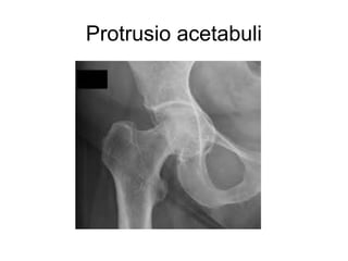
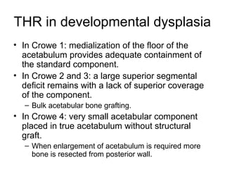
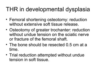
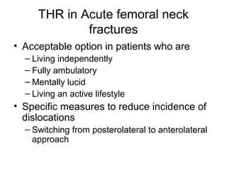
THR in specific hip disorders
Dr.Ramkrishna Dahal
Ortho Resident
Contents
• Arthritic disorders
– OA
– RA
• Osteonecrosis
• Protrusio acetabuli
• Developmental dysplasia
Contents
• Post traumatic disorders
– Acute femoral neck fractures
– Failed hip fracture surgery
– Acetabular fractures
• Failed reconstructive procedure
– Proximal femoral osteotomy and deformity
– Acetabular osteotomy
– Arthrodesis and ankylosis
Contents
• Infectious diorders
– Pyogenic arthritis
– Tuberculosis
• Neuromuscular disorders
• Routine surgical techniques must be
modified to meet the needs of the various
conditions.
THR in Osteoarthritis
• MC indication for THR
• Shortening of extremity
• Hip is flexed, externally
rotated and adducted.
• Additional shortening
due to deformity.
• Difficulty in dislocation due to osteophytes.
• Adequate reaming: due to thick and hard
subchondral bone of the acetabulum.
• Osteophytes may completely cover the
pulvinar and obscure the medial wall.
• Failure to medialize the acetabulum may
leave the superior portion of the cup
unsupported or supported only by
osteophytes.
• Complete removal
of the osteophytes
is necessary to
avoid
impingement,
decreased range
of motion and
dislocation.
THR in Rheumatoid arthritis
• Medical issues
• Corticosteroids/immunosuppressive
medications
• Risk of fracture
• Risk of infection
• Femoral head erosion
• Some degree of acetabular protrusion
THR in Rheumatoid arthritis
• Anasesthetic complications
• Preparation of the femur is easy because
of the wide canal but cortex is thin and
easily fractured.
• Acetabulum: fractured medial wall.
• Severe osteopenia often makes
cementless fixation more difficult.
• Excessive femoral anteversion and
anterior bowing of the proximal femur
• Femoral osteotomy for extreme deformity.
• TKR can be technically difficult in the
presence of a markedly stiff hip.
• A severe flexion contracture of the knee
may predispose to dislocation of THR
• If involvement is equal THR should be
done first.
THR in osteonecrosis
• Patients usually between 24-45 yrs so
results are not as satisfactory as in older
age groups.
• Reactive hyperemia of capsule and
synovial tissue, thus hemorrhage.
• Large synovial effusion, simulate infection.
• Complete intramedullary removal of the
graft for good femoral fit.
Protrusio acetabuli
THR in protrusio acetabuli
• Medial migration of the femoral head beyond the
ilioischial line (Kohler).
• Deformity may progress until GT impinges on
the side of the pelvis.
• A/w varus deformity of the femoral head.
• Principles of reconstruction of protrusio
– Hip center in anatomical location to restore proper
joint biomechanics
– Intact peripheral rim to support acetabular component
– Remaining cavitary and segmental defects in medial
wall reconstructed with bone grafts
THR in protrusio acetabuli
• Risk of sciatic nerve injury.
• Difficult dislocation.
• Removal of small overhanging portion of
the posterior acetabular .
• Osteotomy of femoral neck.
• Adequate capsular release.
• Head extracted with corkscrew/piecemeal.
• Medial reaming is unnecessary.
THR in protrusio acetabuli
• Sloof et al technique of impaction grafting:
– Cancellous bone graft 0.5 to 1 cm tightly impacted
into the medial acetabular defects
– Segment of mesh wire is placed on the top of the
graft.
– A conventional acetabular acetabular component is
cemented into the construct.
• Slight lengthening of the limb due to prior varus
deformity.
• Low level femoral neck resection with enhanced
offset to minimize limb lengthening.
THR in developmental dysplasia
• Hypoplastic acetabulum.
• If more than 80% of the cup was covered
by host bone, morselized graft supero-
laterally.
• If the estimated cup coverage was
between 60% and 80%, structural grafting.
• If cup coverage was less than 60%, the
so-called cotyloplasty technique to reduce
the area of cup supported by the structural
graft.
THR in developmental dysplasia
• Small and deformed femoral head.
• Short and narrow femoral neck.
• Marked anteversion.
• GT is small and posterior.
• Femoral canal is narrow.
• Difficult canal preparation.
THR in developmental dysplasia
• Presence of false acetabulum.
• Poorly developed acetabulum.
• Transverse orientation of acetabulum.
• Extensive capsulotomy and tenotomy of
the psoas, rectus femoris and adductors
required to correct the deformity.
• Sciatic nerve stretch injury.
THR in developmental dysplasia
• In patients with UL dislocations, lengthening of
the affected extremity during surgery is
desirable.
• Shallow dysplastic acetabulum may require a
very small acetabular component (<40 mm).
• Placement of acetabular component in true
acetabulum rather than in false acetabulum.
– Limb lengthening
– Improves abductor function
THR in developmental dysplasia
• In Crowe 1: medialization of the floor of the
acetabulum provides adequate containment of
the standard component.
• In Crowe 2 and 3: a large superior segmental
deficit remains with a lack of superior coverage
of the component.
– Bulk acetabular bone grafting.
• In Crowe 4: very small acetabular component
placed in true acetabulum without structural
graft.
– When enlargement of acetabulum is required more
bone is resected from posterior wall.
THR in developmental dysplasia
• Femoral shortening osteotomy: reduction
without extensive soft tissue release.
• Osteotomy of greater trochanter: reduction
without undue tension on the sciatic nerve
or fracture of the femoral shaft.
• The bone should be resected 0.5 cm at a
time.
• Trial reduction attempted without undue
tension in soft tissue.
Post traumatic disorders
THR in Acute femoral neck
fractures
• Acceptable option in patients who are
– Living independently
– Fully ambulatory
– Mentally lucid
– Living an active lifestyle
• Specific measures to reduce incidence of
dislocations
– Switching from posterolateral to anterolateral
approach
THR in Acute femoral neck
fractures
• If posterior
approach is used,
use of large
diameter head
• Careful repair of
the posterior
capsule and short
external rotators.
THR in Acute femoral neck
fractures
The risk of dislocation:
– the surgical approach,
– the reconstruction of hip biomechanics,
– the head size and offset,
– the quality of capsular closure,
– the experience of the surgeon.
• Treatment of choice because of low revision rate
and better immediate function of hip.
• Abduction pillow for three to four days.
• Avoidance of >90 degree hip flexion and
adduction.
THR in failed hip fracture surgery
• Prolong non ambulatory patients due to
failed hip surgery.
• Dislocations and failure
• High mortality.
• In trochanteric nonunions, length of the
femur cannot be restored with a standard
implants.
• Calcar replacement stem.
THR in Acetabular fractures
• Preop CT
• Bony defect posteriorly in posterior wall
acetabulum fracture.
• Placement of acetabular components in
retroversion with subsequent dislocation.
• Either the acetabulum depeened for cup
support.
• Or the posterior wall grafted.
• For smaller contained defects, morselized
autograft.
• Nonunions of a displaced transverse
acetabular fracture: an antiprotrusio cage
with bone grafting.
• Difficult exposure due to scarring.
• Heterotopic ossification, complicated
exposure and impingement.
THR with antiprotrusio cage
THR in failed reconstructive
procedures
• In the presence of a previous proximal
femoral osteotomy that may have been
performed for slipped capital femoral
epiphysis (SCFE), Legg-Calvé-Perthes
disease, or developmental dysplasia of the
hip.
THR in failed reconstructive
procedures
• Proximal femoral osteotomy and deformity
– Presence of a plate can pose a technical
challenge during THA surgery
– Problem with inserting femoral stem in
deformed femur
– Displacement of fragments, dense cancellous
bone at the healed osteotomy site require
careful reaming.
– High speed burr to remove intramedullary
bone.
– distorted femoral architecture : hip instability.
THR in failed reconstructive
procedures
• Adequate capsular release and hip
dislocation should be done before
hardware removal.
• Acetabular osteotomy
– Distorted anatomy.
– Retroversion of the acetabulum.
– Careful positioning of the acetabular
component.
– Prior acetabular osteotomy had no
compromise in results of arthroplasty.
– Less acetabular augmentation was needed
compared with dysplastic hip.
• Arthrodesis and Ankylosis
– THR is indicated in a fused hip, with severe
persistent LBP, pain IL knee or CL hip, or in
pseudoarthrosis after failed arthrodesis.
– Conversion of arthrodesis into THR is safer
and easier if the trochanter is osteotomized.
– Adductor tenotomy may be required.
– Extremity ususally is lengethened.
THR in Infectious disorders
Pyogenic arthritis
• MRI 100% sensitive in showing the presence of
active infection in patients with prior
osteomyelitis.
• Infection status.
• Shortened limb with flexed and adducted hip.
• Deep scaring and sinuses.
• Complete sinus tract incision.
• Lack of sc tissues over the trochanter and in the
area of the proposed incision may require
rotation of a skin flap before THR.
Tuberculosis
• TB bacilli fewer in number in bone
infections than in infected sputum making
diagnosis TB osteomyelitis difficult.
• TB bacilli has little biofilm and adheres
poorly to implants.
• Reactivation of TB infections after THR
can be treated with debridement and drug
therapy with retention of the prosthesis.
• If GT and subtrochanteric areas resected, hip
will be unstable because reattaching the
abductor muscle is difficult.
• An extra long femoral component is necessary.
• Custom made components and segmental
replacement stem can be used.
• Gluteal muscles are sutured to holes made in
the component for this purpose.
• Acetabulum can be reconstructed with cement
with reinforcement ring or cage.
THR in Neuromuscular disorders
• Combined flexion and adduction
contractures are common, but their
presence may not be appreciated when a
patient has an acute fracture.
• An anterior or anterolateral approach is
preferable in these patients.
• Release of the anterior capsule, psoas,
adductor tenotomy all may be required.
• Placement of acetabular component in
anteversion.
Take Home Messages
• Identifying problems.
– Soft tissue
– Bony
• Preop planning
• Risk assesment
• Possible implant and prosthesis choice
Thank you
Next presentation by Dr Manoj
Hip preserving surgeries

Thr in specific hip disorders

  • 1.
    THR in specifichip disorders Dr.Ramkrishna Dahal Ortho Resident
  • 2.
    Contents • Arthritic disorders –OA – RA • Osteonecrosis • Protrusio acetabuli • Developmental dysplasia
  • 3.
    Contents • Post traumaticdisorders – Acute femoral neck fractures – Failed hip fracture surgery – Acetabular fractures • Failed reconstructive procedure – Proximal femoral osteotomy and deformity – Acetabular osteotomy – Arthrodesis and ankylosis
  • 4.
    Contents • Infectious diorders –Pyogenic arthritis – Tuberculosis • Neuromuscular disorders
  • 5.
    • Routine surgicaltechniques must be modified to meet the needs of the various conditions.
  • 6.
    THR in Osteoarthritis •MC indication for THR • Shortening of extremity • Hip is flexed, externally rotated and adducted. • Additional shortening due to deformity.
  • 7.
    • Difficulty indislocation due to osteophytes. • Adequate reaming: due to thick and hard subchondral bone of the acetabulum. • Osteophytes may completely cover the pulvinar and obscure the medial wall. • Failure to medialize the acetabulum may leave the superior portion of the cup unsupported or supported only by osteophytes.
  • 8.
    • Complete removal ofthe osteophytes is necessary to avoid impingement, decreased range of motion and dislocation.
  • 9.
    THR in Rheumatoidarthritis • Medical issues • Corticosteroids/immunosuppressive medications • Risk of fracture • Risk of infection • Femoral head erosion • Some degree of acetabular protrusion
  • 10.
    THR in Rheumatoidarthritis • Anasesthetic complications • Preparation of the femur is easy because of the wide canal but cortex is thin and easily fractured. • Acetabulum: fractured medial wall. • Severe osteopenia often makes cementless fixation more difficult. • Excessive femoral anteversion and anterior bowing of the proximal femur • Femoral osteotomy for extreme deformity.
  • 11.
    • TKR canbe technically difficult in the presence of a markedly stiff hip. • A severe flexion contracture of the knee may predispose to dislocation of THR • If involvement is equal THR should be done first.
  • 12.
    THR in osteonecrosis •Patients usually between 24-45 yrs so results are not as satisfactory as in older age groups. • Reactive hyperemia of capsule and synovial tissue, thus hemorrhage. • Large synovial effusion, simulate infection. • Complete intramedullary removal of the graft for good femoral fit.
  • 14.
  • 15.
    THR in protrusioacetabuli • Medial migration of the femoral head beyond the ilioischial line (Kohler). • Deformity may progress until GT impinges on the side of the pelvis. • A/w varus deformity of the femoral head. • Principles of reconstruction of protrusio – Hip center in anatomical location to restore proper joint biomechanics – Intact peripheral rim to support acetabular component – Remaining cavitary and segmental defects in medial wall reconstructed with bone grafts
  • 16.
    THR in protrusioacetabuli • Risk of sciatic nerve injury. • Difficult dislocation. • Removal of small overhanging portion of the posterior acetabular . • Osteotomy of femoral neck. • Adequate capsular release. • Head extracted with corkscrew/piecemeal. • Medial reaming is unnecessary.
  • 17.
    THR in protrusioacetabuli • Sloof et al technique of impaction grafting: – Cancellous bone graft 0.5 to 1 cm tightly impacted into the medial acetabular defects – Segment of mesh wire is placed on the top of the graft. – A conventional acetabular acetabular component is cemented into the construct. • Slight lengthening of the limb due to prior varus deformity. • Low level femoral neck resection with enhanced offset to minimize limb lengthening.
  • 18.
    THR in developmentaldysplasia • Hypoplastic acetabulum. • If more than 80% of the cup was covered by host bone, morselized graft supero- laterally. • If the estimated cup coverage was between 60% and 80%, structural grafting. • If cup coverage was less than 60%, the so-called cotyloplasty technique to reduce the area of cup supported by the structural graft.
  • 20.
    THR in developmentaldysplasia • Small and deformed femoral head. • Short and narrow femoral neck. • Marked anteversion. • GT is small and posterior. • Femoral canal is narrow. • Difficult canal preparation.
  • 21.
    THR in developmentaldysplasia • Presence of false acetabulum. • Poorly developed acetabulum. • Transverse orientation of acetabulum. • Extensive capsulotomy and tenotomy of the psoas, rectus femoris and adductors required to correct the deformity. • Sciatic nerve stretch injury.
  • 22.
    THR in developmentaldysplasia • In patients with UL dislocations, lengthening of the affected extremity during surgery is desirable. • Shallow dysplastic acetabulum may require a very small acetabular component (<40 mm). • Placement of acetabular component in true acetabulum rather than in false acetabulum. – Limb lengthening – Improves abductor function
  • 23.
    THR in developmentaldysplasia • In Crowe 1: medialization of the floor of the acetabulum provides adequate containment of the standard component. • In Crowe 2 and 3: a large superior segmental deficit remains with a lack of superior coverage of the component. – Bulk acetabular bone grafting. • In Crowe 4: very small acetabular component placed in true acetabulum without structural graft. – When enlargement of acetabulum is required more bone is resected from posterior wall.
  • 24.
    THR in developmentaldysplasia • Femoral shortening osteotomy: reduction without extensive soft tissue release. • Osteotomy of greater trochanter: reduction without undue tension on the sciatic nerve or fracture of the femoral shaft. • The bone should be resected 0.5 cm at a time. • Trial reduction attempted without undue tension in soft tissue.
  • 25.
  • 26.
    THR in Acutefemoral neck fractures • Acceptable option in patients who are – Living independently – Fully ambulatory – Mentally lucid – Living an active lifestyle • Specific measures to reduce incidence of dislocations – Switching from posterolateral to anterolateral approach
  • 27.
    THR in Acutefemoral neck fractures • If posterior approach is used, use of large diameter head • Careful repair of the posterior capsule and short external rotators.
  • 28.
    THR in Acutefemoral neck fractures The risk of dislocation: – the surgical approach, – the reconstruction of hip biomechanics, – the head size and offset, – the quality of capsular closure, – the experience of the surgeon. • Treatment of choice because of low revision rate and better immediate function of hip. • Abduction pillow for three to four days. • Avoidance of >90 degree hip flexion and adduction.
  • 29.
    THR in failedhip fracture surgery • Prolong non ambulatory patients due to failed hip surgery. • Dislocations and failure • High mortality. • In trochanteric nonunions, length of the femur cannot be restored with a standard implants. • Calcar replacement stem.
  • 30.
    THR in Acetabularfractures • Preop CT • Bony defect posteriorly in posterior wall acetabulum fracture. • Placement of acetabular components in retroversion with subsequent dislocation. • Either the acetabulum depeened for cup support. • Or the posterior wall grafted. • For smaller contained defects, morselized autograft.
  • 31.
    • Nonunions ofa displaced transverse acetabular fracture: an antiprotrusio cage with bone grafting. • Difficult exposure due to scarring. • Heterotopic ossification, complicated exposure and impingement.
  • 32.
  • 33.
    THR in failedreconstructive procedures • In the presence of a previous proximal femoral osteotomy that may have been performed for slipped capital femoral epiphysis (SCFE), Legg-Calvé-Perthes disease, or developmental dysplasia of the hip.
  • 34.
    THR in failedreconstructive procedures • Proximal femoral osteotomy and deformity – Presence of a plate can pose a technical challenge during THA surgery – Problem with inserting femoral stem in deformed femur – Displacement of fragments, dense cancellous bone at the healed osteotomy site require careful reaming. – High speed burr to remove intramedullary bone. – distorted femoral architecture : hip instability.
  • 35.
    THR in failedreconstructive procedures • Adequate capsular release and hip dislocation should be done before hardware removal.
  • 36.
    • Acetabular osteotomy –Distorted anatomy. – Retroversion of the acetabulum. – Careful positioning of the acetabular component. – Prior acetabular osteotomy had no compromise in results of arthroplasty. – Less acetabular augmentation was needed compared with dysplastic hip.
  • 37.
    • Arthrodesis andAnkylosis – THR is indicated in a fused hip, with severe persistent LBP, pain IL knee or CL hip, or in pseudoarthrosis after failed arthrodesis. – Conversion of arthrodesis into THR is safer and easier if the trochanter is osteotomized. – Adductor tenotomy may be required. – Extremity ususally is lengethened.
  • 38.
  • 39.
    Pyogenic arthritis • MRI100% sensitive in showing the presence of active infection in patients with prior osteomyelitis. • Infection status. • Shortened limb with flexed and adducted hip. • Deep scaring and sinuses. • Complete sinus tract incision. • Lack of sc tissues over the trochanter and in the area of the proposed incision may require rotation of a skin flap before THR.
  • 40.
    Tuberculosis • TB bacillifewer in number in bone infections than in infected sputum making diagnosis TB osteomyelitis difficult. • TB bacilli has little biofilm and adheres poorly to implants. • Reactivation of TB infections after THR can be treated with debridement and drug therapy with retention of the prosthesis.
  • 41.
    • If GTand subtrochanteric areas resected, hip will be unstable because reattaching the abductor muscle is difficult. • An extra long femoral component is necessary. • Custom made components and segmental replacement stem can be used. • Gluteal muscles are sutured to holes made in the component for this purpose. • Acetabulum can be reconstructed with cement with reinforcement ring or cage.
  • 42.
    THR in Neuromusculardisorders • Combined flexion and adduction contractures are common, but their presence may not be appreciated when a patient has an acute fracture. • An anterior or anterolateral approach is preferable in these patients. • Release of the anterior capsule, psoas, adductor tenotomy all may be required. • Placement of acetabular component in anteversion.
  • 43.
    Take Home Messages •Identifying problems. – Soft tissue – Bony • Preop planning • Risk assesment • Possible implant and prosthesis choice
  • 44.
    Thank you Next presentationby Dr Manoj Hip preserving surgeries