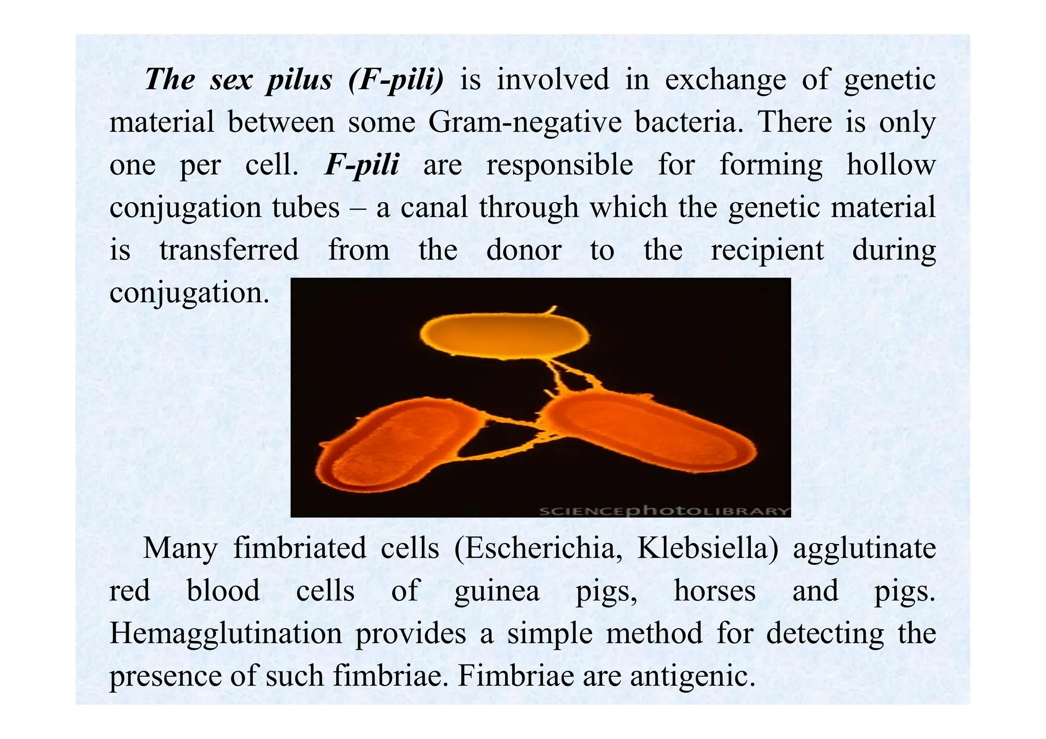
VOLGOGRAD STATE MEDICAL UNIVERSITY
DEPARTMENT OF MICROBIOLOGY
Morphology of
microorganisms.
Bacterial ultrastructure.
Complex staining methods.
Lecturer:
Associate Professor of the Microbiology
Department,
PhD Lyudmila Viktorovna Mikhailova
The Subject and Problems of Microbiology.
Microbiology (Gk. mikros small, bios life, logos science) is the science
of minute organisms, invisible to the naked eye, named microbes. It is
the study of the laws of the life and development of microorganisms, and
also of the changes which they bring about in human, animal and plant
organisms and in non-living matter.
Medical microbiology is the study of interactions between human
organisms & the microorganisms which they co-exist.
Medical microbiology is subdivided into bacteriology – the science of
bacteria, the causative agents of a number of infectious diseases; virology
– the science of viruses, non-cellular living systems capable of causing
infectious diseases in man; mycology – the study of fungi pathogenic for
man, protozoology which deals with pathogenic, unicellular animal
organisms and immunology – the science which is concerned with the
mechanisms of body protection against pathogenic microorganisms and
foreign cells and substances.
Medical microbiology includes the study of the mechanisms of infection
and the methods of specific therapy and prophylaxis of infectious
diseases.
Classification and Morphology of Microorganisms.
Microorganisms constitute a very antique group of living
organisms which appeared on the Earth's surface almost 3000
million years ago. Some scientists assumed that microbes were the
first living organisms of the Earth. Others maintained that non-
cellular forms of living matter (archebionts, photobionts,
protobionts, etc.) appeared prior to the microbes.
It is now generally believed that organisms evolved along the
following lines: viruses containing RNA, viruses containing DNA,
mycoplasmas, chlamydias, rickettsiae, bacteria, blue-green algae,
lower and higher fungi, plants, and animals.
Medical microbiology is mainly concerned with the study of
pathogenic bacteria, actinomycetes, spirochaetes, rickettsiae,
mycoplasmas, chlamydias, viruses, fungi, and protozoa all
grouped under the name of microbes or microorganisms.
The great majority of microbes are invisible to the naked
eye. They comprise multicellular organisms (the blue-green
algae, some fungi and chlamydobacteria, possibly some
corynebacteria, mycobacteria, cocci), unicellular organisms
(bacteria, actinomycetes, spirochaetes, and protozoa), and
non-cellular organisms (viruses).
Based on differences in cellular organization and
biochemistry, microorganisms are divided into two groups:
prokaryotes and eukaryotes.
Bacteria and blue green algae are prokaryotes, while
fungi, other algae, slime moulds and protozoa are
eukaryotes.
Taxonomy and classifications of microbes
• Taxonomy is the science of classification.
• Classification is an orderly arrangement of bacteria in groups.
• Identification is the practical use of classification to isolate and
distinguish desirable organism from undesirable ones.
• Nomenclature is the means through which the characteristics of a
species are defined and communicated among microbiologists.
Cavalier-Smith's six kingdoms classification (1998) is the most
recent and widely taxonomic classification. It divides organisms into
6 kingdoms:
• Bacteria,
• Protozoa,
• Chromista,
• Plantae,
• Fungi and
• Animalia.
In microbiology the binominal system of nomenclature is accepted
where each species has a generic and a specific name. The generic name is
written with a capital letter, and the specific name — with a small letter.
For example, in the case of Staphylococcus aureus. Staphylococcus is
the genus and aureus is the species designation. Some genera with common
characteristics are grouped into families.
The term strain designates a microbial culture obtained from the bodies
of humans or animals and from the environment.
A mixed culture consists of more than one species of microorganisms
isolated from a natural medium (non-sterile body cavities, body tissues,
food products, water, air, soil, washings).
Pure cultures represent a single species of a particular microorganism. It
is a population of bacteria consisting from the microbes of one and the same
species.
Population is an elementary evolutional unit (structure) of a definite
species with no noticeable isolation barriers within them between which
free crossing occurs.
Clone is a group of individuals (cells) arising from one cell.
Morphology and Ultrastructure of Bacteria
Bacteria (Gk. bakterion small staff) are unicellular
prokaryotic microorganisms with reproduction by
binary fission, lacking chlorophyll and absence of true
branching, except in the so-called ‘higher bacteria’
(Actinomycetales).
The size of bacteria is measured in micrometres
(µm). Most pathogenic bacteria measure 0.2 to 10 µm.
Morphologically, bacteria possess four main forms.
They are either spherical (cocci), rod-shaped (bacteria,
bacilli, and clostridia), spiral-shaped (vibrios and
spirilla), and branching filamentous forms.
I. Cocci (Gk. kokkos berry). These forms of bacteria are spherical, ellipsoidal,
bean-shaped, and lanceolate. Cocci are subdivided into six groups according to cell
arrangement, cell division and biological properties.
1. Micrococci (Micrococcus). The cells are arranged singly or irregularly. They
are saprophytes, and live in water and in air (M. agilis, M. roseus, M. luteus, etc.).
2. Diplococci divide in one plane and remain attached in pairs. These include:
meningococcus, causative agent of epidemic cerebrospinal meningitis, and
gonococcus, causative agent of gonorrhoea and blennorrhoea.
3. Streptococci divide in one plane and are arranged in chains of different
length. Some streptococci are pathogenic for humans and are responsible for
various diseases.
4. Tetracocci divide in two planes at right angles to one another and form
groups of fours. They very rarely produce diseases in humans.
5. Sarcinae divide in three planes at right angles to one another and resemble
packets of 8, 16 or more cells. They are frequently found in the air. Virulent species
have not been encountered.
6. Staphylococci divide in several planes resulting in irregular bunches of cells,
sometimes resembling clusters of grapes. Some species of staphylococci cause
diseases in man and animals.
Spherical forms of bacteria
1 - micrococci; 2 - diplococci; 3 - streptococci;
4 - tetracocci; 5 - sarcinae; 6 – staphylococci
II. Rods. Rod-shaped or cylindrical forms are subdivided into bacteria,
bacilli, and clostridia.
- Bacteria include those microorganisms which do not produce spores
(E.coli, and organisms responsible for enteric fever, paratyphoids,
dysentery).
- Bacilli and clostridia include organisms which produce spores (bacilli
responsible for anthrax, tetanus, anaerobic infections, etc.).
Rod-shaped bacteria exhibit differences in form:
- short bacillus - coccobacillus (Francisella tularaensis);
- long bacillus (Bacillus anthracis).
In bacilli and clostridia, spores are located:
(1) centrally, in the centre of the cell (causative agent of
anthrax);
(2) terminally, at the ends of the rod (causative agent of
tetanus);
(3) subterminally, towards the ends (causative agents of
botulism).
III. Spiral-shaped bacteria.
1. Vibrios are comma shaped, curved rods. (Vibrio
cholera - the causative agent of cholera, and aquatic
vibriones which are widely distributed in fresh water
reservoirs.
2. Spirilla are coiled forms of bacteria exhibiting twists
with one or more turns.
3. Spirochetes are flexuous spiral forms.
Spiral-shaped bacteria
1— vibrios; 2— spirilla; 3 – spirochetes
IV. Branching filamentous forms: Actinomycetes
are branching filamentous bacteria, so called
because of resemblance to the radiating rays of the
sun when seen in tissue lesions (from actis meaning
ray and mykes meaning fungus). The characteristic
shape is due to the presence of a rigid cell wall.
Bacteria are the first and smallest organisms capable of
independent existence.
Bacteria (prokaryotes) differ essentially from plant and
animal cells (eukaryotes) in structure.
The major structures of the cell are the multilayered
envelope (cell wall and cytoplasmic membrane), the
nucleoid (or nuclear body) and the cytosol (the cytoplasm).
There is no nucleus, and the genetic material is not
separated from the cytoplasm. The general chemical nature of
the bacterial design includes DNA, RNA, protein,
carbohydrate, phospholipid and some molecules unique to
bacteria such as the peptidoglycan and lipopolysaccharide of
bacterial cell walls.
Schematically representation of the structure of a bacterial cell
1 - capsule; 2 - cell wall; 3 - cell septum; 4 - flagellum;
5 - cytoplasmic membrane; 6 - nucleoid; 7 - plasmid;
8 - ribosomes; 9 - inclusions; 10 - spore
NUCLEOID
The bacterial genome resides on a single chromosome and
typically consists of about 4000 genes encoded in one, large,
circular molecule of double stranded DNA containing about
5 million nucleotide base pairs.
The nucleoid of bacteria is diffuse in character and is filled
with DNA fibrils which are arranged to form a closed loop. It
is located in the central part of the cell cytoplasm and comes
in contact with the cytoplasmic membrane, mesosomes,
polysomes or some central structure at a large number of
points. In the different stages of the development of the
bacterial cell, the nucleoid DNA may occur in the form of a
circle, threads, strands, a knotted or fine network or coarse
clusters.
The double-helical DNA chain is twisted into supercoils; it
has no nucleolus and no nuclear membrane separating it from
the cytoplasm. DNA is not associated with the basic protein.
The bacterial chromosome is haploid and replicates by simple
fission instead of by mitosis as in higher cells.
The number of nuclear bodies varies as a function of
growth rate; resting cells have only one, and rapidly growing
cells may have as many as four.
The absence of a nuclear membrane confers on the
prokaryotic cell a great advantage for rapid growth in
changing environments.
The refraction index of the bacterial nucleoid in actively
growing cells is the same as that of the cytoplasm. The
genome substance to cytoplasm ratio varies between 1:2 and
1:10.
CYTOPLASM (CYTOSOL)
The cytoplasm of bacteria is a dispersed colloid mixture of
water, proteins, carbohydrates, lipids, mineral compounds, and
other substances. The cytoplasm contains ribosomes, mesosomes,
inclusions and vacuoles. All of the metabolic reactions of the cell
take place in the cytosol.
The bacterial cytoplasm is immobile and is marked by high
density. The dense cytosol is bounded by the cell membrane. It
appears granular because it is densely packed with ribosomes.
These small granules 1000-2000 nm in diameter are
ribonucleoproteins. They are the site of protein synthesis. Each
ribosome consists of three species of rRNA (5S, 16S, and 23S) and
over 50 proteins. The overall subunit structure (one 50S plus one
30S particle) of the 70S bacterial ribosome resembles that of
eukaryotic ribosomes, but is smaller. The number of ribosomes
varies directly with the growth rate of the cell.
A group of 50 to 55 ribosomes form a polysome. The
ribosomes and polysomes are attached to the membrane and
fibrillar structures.
Numerous inclusions are located in the cytoplasm
comprising volutin granules, lipoprotein bodies, glycogen,
amylose, accumulations of pigment, sulphur, calcium, etc.
Volutin granules contain polymetaphosphate and stain
more intensely than the cytoplasm. A characteristic feature of
the granules of volutin is their metachromatic stain. They are
stained purple, with methylene blue while the cytoplasm is
stained blue.
Volutin was first discovered in the cell of Spirillum
volutans, then in Corynebacterium diphtheriae and other
organisms. The presence of volutin is found by Neisser's
method.
Lipoprotein bodies are found as droplets of fat in bacilli and
spirilla. They disappear when the cells are deprived of nutrients,
and appear when bacteria are grown on nutrient media of a high
carbohydrate content. They are discernible if stained with Sudan
or fuchsine.
The presence of volutin granules and lipoprotein bodies is
biologically important since they serve as sources of stored food
for the bacterium.
Glycogen and granulose are intracellular inclusions which can
be identified by treating the cell with Lugol's solution (iodine).
Glycogen stains reddish-brown and granulose grey-blue. Glycogen
granules are prominent in aerobic bacilli. Granulose is frequently
found in butyric-acid bacteria, and in Clostridium.
Some bacteria contain protein crystals which are extremely
toxic; sulphur and granules of amorphic calcium carbonate.
The cytoplasm contains cytoplasmatic structures in the
form of small DNA molecules (plasmids, episomes) which
determine the synthesis of various substances.
PLASMIDS
Many bacteria contain small, usually circular, covalently
closed, double-stranded DNA molecules separate from the
chromosome. More than one type of plasmid or several
copies of a single plasmid may be present in the cell. They
are not essential for the life of the cell they inhabit but may
confer on it certain properties like toxigenicity and drug
resistance which may constitute a survival advantage.
Many plasmids carry genes coding for the production of
enzymes that protect the cell from antibiotics (antibiotic
resistance – R-plasmid ). Many attributes of virulence, such
as production of some pili (F-plasmid) and of some exotoxins
(Hly-, Ent- plasmids), are also determined by plasmid genes.
ENVELOPE= the outer layer (bacterial wall)
The bacterial wall consists of a cell wall and a
cytoplasmic membrane. In some species, the
bacterial cells are surrounded by a capsule. Bacteria
envelope protects the cell against chemical and
biologic threats in its environment, it is responsible
for many metabolic processes, and it mediates
attachment to human cell surfaces.
The bacterial wall may be demonstrated by
plasmolysis. When placed in a hypertonic solution,
the cytoplasm loses water by osmosis and shrinks,
while the wall retains its original shape and size
(bacterial ghost).
CELL WALL
A rigid cell wall surrounds all bacterial cells except wall-less
bacteria such as the mycoplasmas and Chlamydia.
The bacterial wall protects the cell from mechanical disruption
and from bursting caused by the turgor pressure resulting from the
hypertonicity of the cell interior relative to the environment.
It also provides a barrier against certain toxic chemical and
biologic agents.
Its form is responsible for the shape of the cell and confers on it
rigidity and ductility.
The cell wall protects the bacteria from harmful environmental
factors and takes part in the growth and division of the cell.
It protects the cells from chemical and physical assault while
still permitting the rapid exchange of nutrients and metabolic
byproducts required for rapid growth.
The cell wall carries bacterial antigens that are important in
virulence and immunity.
Bacterial cell wall has a layered structure. It is
composed of three layers: outer (lipoprotein), middle
(lipopolysaccharide) and inner (rigid, containing
mucopolymers) layer.
The main polymer of the cell wall is mucopeptide
(peptidoglycan or murein) which forms the rigid base
of the membrane; the wall may be separated from the
cytoplasmic membrane and obtained in its pure form.
The cell walls of some bacilli are dissolved by
exposure to lysozyme and naked protoplasts are freed
as the result.
Two types of the bacterial cell wall are identified.
The separation derives from their reaction to a
particular staining by Gram’s method.
Gram-Positive Cell Wall
The Gram-positive cell wall contains two major
components: peptidoglycan and teichoic acids, plus additional
carbohydrates and proteins, depending on the species. The
chief component is peptidoglycan, which is found nowhere
except in prokaryotes.
Peptidoglycan consists of a linear glycan chain of two
alternating sugars, N-acetylglucosamine (NAG) and N-
acetylmuramic acid (NAM). Each muramic acid residue bears
a tetrapeptide of alternating L- and D-amino acids. Glycan
chains are cross-linked into sheets by peptide chains between
the third amino acid of one tetrapeptide and the terminal D-
alanine of another. The same cross-links between other
tetrapeptides connect the sheets to form a three-dimensional,
rigid matrix. The cross-linking extends around the cell,
producing a scaffold-like giant molecule.
A second component of the Gram-positive cell wall is a
teichoic acid. These compounds are polymers of either
glycerol phosphate or ribitol phosphate, with various
sugars, amino sugars, and amino acids.
Lipoteichoic acids are the type of teichoic acids made of
polyglycerol phosphate, linked to a glycolipid in the
underlying cell membrane.
Gram-Negative Cell Wall
The architecture of cell walls of Gram-negative bacteria is
fundamentally different. In Gram-negative cells, the amount
of peptidoglycan is reduced, forming a single-layered sheet
around the cell. If part of the cell wall is dissolved, due to the
action of lysozyme or other factors, then the rod-shaped cells,
predominantly Gram-negative, transform into spherical
bodies called spheroplasts.
The outer layer of Gram negative bacterial cell wall is
called the outer membrane, which contains various proteins
(outer membrane proteins): enzymes with hydrolytic
functions, antibiotic-inactivating enzymes, binding proteins
with roles in chemotaxis and in the active transport of
solutions into the cell. They also serve as specific receptors
for some bacteriophages.
The outer membrane is important in evading
phagocytosis and the action of complement and
providing a permeability barrier against such dangerous
molecules as host lysozyme, bile salts, digestive
enzymes, and many antibiotics.
The outer membrane is a permeability barrier and
Gram-negative bacteria must make provision for the
entry of nutrients. Special structural proteins, called
porins, form transmembrane pores that serve as
diffusion channels for small molecules, which diffuse
through it and into the periplasm.
The periplasm is an intermembrane structure, lying
between the cell membrane and the outer membrane.
The periplasm holds digestive and protective enzymes
and proteins important in transport and chemotaxis.
Gram-negative outer membrane is phospholipoprotein
bilayer, of which the outer leaflet is lipopolysaccharide
(LPS). The lipopolysaccharides present on the cell walls of
Gram negative bacteria account for their endotoxic activity
and О antigen specificity.
The LPS consists of three regions.
Region I is the polysaccharide portion determining the О
antigen specificity (O antigen polysaccharide side chains).
Region II is the core polysaccharide.
Region III is the glycolipid portion (a toxic lipid A) and is
responsible for the endotoxic activities, pyrogenicity, lethal
effect, tissue necrosis, anticomplementary activity, В cell
mitogenicity, immunoadjuvant property and antitumour
activity.
CYTOPLASMIC MEMBRANE
The cytoplasmic (plasma) membrane is a thin (5-10 nm)
layer lining the inner-surface of the cell wall and separating
it from the cytoplasm. It is a complex highly organized and
highly specialized structure composed of three layers:
phospholipid, protein, and polysaccharide. Sterols are
absent, except in mycoplasma.
The bacterial chromosome is attached to the cytoplasmic
membrane, which plays a role in segregation of daughter
chromosomes at cell division. The cytoplasmic membrane
is the site of synthesis of DNA, cell wall polymers,
membrane lipids, protein, toxins, enzymes, and other
substances and in oxidative phosphorylation.
On invagination into the cytoplasm, the cytoplasmic membrane
forms mesosomes. It contains the entire electron transport system of
the cell and is functionally analogous to the mitochondria of
eukaryotes. The mesosomes play a definite role in the growth of the
cell walls and in cell division. Bacterial cell properties such as
osmotic pressure are associated with the cytoplasmic membranes.
It contains receptor proteins that function in chemotaxis, by means
of which the cells recognize and convert signals arriving from the
environment and differentiate nutrients and different antibacterial
compounds.
It is a permeability barrier and contains proteins involved in
selective and active transport of solutes. It is involved in secretion to
the exterior of proteins (exoproteins), including exotoxins and
hydrolytic enzymes. The cytoplasmic membrane is the functional
equivalent of most of the organelles of the eukaryotic cell and is vital
to the growth and maintenance of the cell.
ТHE CAPSULE.
Many bacterial cells surround themselves with one or another kind
of hydrophilic gel. This layer is often thick and transparent.
If the material forms a reasonably discrete layer, it is called a
capsule;
if it is amorphous in appearance, it is referred to as a slime layer.
A capsule is not a necessary part of the cell.
Most capsules are polysaccharides; a few are simple polypeptides.
Hydrophilic capsules are usually polysaccharides. The presence of a
slime layer protects the capsulated microbes from desiccation.
The pathogenic microbes produce capsules within the bodies of
animals or humans (Streptococcus pneumoniae, anthrax bacillus,
Clostridium perfringens). Some bacteria (staphylococci, streptococci,
and others) form microcapsules demonstrated by electron microscopy
as mucopolysaccharide microfibrils tightly attached to the cell wall.
Capsules provide some general protection for bacteria, but
their major function in pathogenic bacteria is protection from
the immune system. Pathogenic capsulated microbes are
resistant to phagocytosis and to the effect of antibodies.
Capsules do not contribute to growth and multiplication and
are not essential for cell survival in artificial culture. Capsule
synthesis depends on growth conditions. The majority of
microbes can produce capsules, particularly when cultivated in
nutrient media of a high carbohydrate content.
The capsule has a weak affinity to dyes and stains poorly.
Bacterial capsule surrounding cells of Klebsiella pneumoniae
FLAGELLA.
Motile bacteria are subdivided into creeping and swimming bacteria.
Creeping bacteria move slowly (creep) on a supporting surface as a result of
wave-like contractions of their bodies, which cause periodic alterations in the
shape of the cell.
Swimming bacteria move freely in a liquid medium. They possess flagella –
unbranched, long, sinuous filaments, which are the organs of locomotion.
Flagella are found in many species of bacteria, both Gram-positive and
Gram-negative.
Each flagellum consists of three distinct parts: the filament, the hook and
the basal body.
The basal body consists of several proteins organized as rings on a central
rod. The hook acts as a universal joint and ring-like bushings.
The hook-basal body portion is embedded in the cell envelope. The hook
and basal body are antigenically different.
The filament, which consists of polymerized molecules of a single protein
flagellin (similar to keratin or myosin), is external to the cell and connected to
the hook at the cell surface.
The flagella of different genera of bacteria are antigenically different.
Flagella motile microbes can be divided into 4 groups:
(1)monotrichates, bacteria having a single (polar) flagellum at
one pole of the cell (cholera vibrio),
(2) lophotrichates, bacteria with a tuft of flagella at one pole,
(3) amphitrichates, bacteria with two polar flagella or with a
tuft of flagella at both poles (Spirillum volutans),
(4) peritrichates, bacteria having flagella distributed over the
whole surface of their cells (E.coli).
Pili (also called fimbriae) are molecular hair-like projections
found on the surface of cells of many species. They are
composed of molecules of a protein called pilin. They serve to
attach the microbial cells to the surface of some substrates.
The pili contribute to the nutrition of bacteria since they
greatly increase the surface area of the bacterial cell.
There are two general classes, common pili and sex pili.
Common pili cover the surface of the cell. They are adhesins,
which are responsible for the ability of bacteria to colonize
surfaces and cells.
The sex pilus (F-pili) is involved in exchange of genetic
material between some Gram-negative bacteria. There is only
one per cell. F-pili are responsible for forming hollow
conjugation tubes – a canal through which the genetic material
is transferred from the donor to the recipient during
conjugation.
Many fimbriated cells (Escherichia, Klebsiella) agglutinate
red blood cells of guinea pigs, horses and pigs.
Hemagglutination provides a simple method for detecting the
presence of such fimbriae. Fimbriae are antigenic.
SPORES AND SPORULATION.
Endospores are small spherical or oval dehydrated,
metabolically quiescent bodies formed within the cell in
response to nutrient limitation or under unfavourable conditions.
Some microorganisms, principally rod-shaped (bacilli and
clostridia), are capable of sporulation. These include the
causative agents of anthrax, gas gangrene, tetanus, and
botulism.
The bacterial endospore is not a reproductive structure. One
cell forms one spore under adverse conditions (the process is
called sporulation). The spore may persist for a long time
(many years) and then, on appropriate stimulation, give rise to a
single bacterial cell (germination). Spores are survival devices.
Complex methods of staining:
GRAM'S METHOD.
The Gram stain known since 1884 has not lost its practical significance.
All bacteria stained by the Gram method can be subdivided according to
colour into Gram-positive and Gram-negative.
- Flame-fixed smears are stained first by gentian violet (crystal violet,
methyl violet) for 1-2 minutes,
- then treated with Lugol's iodine solution, leave for 60 seconds and pour off
the excess.
- Decoloration with alcohol for 30-40 seconds.
- The smears are washed with water
- Counterstain with dilute water fuchsine for 1-2 minutes.
- Wash with water and dry.
Gram-positive organisms retain the violet stain following treatment with
ethanol and are a deep violet (staphylococci, and streptococci).
Gram-negative organisms lose the violet stain in the decolorization
process, but take up the counterstain and are pink or red (gonococci,
meningococci, brucella, E.coli, salmonellae, cholera vibrio, etc.).
ZIEHL-NEELSEN (ZN) STAIN
for acid-fast bacilli and spores.
The Ziehl-Neelsen stain is employed for differentiating acid-fast
bacteria (bacilli of tuberculosis, leprosy, actinomycetes) which stain
with difficulty.
1. Cover the heat-fixed smear with strong carbol fuchsine, heat with
a flame until it streams (but not boils), and keep it streaming for 5-10
minutes, replenishing the stain if necessary.
2. Treated with a 3-5% sulphuric acid solution, or acid alcohol (3%
HCl in 95% ethanol or 5%H2SO4) and leave for 5-10 minutes.
3. Wash with water.
4. Counterstrain with methylene blue for 3 minutes.
5. Wash with water and dry.
Acid-fast bacteria and spores retain the red stain while all non fast
bacteria are stained blue.
Acid-fastness in bacteria is related to the presence of a large
amount of lipids, waxes, arid oxyacids.
NEISSER'S METHOD.
The granules of volutin consist of polyphosphates and are
detected at Neisser's staining. They are the feature of the
diphtheria bacteria.
1. A smear is stained with acetic methylene blue for 1 minute.
2. The dye is poured off; a smear is washed with water.
3. Flood with Lugol’s iodine for 30 seconds.
4. Stain vesuvin for 1-3 minutes.
5. A preparation is washed with water and dried up.
By Neisser’s staining volutin granules are stained blue or
black and the cytoplasm of bacteria – yellow.
BURRI-GINS'S METHOD.
1. A drop of the Indian ink is put on a slide.
2. A culture of a microbe is introduced with a sterile loop and
carefully admixed with the dye.
3. Then a smear is uniformly distributed to a thin layer with the
help of the second glass.
4. A preparation is dried up, flooded in 1% solution of muriatic
alcohol for some seconds.
5. After exsiccation or the burning of alcohol above the flame a
smear is counterstained with water fuchsine for 1-3 minutes.
6. A preparation is washed with water, dried up and
microscoped.
The red rods surrounded by colourless capsules are visible
against the dark background.

Bacterial ultrastructure. Complex methods of staining (1).pdf

  • 1.
    VOLGOGRAD STATE MEDICALUNIVERSITY DEPARTMENT OF MICROBIOLOGY Morphology of microorganisms. Bacterial ultrastructure. Complex staining methods. Lecturer: Associate Professor of the Microbiology Department, PhD Lyudmila Viktorovna Mikhailova
  • 2.
    The Subject andProblems of Microbiology. Microbiology (Gk. mikros small, bios life, logos science) is the science of minute organisms, invisible to the naked eye, named microbes. It is the study of the laws of the life and development of microorganisms, and also of the changes which they bring about in human, animal and plant organisms and in non-living matter. Medical microbiology is the study of interactions between human organisms & the microorganisms which they co-exist. Medical microbiology is subdivided into bacteriology – the science of bacteria, the causative agents of a number of infectious diseases; virology – the science of viruses, non-cellular living systems capable of causing infectious diseases in man; mycology – the study of fungi pathogenic for man, protozoology which deals with pathogenic, unicellular animal organisms and immunology – the science which is concerned with the mechanisms of body protection against pathogenic microorganisms and foreign cells and substances. Medical microbiology includes the study of the mechanisms of infection and the methods of specific therapy and prophylaxis of infectious diseases.
  • 3.
    Classification and Morphologyof Microorganisms. Microorganisms constitute a very antique group of living organisms which appeared on the Earth's surface almost 3000 million years ago. Some scientists assumed that microbes were the first living organisms of the Earth. Others maintained that non- cellular forms of living matter (archebionts, photobionts, protobionts, etc.) appeared prior to the microbes. It is now generally believed that organisms evolved along the following lines: viruses containing RNA, viruses containing DNA, mycoplasmas, chlamydias, rickettsiae, bacteria, blue-green algae, lower and higher fungi, plants, and animals. Medical microbiology is mainly concerned with the study of pathogenic bacteria, actinomycetes, spirochaetes, rickettsiae, mycoplasmas, chlamydias, viruses, fungi, and protozoa all grouped under the name of microbes or microorganisms.
  • 4.
    The great majorityof microbes are invisible to the naked eye. They comprise multicellular organisms (the blue-green algae, some fungi and chlamydobacteria, possibly some corynebacteria, mycobacteria, cocci), unicellular organisms (bacteria, actinomycetes, spirochaetes, and protozoa), and non-cellular organisms (viruses). Based on differences in cellular organization and biochemistry, microorganisms are divided into two groups: prokaryotes and eukaryotes. Bacteria and blue green algae are prokaryotes, while fungi, other algae, slime moulds and protozoa are eukaryotes.
  • 5.
    Taxonomy and classificationsof microbes • Taxonomy is the science of classification. • Classification is an orderly arrangement of bacteria in groups. • Identification is the practical use of classification to isolate and distinguish desirable organism from undesirable ones. • Nomenclature is the means through which the characteristics of a species are defined and communicated among microbiologists. Cavalier-Smith's six kingdoms classification (1998) is the most recent and widely taxonomic classification. It divides organisms into 6 kingdoms: • Bacteria, • Protozoa, • Chromista, • Plantae, • Fungi and • Animalia.
  • 6.
    In microbiology thebinominal system of nomenclature is accepted where each species has a generic and a specific name. The generic name is written with a capital letter, and the specific name — with a small letter. For example, in the case of Staphylococcus aureus. Staphylococcus is the genus and aureus is the species designation. Some genera with common characteristics are grouped into families. The term strain designates a microbial culture obtained from the bodies of humans or animals and from the environment. A mixed culture consists of more than one species of microorganisms isolated from a natural medium (non-sterile body cavities, body tissues, food products, water, air, soil, washings). Pure cultures represent a single species of a particular microorganism. It is a population of bacteria consisting from the microbes of one and the same species. Population is an elementary evolutional unit (structure) of a definite species with no noticeable isolation barriers within them between which free crossing occurs. Clone is a group of individuals (cells) arising from one cell.
  • 7.
    Morphology and Ultrastructureof Bacteria Bacteria (Gk. bakterion small staff) are unicellular prokaryotic microorganisms with reproduction by binary fission, lacking chlorophyll and absence of true branching, except in the so-called ‘higher bacteria’ (Actinomycetales). The size of bacteria is measured in micrometres (µm). Most pathogenic bacteria measure 0.2 to 10 µm. Morphologically, bacteria possess four main forms. They are either spherical (cocci), rod-shaped (bacteria, bacilli, and clostridia), spiral-shaped (vibrios and spirilla), and branching filamentous forms.
  • 8.
    I. Cocci (Gk.kokkos berry). These forms of bacteria are spherical, ellipsoidal, bean-shaped, and lanceolate. Cocci are subdivided into six groups according to cell arrangement, cell division and biological properties. 1. Micrococci (Micrococcus). The cells are arranged singly or irregularly. They are saprophytes, and live in water and in air (M. agilis, M. roseus, M. luteus, etc.). 2. Diplococci divide in one plane and remain attached in pairs. These include: meningococcus, causative agent of epidemic cerebrospinal meningitis, and gonococcus, causative agent of gonorrhoea and blennorrhoea. 3. Streptococci divide in one plane and are arranged in chains of different length. Some streptococci are pathogenic for humans and are responsible for various diseases. 4. Tetracocci divide in two planes at right angles to one another and form groups of fours. They very rarely produce diseases in humans. 5. Sarcinae divide in three planes at right angles to one another and resemble packets of 8, 16 or more cells. They are frequently found in the air. Virulent species have not been encountered. 6. Staphylococci divide in several planes resulting in irregular bunches of cells, sometimes resembling clusters of grapes. Some species of staphylococci cause diseases in man and animals.
  • 9.
    Spherical forms ofbacteria 1 - micrococci; 2 - diplococci; 3 - streptococci; 4 - tetracocci; 5 - sarcinae; 6 – staphylococci
  • 10.
    II. Rods. Rod-shapedor cylindrical forms are subdivided into bacteria, bacilli, and clostridia. - Bacteria include those microorganisms which do not produce spores (E.coli, and organisms responsible for enteric fever, paratyphoids, dysentery). - Bacilli and clostridia include organisms which produce spores (bacilli responsible for anthrax, tetanus, anaerobic infections, etc.). Rod-shaped bacteria exhibit differences in form: - short bacillus - coccobacillus (Francisella tularaensis); - long bacillus (Bacillus anthracis).
  • 11.
    In bacilli andclostridia, spores are located: (1) centrally, in the centre of the cell (causative agent of anthrax); (2) terminally, at the ends of the rod (causative agent of tetanus); (3) subterminally, towards the ends (causative agents of botulism).
  • 12.
    III. Spiral-shaped bacteria. 1.Vibrios are comma shaped, curved rods. (Vibrio cholera - the causative agent of cholera, and aquatic vibriones which are widely distributed in fresh water reservoirs. 2. Spirilla are coiled forms of bacteria exhibiting twists with one or more turns. 3. Spirochetes are flexuous spiral forms. Spiral-shaped bacteria 1— vibrios; 2— spirilla; 3 – spirochetes
  • 13.
    IV. Branching filamentousforms: Actinomycetes are branching filamentous bacteria, so called because of resemblance to the radiating rays of the sun when seen in tissue lesions (from actis meaning ray and mykes meaning fungus). The characteristic shape is due to the presence of a rigid cell wall.
  • 14.
    Bacteria are thefirst and smallest organisms capable of independent existence. Bacteria (prokaryotes) differ essentially from plant and animal cells (eukaryotes) in structure. The major structures of the cell are the multilayered envelope (cell wall and cytoplasmic membrane), the nucleoid (or nuclear body) and the cytosol (the cytoplasm). There is no nucleus, and the genetic material is not separated from the cytoplasm. The general chemical nature of the bacterial design includes DNA, RNA, protein, carbohydrate, phospholipid and some molecules unique to bacteria such as the peptidoglycan and lipopolysaccharide of bacterial cell walls.
  • 15.
    Schematically representation ofthe structure of a bacterial cell 1 - capsule; 2 - cell wall; 3 - cell septum; 4 - flagellum; 5 - cytoplasmic membrane; 6 - nucleoid; 7 - plasmid; 8 - ribosomes; 9 - inclusions; 10 - spore
  • 16.
    NUCLEOID The bacterial genomeresides on a single chromosome and typically consists of about 4000 genes encoded in one, large, circular molecule of double stranded DNA containing about 5 million nucleotide base pairs. The nucleoid of bacteria is diffuse in character and is filled with DNA fibrils which are arranged to form a closed loop. It is located in the central part of the cell cytoplasm and comes in contact with the cytoplasmic membrane, mesosomes, polysomes or some central structure at a large number of points. In the different stages of the development of the bacterial cell, the nucleoid DNA may occur in the form of a circle, threads, strands, a knotted or fine network or coarse clusters.
  • 17.
    The double-helical DNAchain is twisted into supercoils; it has no nucleolus and no nuclear membrane separating it from the cytoplasm. DNA is not associated with the basic protein. The bacterial chromosome is haploid and replicates by simple fission instead of by mitosis as in higher cells. The number of nuclear bodies varies as a function of growth rate; resting cells have only one, and rapidly growing cells may have as many as four. The absence of a nuclear membrane confers on the prokaryotic cell a great advantage for rapid growth in changing environments. The refraction index of the bacterial nucleoid in actively growing cells is the same as that of the cytoplasm. The genome substance to cytoplasm ratio varies between 1:2 and 1:10.
  • 18.
    CYTOPLASM (CYTOSOL) The cytoplasmof bacteria is a dispersed colloid mixture of water, proteins, carbohydrates, lipids, mineral compounds, and other substances. The cytoplasm contains ribosomes, mesosomes, inclusions and vacuoles. All of the metabolic reactions of the cell take place in the cytosol. The bacterial cytoplasm is immobile and is marked by high density. The dense cytosol is bounded by the cell membrane. It appears granular because it is densely packed with ribosomes. These small granules 1000-2000 nm in diameter are ribonucleoproteins. They are the site of protein synthesis. Each ribosome consists of three species of rRNA (5S, 16S, and 23S) and over 50 proteins. The overall subunit structure (one 50S plus one 30S particle) of the 70S bacterial ribosome resembles that of eukaryotic ribosomes, but is smaller. The number of ribosomes varies directly with the growth rate of the cell.
  • 19.
    A group of50 to 55 ribosomes form a polysome. The ribosomes and polysomes are attached to the membrane and fibrillar structures. Numerous inclusions are located in the cytoplasm comprising volutin granules, lipoprotein bodies, glycogen, amylose, accumulations of pigment, sulphur, calcium, etc. Volutin granules contain polymetaphosphate and stain more intensely than the cytoplasm. A characteristic feature of the granules of volutin is their metachromatic stain. They are stained purple, with methylene blue while the cytoplasm is stained blue. Volutin was first discovered in the cell of Spirillum volutans, then in Corynebacterium diphtheriae and other organisms. The presence of volutin is found by Neisser's method.
  • 20.
    Lipoprotein bodies arefound as droplets of fat in bacilli and spirilla. They disappear when the cells are deprived of nutrients, and appear when bacteria are grown on nutrient media of a high carbohydrate content. They are discernible if stained with Sudan or fuchsine. The presence of volutin granules and lipoprotein bodies is biologically important since they serve as sources of stored food for the bacterium. Glycogen and granulose are intracellular inclusions which can be identified by treating the cell with Lugol's solution (iodine). Glycogen stains reddish-brown and granulose grey-blue. Glycogen granules are prominent in aerobic bacilli. Granulose is frequently found in butyric-acid bacteria, and in Clostridium. Some bacteria contain protein crystals which are extremely toxic; sulphur and granules of amorphic calcium carbonate.
  • 21.
    The cytoplasm containscytoplasmatic structures in the form of small DNA molecules (plasmids, episomes) which determine the synthesis of various substances. PLASMIDS Many bacteria contain small, usually circular, covalently closed, double-stranded DNA molecules separate from the chromosome. More than one type of plasmid or several copies of a single plasmid may be present in the cell. They are not essential for the life of the cell they inhabit but may confer on it certain properties like toxigenicity and drug resistance which may constitute a survival advantage. Many plasmids carry genes coding for the production of enzymes that protect the cell from antibiotics (antibiotic resistance – R-plasmid ). Many attributes of virulence, such as production of some pili (F-plasmid) and of some exotoxins (Hly-, Ent- plasmids), are also determined by plasmid genes.
  • 22.
    ENVELOPE= the outerlayer (bacterial wall) The bacterial wall consists of a cell wall and a cytoplasmic membrane. In some species, the bacterial cells are surrounded by a capsule. Bacteria envelope protects the cell against chemical and biologic threats in its environment, it is responsible for many metabolic processes, and it mediates attachment to human cell surfaces. The bacterial wall may be demonstrated by plasmolysis. When placed in a hypertonic solution, the cytoplasm loses water by osmosis and shrinks, while the wall retains its original shape and size (bacterial ghost).
  • 23.
    CELL WALL A rigidcell wall surrounds all bacterial cells except wall-less bacteria such as the mycoplasmas and Chlamydia. The bacterial wall protects the cell from mechanical disruption and from bursting caused by the turgor pressure resulting from the hypertonicity of the cell interior relative to the environment. It also provides a barrier against certain toxic chemical and biologic agents. Its form is responsible for the shape of the cell and confers on it rigidity and ductility. The cell wall protects the bacteria from harmful environmental factors and takes part in the growth and division of the cell. It protects the cells from chemical and physical assault while still permitting the rapid exchange of nutrients and metabolic byproducts required for rapid growth. The cell wall carries bacterial antigens that are important in virulence and immunity.
  • 24.
    Bacterial cell wallhas a layered structure. It is composed of three layers: outer (lipoprotein), middle (lipopolysaccharide) and inner (rigid, containing mucopolymers) layer. The main polymer of the cell wall is mucopeptide (peptidoglycan or murein) which forms the rigid base of the membrane; the wall may be separated from the cytoplasmic membrane and obtained in its pure form. The cell walls of some bacilli are dissolved by exposure to lysozyme and naked protoplasts are freed as the result. Two types of the bacterial cell wall are identified. The separation derives from their reaction to a particular staining by Gram’s method.
  • 25.
    Gram-Positive Cell Wall TheGram-positive cell wall contains two major components: peptidoglycan and teichoic acids, plus additional carbohydrates and proteins, depending on the species. The chief component is peptidoglycan, which is found nowhere except in prokaryotes. Peptidoglycan consists of a linear glycan chain of two alternating sugars, N-acetylglucosamine (NAG) and N- acetylmuramic acid (NAM). Each muramic acid residue bears a tetrapeptide of alternating L- and D-amino acids. Glycan chains are cross-linked into sheets by peptide chains between the third amino acid of one tetrapeptide and the terminal D- alanine of another. The same cross-links between other tetrapeptides connect the sheets to form a three-dimensional, rigid matrix. The cross-linking extends around the cell, producing a scaffold-like giant molecule.
  • 26.
    A second componentof the Gram-positive cell wall is a teichoic acid. These compounds are polymers of either glycerol phosphate or ribitol phosphate, with various sugars, amino sugars, and amino acids. Lipoteichoic acids are the type of teichoic acids made of polyglycerol phosphate, linked to a glycolipid in the underlying cell membrane.
  • 27.
    Gram-Negative Cell Wall Thearchitecture of cell walls of Gram-negative bacteria is fundamentally different. In Gram-negative cells, the amount of peptidoglycan is reduced, forming a single-layered sheet around the cell. If part of the cell wall is dissolved, due to the action of lysozyme or other factors, then the rod-shaped cells, predominantly Gram-negative, transform into spherical bodies called spheroplasts. The outer layer of Gram negative bacterial cell wall is called the outer membrane, which contains various proteins (outer membrane proteins): enzymes with hydrolytic functions, antibiotic-inactivating enzymes, binding proteins with roles in chemotaxis and in the active transport of solutions into the cell. They also serve as specific receptors for some bacteriophages.
  • 28.
    The outer membraneis important in evading phagocytosis and the action of complement and providing a permeability barrier against such dangerous molecules as host lysozyme, bile salts, digestive enzymes, and many antibiotics. The outer membrane is a permeability barrier and Gram-negative bacteria must make provision for the entry of nutrients. Special structural proteins, called porins, form transmembrane pores that serve as diffusion channels for small molecules, which diffuse through it and into the periplasm. The periplasm is an intermembrane structure, lying between the cell membrane and the outer membrane. The periplasm holds digestive and protective enzymes and proteins important in transport and chemotaxis.
  • 29.
    Gram-negative outer membraneis phospholipoprotein bilayer, of which the outer leaflet is lipopolysaccharide (LPS). The lipopolysaccharides present on the cell walls of Gram negative bacteria account for their endotoxic activity and О antigen specificity. The LPS consists of three regions. Region I is the polysaccharide portion determining the О antigen specificity (O antigen polysaccharide side chains). Region II is the core polysaccharide. Region III is the glycolipid portion (a toxic lipid A) and is responsible for the endotoxic activities, pyrogenicity, lethal effect, tissue necrosis, anticomplementary activity, В cell mitogenicity, immunoadjuvant property and antitumour activity.
  • 30.
    CYTOPLASMIC MEMBRANE The cytoplasmic(plasma) membrane is a thin (5-10 nm) layer lining the inner-surface of the cell wall and separating it from the cytoplasm. It is a complex highly organized and highly specialized structure composed of three layers: phospholipid, protein, and polysaccharide. Sterols are absent, except in mycoplasma. The bacterial chromosome is attached to the cytoplasmic membrane, which plays a role in segregation of daughter chromosomes at cell division. The cytoplasmic membrane is the site of synthesis of DNA, cell wall polymers, membrane lipids, protein, toxins, enzymes, and other substances and in oxidative phosphorylation.
  • 31.
    On invagination intothe cytoplasm, the cytoplasmic membrane forms mesosomes. It contains the entire electron transport system of the cell and is functionally analogous to the mitochondria of eukaryotes. The mesosomes play a definite role in the growth of the cell walls and in cell division. Bacterial cell properties such as osmotic pressure are associated with the cytoplasmic membranes. It contains receptor proteins that function in chemotaxis, by means of which the cells recognize and convert signals arriving from the environment and differentiate nutrients and different antibacterial compounds. It is a permeability barrier and contains proteins involved in selective and active transport of solutes. It is involved in secretion to the exterior of proteins (exoproteins), including exotoxins and hydrolytic enzymes. The cytoplasmic membrane is the functional equivalent of most of the organelles of the eukaryotic cell and is vital to the growth and maintenance of the cell.
  • 32.
    ТHE CAPSULE. Many bacterialcells surround themselves with one or another kind of hydrophilic gel. This layer is often thick and transparent. If the material forms a reasonably discrete layer, it is called a capsule; if it is amorphous in appearance, it is referred to as a slime layer. A capsule is not a necessary part of the cell. Most capsules are polysaccharides; a few are simple polypeptides. Hydrophilic capsules are usually polysaccharides. The presence of a slime layer protects the capsulated microbes from desiccation. The pathogenic microbes produce capsules within the bodies of animals or humans (Streptococcus pneumoniae, anthrax bacillus, Clostridium perfringens). Some bacteria (staphylococci, streptococci, and others) form microcapsules demonstrated by electron microscopy as mucopolysaccharide microfibrils tightly attached to the cell wall.
  • 33.
    Capsules provide somegeneral protection for bacteria, but their major function in pathogenic bacteria is protection from the immune system. Pathogenic capsulated microbes are resistant to phagocytosis and to the effect of antibodies. Capsules do not contribute to growth and multiplication and are not essential for cell survival in artificial culture. Capsule synthesis depends on growth conditions. The majority of microbes can produce capsules, particularly when cultivated in nutrient media of a high carbohydrate content. The capsule has a weak affinity to dyes and stains poorly. Bacterial capsule surrounding cells of Klebsiella pneumoniae
  • 34.
    FLAGELLA. Motile bacteria aresubdivided into creeping and swimming bacteria. Creeping bacteria move slowly (creep) on a supporting surface as a result of wave-like contractions of their bodies, which cause periodic alterations in the shape of the cell. Swimming bacteria move freely in a liquid medium. They possess flagella – unbranched, long, sinuous filaments, which are the organs of locomotion. Flagella are found in many species of bacteria, both Gram-positive and Gram-negative. Each flagellum consists of three distinct parts: the filament, the hook and the basal body. The basal body consists of several proteins organized as rings on a central rod. The hook acts as a universal joint and ring-like bushings. The hook-basal body portion is embedded in the cell envelope. The hook and basal body are antigenically different. The filament, which consists of polymerized molecules of a single protein flagellin (similar to keratin or myosin), is external to the cell and connected to the hook at the cell surface. The flagella of different genera of bacteria are antigenically different.
  • 35.
    Flagella motile microbescan be divided into 4 groups: (1)monotrichates, bacteria having a single (polar) flagellum at one pole of the cell (cholera vibrio), (2) lophotrichates, bacteria with a tuft of flagella at one pole, (3) amphitrichates, bacteria with two polar flagella or with a tuft of flagella at both poles (Spirillum volutans), (4) peritrichates, bacteria having flagella distributed over the whole surface of their cells (E.coli).
  • 36.
    Pili (also calledfimbriae) are molecular hair-like projections found on the surface of cells of many species. They are composed of molecules of a protein called pilin. They serve to attach the microbial cells to the surface of some substrates. The pili contribute to the nutrition of bacteria since they greatly increase the surface area of the bacterial cell. There are two general classes, common pili and sex pili. Common pili cover the surface of the cell. They are adhesins, which are responsible for the ability of bacteria to colonize surfaces and cells.
  • 37.
    The sex pilus(F-pili) is involved in exchange of genetic material between some Gram-negative bacteria. There is only one per cell. F-pili are responsible for forming hollow conjugation tubes – a canal through which the genetic material is transferred from the donor to the recipient during conjugation. Many fimbriated cells (Escherichia, Klebsiella) agglutinate red blood cells of guinea pigs, horses and pigs. Hemagglutination provides a simple method for detecting the presence of such fimbriae. Fimbriae are antigenic.
  • 38.
    SPORES AND SPORULATION. Endosporesare small spherical or oval dehydrated, metabolically quiescent bodies formed within the cell in response to nutrient limitation or under unfavourable conditions. Some microorganisms, principally rod-shaped (bacilli and clostridia), are capable of sporulation. These include the causative agents of anthrax, gas gangrene, tetanus, and botulism. The bacterial endospore is not a reproductive structure. One cell forms one spore under adverse conditions (the process is called sporulation). The spore may persist for a long time (many years) and then, on appropriate stimulation, give rise to a single bacterial cell (germination). Spores are survival devices.
  • 39.
    Complex methods ofstaining: GRAM'S METHOD. The Gram stain known since 1884 has not lost its practical significance. All bacteria stained by the Gram method can be subdivided according to colour into Gram-positive and Gram-negative. - Flame-fixed smears are stained first by gentian violet (crystal violet, methyl violet) for 1-2 minutes, - then treated with Lugol's iodine solution, leave for 60 seconds and pour off the excess. - Decoloration with alcohol for 30-40 seconds. - The smears are washed with water - Counterstain with dilute water fuchsine for 1-2 minutes. - Wash with water and dry. Gram-positive organisms retain the violet stain following treatment with ethanol and are a deep violet (staphylococci, and streptococci). Gram-negative organisms lose the violet stain in the decolorization process, but take up the counterstain and are pink or red (gonococci, meningococci, brucella, E.coli, salmonellae, cholera vibrio, etc.).
  • 40.
    ZIEHL-NEELSEN (ZN) STAIN foracid-fast bacilli and spores. The Ziehl-Neelsen stain is employed for differentiating acid-fast bacteria (bacilli of tuberculosis, leprosy, actinomycetes) which stain with difficulty. 1. Cover the heat-fixed smear with strong carbol fuchsine, heat with a flame until it streams (but not boils), and keep it streaming for 5-10 minutes, replenishing the stain if necessary. 2. Treated with a 3-5% sulphuric acid solution, or acid alcohol (3% HCl in 95% ethanol or 5%H2SO4) and leave for 5-10 minutes. 3. Wash with water. 4. Counterstrain with methylene blue for 3 minutes. 5. Wash with water and dry. Acid-fast bacteria and spores retain the red stain while all non fast bacteria are stained blue. Acid-fastness in bacteria is related to the presence of a large amount of lipids, waxes, arid oxyacids.
  • 41.
    NEISSER'S METHOD. The granulesof volutin consist of polyphosphates and are detected at Neisser's staining. They are the feature of the diphtheria bacteria. 1. A smear is stained with acetic methylene blue for 1 minute. 2. The dye is poured off; a smear is washed with water. 3. Flood with Lugol’s iodine for 30 seconds. 4. Stain vesuvin for 1-3 minutes. 5. A preparation is washed with water and dried up. By Neisser’s staining volutin granules are stained blue or black and the cytoplasm of bacteria – yellow.
  • 42.
    BURRI-GINS'S METHOD. 1. Adrop of the Indian ink is put on a slide. 2. A culture of a microbe is introduced with a sterile loop and carefully admixed with the dye. 3. Then a smear is uniformly distributed to a thin layer with the help of the second glass. 4. A preparation is dried up, flooded in 1% solution of muriatic alcohol for some seconds. 5. After exsiccation or the burning of alcohol above the flame a smear is counterstained with water fuchsine for 1-3 minutes. 6. A preparation is washed with water, dried up and microscoped. The red rods surrounded by colourless capsules are visible against the dark background.