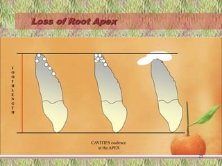
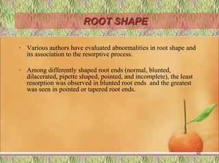
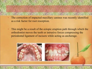
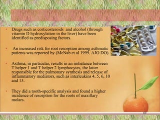
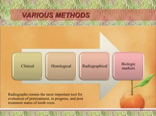
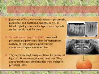
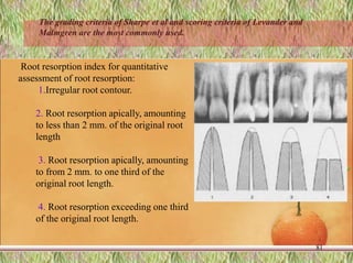
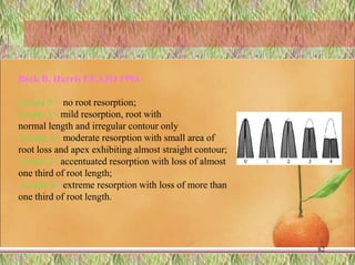
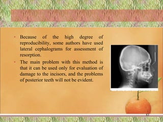
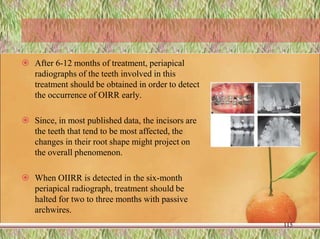
SlideShare a Scribd company logo
Root Resorption
1
Presented by:
Dr.Pooja Kale
PG IIIrd YEAR
P.M.N.M.DENTAL COLLEGE AND
HOSPITAL,BAGALKOT
Contents
• INTRODUCTION
• CLASSIFICATION
• NATURAL PROTECTION MECHANISM
• BIOLOGY
• FACTORS AFFECTING ROOT RESORPTION
• PREDISPOSING FACTORS
• METHODS OF ASSESSMENT
2
• LONG TERM EFFECTS
• GENETICS AND RESORPTION
• MANAGEMENT OF ROOT RESORPTION
• CONCLUSION
• REFERENCES
3
INTRODUCTION
 Root resorption is a condition characterized by a partial loss
of root cementum and dentin.
 Root resorption of the deciduous dentition is a physiological
process and it is a necessary precursor to the eruption of
permanent teeth.
 Permanent teeth root resorption is a pathological
inflammatory process and it can be affected by several factors
4
• Apical root resorption can be also
related to an orthodontic treatment and
it can be present during the treatment
or at the end of it.
• This root resorption is called
orthodontically- induced inflammatory
root resorption (OIRR) and it is
considered a distinct pathologic
process.
• Patient-related and treatment-related
factors are involved in the onset and
progression of this root resorption.
5
Internal root
resorption
Pulpal infection
External root
resorption
Pulpal infection
Dental trauma
Bleaching procedures
Periodontal procedures
Impacted teeth/cysts
and tumors
Orthodontic treatment
Ankylotic root
resorption
Severe dental
trauma
ROOT RESORPTION
6
There are three degrees of severity of OIIRR
(Naphtali Brezniak et al(2002) Angle Orthod
• 1. Cemental or surface resorption with remodeling. In this
process, only the outer cemental layers are resorbed, and they
are later fully regenerated or remodeled. This process resembles
trabecular bone remodeling.
• 2. Dentinal resorption with repair (deep resorption). In this
process, the cementum and the outer layers of the dentin are
resorbed and usually repaired with cementum material. The final
shape of the root after this resorption and formation process may
or may not be identical to the original form.
7
Brezniak N, Wasserstein A.-- Orthodontically induced inflammatory root resorption. Part I: The basic science aspects.
Angle Orthod 2002;72:175-9
• 3. Circumferential apical root resorption. In this process,
full resorption of the hard tissue components of the root
apex occurs, and root shortening is evident.
8
Protection mechanism
• Orthodontic forces applied to the biologic system act similarly on bone
and cementum, which are separated by the periodontal membrane. If
there are no differences in the biologic behavior of these two organs,
both would resorb equally.
• Since cementum is more resistant to resorption compared with the
more vulnerable bone, applied forces usually cause bone resorption,
which leads to tooth movement. However, resorption of the cementum
and dentin may also occur
9
• Several theories explaining the resistance of the dental tissues,
especially cemental resistance to resorption, exist.
• It is documented that the uncalcified mineral tissues, osteoid,
precementum, and predentin are resistant to resorption and may
initially prevent loss of root tissue.
• These layers might contain noncollagenic materials, eg, the cells
themselves, that possess potent anticollagenase properties.
10
 After extensive research in this field, mainly with tooth replantation
models, Andreasen, relates surface resistance to the innermost cellular
layer of the periodontal ligament.
 This layer supplies the protective mechanism to the root, as well as the
potential for a repair.
 The cementoblasts, fibroblasts, osteoblasts, endothelial, and
perivascular cells are included in this layer
 However, continuous pressure will eventually lead to resorption of
these areas
11
• Root resorption occurs when pressure on the cementum
exceeds its reparative capacity and dentin is exposed, allowing
multinucleated odontoclasts to degrade the root substance.
• Acc to Rudolph ,Resorption typically attacks the root tip and
travels coronally.The portion of the root nearest the pulp
appears to be the last to give way. This process is exactly
opposite to that of tooth formation
12
• Andreasen defines three external root resorption types:
– Surface resorption, which is a self-limiting process, usually
involving small outlining areas followed by spontaneous repair
from adjacent intact parts of the periodontal ligament.
– Inflammatory resorption, where initial root resorption has
reached dentinal tubules of an infected necrotic pulpal tissue or
an infected leukocyte zone.
– Replacement resorption, where bone replaces the resorbed
tooth material that leads to ankylosis.
Andreasen FM. Transient root resorption after dental trauma:the clinician's dilemma. J Esthet Restor
Dent. 2003;15(2):80–92
13
 According to Tronstad, inflammatory resorption is related to
the presence of multinucleated cells that colonize the
mineralized or denuded cemental surface.
 He characterizes two kinds of inflammatory resorption.
 Transient inflammatory resorption occurs when the stimulation to the
damage is minimal and for a short period. This defect is usually
undetected radiographically and is repaired by a cementum-like tissue.
 When stimulation is for a long period, Tronstad suggests the term
progressive inflammatory resorption. Ankylosis is the result of an
extensive necrosis of the periodontal ligament with formation of bone
onto a denuded area of the root surface.
Tronstad L. Root resorption--etiology, terminology and clinical manifestations. Endod Dent
Traumatol. 1988;4(6):241–52.
14
 Root resorption after orthodontic treatment is surface
resorption, or transient inflammatory resorption.
15
Biology of root resoprtion
 Orthodontic force initiation
stimulate the remodeling of
alveolar bone , which results in
tooth movement.
 Before this remodeling, initial
changes in response to a local
compression of PDL include
reduction in width and vascular
changes.
 PDL changes occur most noticeably
at pressure sites during tooth
movement.
16
 Tissue necrosis is evident at all stages and at varying degrees of
progression during orthodontic tooth movement.
 These area are devoid of cellular elements as seen in histologic
sections, commonly referred as HYLANIZATION.
 Results of histologic studies strongly suggest that the root resorption
occurs in response to damage initiated by orthodontic treatment to
periodontal ligament.
 Rygh and co-workers have shown that cementum adjacent to
hyalinized ( necrotic ) areas in PDL is “marked” by this contact and
that osteoclast cells attack this marked cementum when the PDL area
is repaired.
17
Rygh P. Orthodontic root resorption studied by electron microscopy. Angle Orthod. 1977;47:1–16.
 Osteoclast cells resorb not only necrotic tissue, but also the root
surface indiscriminately.
 Root surface change does not become apparent before cellular
changes in adjacent PDL and Bone.
 In human teeth, changes are observed in 3-5 week after initiation of a
light force.
 Initial stages mononuclear giant cells along the root surface associated
with areas of damaged PDL are seen.
• Advanced stages are typified by Odontoclast - The odontoclast is the
root-resorbing cell. It is a large pleomorphic, usually multinucleated,
cell formed by monocyte precursors like cells with ruffled borders.
18
 The residual matrix covering the surface of the resorption defects consists
of exposed collagen fibrils.
 Both the healing process and early stages of root resorption involve a high
percentage of Mononuclear giant cells.
 Mononuclear giant cells may also be induced to transform into
odontoclasts by the exposed mineralized root surface.
 Hence, it is suggested that Mononuclear giant cells non-selectively remove
the precementum layer together with adjacent necrotic PDL.
19
• The organic matrix degradation :
• According to Jones and Boyde, the osteoclast is credited
for both demineralization of the calcified tissue and
degradation of the organic matrix after demineralization.
• Cysteine proteinases of osteoclastic origin were found to be
important in the removal of organic matrices.
20
 Some articles state a resorbing activity, as a response to mechanical or
chemical stimuli by the periodontal ligament cells.
 This process is regulated by hormones (parathyroid and calcitonin),
neurotransmitters (substance P, vasoactive intestinal peptide, and
calcitonin gene related peptide), and cytokines or monokines
(interleukin-1 alpha, interleukin-1 beta, interleukin-2, tumor necrosis
factor, and interferon-gamma). It was also suggested that the
osteoclasts are controlled by osteoblasts in many ways.
21
Initiation, Cessation and Repair
 First histologic detection of root resorption vary from 1 week to
a few weeks.
 Progression of root resorption in terms of depth and number of
lacunae has been fond to increase with continuation of force.
 Cessation of process : few investigations conclude that
resorption ceases with termination of force application ,
whereas other studies show that process continues even after
force application.
 Presently cessation is more likely linked to complete removal
of necrotic tissue than force application.
22
• Repair of resorption lacunae has been observed as early as 3-
5 weeks after the initiation of light orthodontic tooth
movement.
• Examination of the root surfaces of teeth that have been
moved reveals repaired areas of resorption of both cementum
and dentin of the root.
23
Breznlak N and Wassersteln A .Root resorption after orthodontic treatment: Part 1. Literature review.
Am. J. Orthod. Dentofac. Orthop. 1993;103(1):62-66.
 Cementum (and Dentin , if resorption penetrates through the cementum) removed
form the root surface is restored by the formation of Dentinocemental junction
corresponding to that formed at Odontogenesis.
 Cemental lacunae become fully anatomically reconstructed. Deep dentinal lacunae
are repaired by a thin cemental layer resulting in an irregular root shape.
 After both types of repair, the periodontal ligament width is usually normal. Root
contour is frequently followed by bone contour, which increases tooth anchorage
without compromising function.
 Root remodeling is a constant feature of orthodontic tooth movement, but
permanent loss of root structure would occur only if repair did not replace the
initially resorbed cementum.
24
Loss of Root Apex
25
T
O
O
T
H
L
E
N
G
T
H
CAVITIES coalesce
at the APEX
FACTORS AFFECTING ROOT
RESORPTION
26
Predisposing Patient [Biologic] Factors
› Individual susceptibility
› Genetics
› Systemic factors
› Nutrition
› Gender of the patient
› Chronologic age
› Dental age
› Tooth structure
› Traumatized teeth
› Presence of root resorption before orthodontic treatment
› Endodontically treated teeth
› Adverse habits as nail-biting, tongue thrusting
› Specific tooth vulnerability to tooth resorption
27
• Treatment Related [Mechanical] Factors
– Type of orthodontic appliances used
– Magnitude of applied forces
– Direction of tooth movement
– Amount of tooth movement
– Jiggling and Occlusal trauma
• Combined Factors
– Treatment duration
– Root resorption detected radiographically during orthodontic
treatment
– Root resorption after appliance removal
– Relapse
28
MECHANOTHERAPY AND ROOT RESORPTION:
• Root resorption after orthodontic mechanotherapy is a well-known fact. The
dependence of this iatrogenic sequela on various mechanotherapeutics
performed has been studied extensively, but the results have been conflicted
• Many parameters such as:
– the type of malocculsion,
– extraction versus nonextraction,
– the type of appliance used
(removable, fixed, or functional appliances),
– the type of tooth movement performed,
– the duration of force application
29
• The magnitude of force, as well as rigid fixation of the arch-wire with
brackets or the use of full-size rectangular wires in bracket slots, could
be the most important factors predisposing a tooth to the resorptive
process
30
Brin I. External apical root resorption in Class II malocclusion: A retrospective review of 1-versus 2-phase
treatment. Am J Orthod Dentofac Orthop. 2003; 124:151-6
Root resorption among different
malocclusions
 There is a statistically significant difference
between Class I and Class II Division 1
malocclusions, with the latter exhibiting
more resorption.
 Janson et al (2007)reported a higher
resorption potential for Class II Division 2
cases in comparison with Class I, Class II
Division 1, and Class III patients.
 The rationale was that the intrusion
mechanics necessary to correct deep overbite
in these cases, as well as the excessive labial
torque needed to correct the palatal
inclination of the incisors, were the cause
31
• However it can be inferred from the published literature that
all types of malocclusion are prone to root resorption when
exposed to orthodontic treatment.
• Harris D et al (2006): showed that the volume of the root
resorption craters after intrusion was found to be directly
proportional to the magnitude of the intrusive force applied.
The mesial and distal surfaces had the greatest resorption
volume, with no statistically significant difference between
the 2 surfaces.
Harris D et al .Physical properties of root cementum: Part 8. Volumetric analysis of root resorption craters after application of
controlled intrusive light and heavy orthodontic forces: A microcomputed tomography scan study.Am J Orthod dentofacial
Orthop 2006;130:639-47 32
Extraction versus Nonextraction
modalities.
 Most studies revealed that both techniques have the
potential to produce damage, with extraction therapy
being potentially more detrimental.
 Some authors observed a definite increase in root
resorption following extraction therapy.
 Others reported a lack of correlation between root
resorption and treatment with extraction or nonextraction.
33
Graber L,Vanarsdall R,Vig k and Huang G.orthodontics current principles and techniques.2017.1st ed.
TYPE OF APPLIANCE
• The challenge for research these days is to determine
which of the various fixed appliance techniques—standard
edgewise, Begg, straight-wire, bioefficient, or Speed—
causes the most resorption
• Studies have reported different values for resorption
following the different types of appliance therapy
34
Janson GRP, De Luca Canto GDL, et al. A radiographic comparison of apical root resorption after orthodontic
treatment with 3 different fixed appliance techniques: Am J Orthod Dentofacial Orthop 2000:118:262-273.
• It was recently reported that a lower incidence of
resorption, as well as amount of root resorption, in patients
treated with the BIOEFFICENT THERAPY.
35
 The incidence rate of root resorption was 3.72 times
higher when extractions were performed as part of Begg
appliance therapy.
 An increased amount of root resorption with the Begg
appliance has also been extensively reported in the
previous literature.
 L'Abee and Saderink observed root resorption in all three
stages of Begg mechanics, with the second stage
exhibiting the least severity.
36
 Mayoral,AJO 1982 however, reported a low incidence of root
resorption following Begg mechanics and emphasized the
importance of using light forces to diminish damage to the roots.
 This report stands alone at present, but the use of light forces to
prevent root damage seems to be an interesting subject and
potentially fruitful approach for future research.
37
 A comparison of standard edgewise with straight-wire
appliances reveals statistically insignificant results except for
one study, (Mavragani M et al EJO 2000 )which reported
increased resorption for central incisors in the edgewise group.
 When sectional mechanics were compared with continuous
arch mechanics reported the same resorption potential
38
Root resorption and Invisalign®
• Gay G et al (2017) - investigated the incidence
and severity of RR in adult patients treated with
aligners during class I treatments. They evaluated
that every patient showed a minimum of one tooth
with root length reduction.
• They also concluded that Severe RR affected
mostly the upper lateral incisors and lower lateral
and central incisors.
Gay G et al. Root resorption during orthodontic treatment with Invisalign®: a radiometric study.Progress
in orthod 2017;18(12):1-9.
39
Type of tooth movements
 Of the various tooth movements, intrusion and torque are most
commonly associated with the resorption process. This is
evident when studying Class II Division 2 correction as well as
Begg mechanics.
 The intrusion performed in the first stage and torquing in the
third stage make the Begg technique more vulnerable to
resorption.
40
Brezniak N. Root resorption after orthodontic treatment. Part II. Literature review. Am J Orthod Dentofac
Orthop.1993; 103:138-46.
 Displacement of the root apex horizontally or torquing has
been proven beyond doubt to produce root resorption.
 The highest incidence of root resorption is reported to
occur when 3 to 4.5 mm of torquing movement were
performed.
 Resorption with tads mechanics were recently evident
when the studies was done.
41
Treatment time
 The length of treatment time and root resorption have
been positively correlated by almost all studies, which
have shown that increased treatment time makes tooth
roots more prone to the iatrogenic response
42
Aryal N & Jing M. Root Resorption in Orthodontic Treatment: Scoping Review 2017;7(2):47-52.
 Acar et al AO 1999 evaluated the effect of the type of force
applied— continuous versus interrupted— on the resorption
pattern and observed less severe apical blunting and smaller
resorption-affected areas when interrupted force was applied.
 Their findings are in agreement with study in this regard Maltha
JC et al, EJO 1996 and emphasize the use of less detrimental
discontinuous forces (in the form of elastic usage, instead of
elastomeric chains) during space-closure stages of orthodontic
mechanotherapy
43
Magnitude of applied force
• Ballard D et al.(2009) studied the Continuous vs intermittent controlled
orthodontic forces on root resorption.They concluded that : Intermittent
force produced less root resorption than continuous force
• Buccally directed intermittent forces for 8 weeks (after 14 days of
initial continuous force application, the intermittent force application
was obtained with a 3-day resting period followed by a 4-day force
application period) produce significantly less total root resorption than
a similarly directed continuous force of the same magnitude and
duration.
Ballard D. Allan S. Petocz P and Darendeliler M. Physical properties of root cementum: Part 11. Continuous vs intermittent controlled
orthodontic forces on root resorption. A microcomputed-tomography study. Am J Orthod Dentofacial Orthop 2009;136:8.e1-8.e8
44
• Gonzales C. Hotokezaka H. Darendeliler M and
Yoshida N concluded that the resorption and repair processes
during the early stages of retention are balanced, and most of the
reparative process occurs after 4 weeks of passive retention after
the application of orthodontic force. Frequent orthodontic
reactivations should be avoided to allow recovery and repair of
root surface damage.
Gonzales C .Hotokezaka H..Darendeliler M and Yoshida N .Repair of root resorption 2 to 16 weeks after the application
of continuous forces on maxillary first molars in rats: A 2- and 3-dimensional quantitative evaluation. Am J Orthod
Dentofacial Orthop 2010;137:477-85
45
TOOTH SPECIFICITY
 Evaluation of the vulnerability of specific teeth to the resorption
process in the literature has resulted in common agreement among
authors that the maxillary incisors are the teeth that are the most
susceptible to the process
 But controversy still exists regarding which incisors resorb the most:
the centrals or the laterals The majority of studies published reported
that central incisors were more susceptible to the process, except for
two recent studies, which favored the lateral incisors are equally prone
for resorption.
46
Graber L,Vanarsdall R,Vig k and Huang G.orthodontics current principles and techniques.2017.1st ed.
• Following the incisors in susceptibility to resorption in the maxillary
arch are the molars, followed by the canines. In the mandibular arch,
the most resorption vulnerable tooth is the canine, followed by the
lateral and central incisors.
• Among posterior teeth, the most resorbed are the mandibular molars
(with the distal root exhibiting more resorption), followed by
maxillary molars, mandibular premolars, maxillary first premolars, and
maxillary second premolars
47
 Beck and Harris, described the
relationship of mechanotherapy to root
resorption in the distal roots of molars.
 According to them, anchorage
archwire bends at the mesial of molars,
for bite opening, cause the distal roots
to be compressed in the tooth sockets,
thereby initiating root resorption.
48
 It would be of interest to quantify the consequences of buccal tooth
root lengths with the bioprogressive technique in which the buccal
molar roots are moved toward the cortical plates for anchorage.
49
ROOT SHAPE
• Various authors have evaluated abnormalities in root shape and
its association to the resorptive process.
• Among differently shaped root ends (normal, blunted,
dilacerated, pipette shaped, pointed, and incomplete), the least
resorption was observed in blunted root ends and the greatest
was seen in pointed or tapered root ends.
50
• This phenomenon is explained by the fact that the pressure from
the axial component of orthodontic forces is felt most at the root
apex regions, which are abnormal in shape.
• This results in localized ischemic necrosis, which denudes the
precementum and cementoblasts, permitting colonization of
dentinoclasts.
51
 In comparison to the normal root shape, dilacerated roots show the
most resorption, followed by pipette-shaped and incomplete roots.
 Levander and Malmgre(1988) noted that blunt shaped roots are at
greater risk of resorption.
 Therefore any abnormal root shape observed in pretreatment
diagnostic records should be observed with caution and should be
monitored throughout the treatment period for any iatrogenic damage.
52
Root length and root resorption
• Root length and resorption were found to
have a positive correlation. The studies in
this regard report that longer roots are more
prone than shorter ones to resorption,
because of the greater displacement
required to produce an equal amount of
torque, versus shorter roots.
53
ROOT CANAL TREATMENT AND ROOT
RESORPTION
54
The increase in dentin density following
endodontic treatment produces increased
resistance toward the resorption process,
in comparison to untreated teeth
However some studies found no
difference b/w the endodontically treated
tooth and normal tooth(Walker SL et al
(2017 EJO).
Trauma and root resorption
• Previous history of trauma and the presence
of pretreatment root resorption have been
positively correlated with root resorption
seen after orthodontic treatment.
• There exists a relationship between cortical
plate proximity and increased root
resorption.
• All these findings point toward the
importance of obtaining pretreatment
diagnostic records and proper evaluation,
so that any risk elements can be identified
and described 55
 Studies to date have agreed with a positive
correlation between an increase in overjet
and root resorption.
 The main reasons attributed to this
phenomenon are the greater amount of
torque and greater root displacements
required to correct excess overjet. The use of
interrupted rather than continuous force is
one way to reduce this problem
56
Baumrind S AJO 1996, Horiuchi A et al AJO 1998 ,Sameshima GT, AJO
2001& McNab S et al AJO 2000
OVER BITE AND OVER JET
Crestal alveolar bone levels.
 Sharpe et al (1987) conducted extensive research on relapse, apical
root resorption, and crestal alveolar bone levels. They found
possible relationships between increased root resorption, decreased
crestal bone levels, and orthodontic relapse.
 Root length and crestal alveolar bone height are considered to
influence the total area of subcrestal periodontal support for a tooth.
 A reduction in root length as well as in the crestal bone level will
predispose a tooth toward relapse because of the decreased
resistance against the forces causing relapse
57
 At some stage in tooth movement during relapse, these teeth
might undergo further root resorption and crestal bone loss.
 A number of authors have researched the importance of
periodontal support and root length in relation to orthodontic
tooth movement.
 They all agreed that the periodontal condition of patients
should be monitored throughout treatment and during the
posttreatment period to prevent relapse tendencies and enhance
the longevity of the dentition.
58
Krishnan V .Root Resorption with Orthodontic Mechanics: Pertinent Areas Revisited.Aust Dent J.2017 ;62(1):71-77
AGE, GENDER, AND ETHNICITY
• Biologic factors, such as age at the start of treatment and
gender, have long been associated with risk factors for the
initiation of root resorption.
• Age at the start of orthodontic treatment and incidence of root
resorption have been poorly correlated in almost all recent
studies
59
Graber L,Vanarsdall R,Vig k and Huang G.orthodontics current principles and techniques.2017.1st ed.
 Sameshima and Sinclair (2001) claim that mandibular
anterior teeth demonstrate significant root resorption when
orthodontic treatment is carried out in adults. Otherwise,
their study agrees with previous findings of a lack of
correlation between age and root resorption
60
 Conflicting results have been seen when gender is considered.
Two studies supported the view that female patients are more
prone to the process, while others cite evidence for men.
 Sameshima and Sinclair state that male subjects are more prone to
the process but the results are statistically insignificant. The
majority of the studies support a lack of correlation between
gender and resorption.
61
• The relationship between ethnicity and root resorption was
evaluated . The results showed less severity among Asians in
comparison to Caucasians and Hispanics (Sameshima GT AJO
2001 et al).
62
Micro-osteoperforations & orthodontic
root resorption
• Chan E et al.(2018) conducted a study to investgate the the effects of
micro-osteoperforations on orthodontic root resorption with
microcomputed tomography. They concluded that : Premolars treated
with micro-osteoperforation exhibited significantly greater average
total amounts of root resorption
• The total average volumetric root loss of premolars treated with micro-
osteoperforation was 42% greater than that of the control teeth.
Chan E et al. Physical properties of root cementum: Part 26. Effects of micro-osteoperforations on orthodontic root resorption: A
microcomputed tomography study. Am J Orthod Dentofacial Orthop 2018;153:204-13
63
occlusal trauma
• 1. Restorative buildups, used to increase the vertical dimension
by 2 mm for 4 weeks, caused root resorption along the sides of
the teeth during the active bite-increase period.
• 2. The level of pain was not correlated to the amount of root
resorption.
• 3. To improve our current understanding of the detrimental
effects of bite raisers, they should be tested in different heights
and different experimental durations.
Cakmak F et al. Physical properties of root cementum: Part 24. Root resorption of the first premolars after 4
weeks of occlusal trauma. Am J Orthod Dentofacial Orthop 2014;145:617-25 64
OTHER PREDISPOSING FACTORS
FOR ROOT RESORPTION
• A review of the published literature reveals numerous reports with
positive as well as negative associations of various factors predisposing a
patient to the resorption process evaluated the individual variation
expressed in patients.
• They reported that long, narrow, and deviated roots increased the risk of
resorption.
65
Tekale P &Vakil K. Orthodontics and root resorption: A review. Europ J Parma:2015;vol 2(2) pp-589-595.
 Some reports describe a positive correlation between
various habits, eg, lip/ tongue dysfunction with a history
of thumb sucking and nail biting
66
• Dental anomalies such as tooth agenesis,
peg-shaped laterals, dens invaginatus,
taurodontism, ectopic eruption, and
abnormally short roots have been evaluated
recently.
• More recently a study reported that
invaginated teeth may have malformed
roots more often than non invaginated teeth
however dental invagination type I (milder
form ) can not be considered as a risk
factor for apical root resorption during the
treatment.
67
Kook YA et al. peg shaped and small lateral incisors not at risk of root resorption .Am J Orthod .2003;123(3):253-258.
• The correction of impacted maxillary canines was recently identified
as a risk factor for root resorption.
• This might be a result of the ectopic eruption path through which the
orthodontist moves the teeth or intrusive forces compressing the
periodontal ligament of incisors while acting as anchorage.
68
• Drugs such as corticosteroids and alcohol (through
vitamin D hydroxylation in the liver) have been
identified as predisposing factors.
• An increased risk for root resorption among asthmatic
patients was reported by (McNab et al 1999. AJO DO).
• Asthma, in particular, results in an imbalance between
T helper 1 and T helper 2 lymphocytes, the latter
responsible for the pulmonary synthesis and release of
inflammatory mediators, such as interleukins 4, 5, 6, 10
and 13.
• They did a tooth-specific analysis and found a higher
incidence of resorption for the roots of maxillary
molars.
69
• A concept of the "hypofunctional periodontium," associated
with non-occluding teeth, as a risk factor for root resorption
following orthodontic treatment. (Sringkarnboriboon S,et al J
Dent Res 2003 )
70
METHODS OF ASSESSMENT
• Quantitative as well as qualitative
analyses of the resorption process
are required to prevent the
occurrence of the most common
iatrogenic damage following
orthodontic tooth movement
71
VARIOUS METHODS
Clinical Histological Radiographical
Biologic
markers
72
Radiographs remain the most important tool for
evaluation of pretreatment, in progress, and post
treatment status of tooth roots.
 Radiology offers a variety of choices— periapicals,
panoramic, and digital radiographs, as well as
lateral cephalograms and the type chosen depends
on the specific tooth location.
 Sameshima and Asgarifar (2002) compared
periapical and panoramic films for pretreatment
analysis of root shape and posttreatment
assessment of apical root resorption.
 They recommended periapical films, for patients at
high risk for root resorption and bone loss. They
also found that root abnormalities were clearer in
periapical films.
73
• Armstrong D, Kharbanda OP, Petocz P, Darendeliler MA (2003)
did a study to determine if apical root resorption is related to the type of
appliance used and/or the direction and amount of tooth movement.
• The pre and post-treatment tooth lengths of the maxillary and
mandibular first molars and incisors were measured on panoramic
radiographs of 114 subjects. with pre- and post-treatment
cephalometric radiographs.
74
 Within the groups four teeth decreased significantly in
length when the pre-adjusted appliance was used and four
teeth when the Speed appliance was . Only one tooth was
shorter when the Tip-Edge appliance was used.
 Lower incisors were significantly shorter post treatment if
the apices were moved close to the lingual cortex.
75
 They reported that when panoramic radiographs are used to
assess treatment-induced changes in the lengths of the incisors,
apical resorption is only one factor that should be considered.
 The images of lower incisors proclined during treatment may
be foreshortened and/or the apices may lie outside the focal
plane: both may result in 'shorter' teeth post-treatment.
 Because of the confounding factors panoramic radiographs
may not be a reliable method of determining apical root
resorption.
76
• Glenn T. Sameshima, Kati O. Asgarifar AO 2001
reported that, in cases where the apices are obscured or
other factors are present that might suggest higher risk for
root resorption or vertical bone loss, periapical films
should be ordered.
• The use of panoramic films to measure pre- and
posttreatment root resorption may overestimate the amount
of root loss by 20% or more.
77
• However, periapicals include projection errors and are not
reproducible.
• To overcome the problem, Dermaut and De Munck
published formulae that correct for angulation of a tooth
relative to the x-ray film, at least as compared to a prior
film:
78
• (Crown A X Root B) / (Root A X Crown B) =
Root B/Root A
• in which "crown" is the distance from the
incisive edge to the cementoenamel junction,
"root“ is the distance from the CEJ to the root
apex, and A and B are two examinations, such
as pretreatment and posttreatment.
79
• Pretreatment to posttreatment comparison of tooth length
(incisal edge to root apex) or root length (cementoenamel
junction to root apex) is still the main measurement method
for assessment of root resorption
80
Root resorption index for quantitative
assessment of root resorption:
1.Irregular root contour.
2. Root resorption apically, amounting
to less than 2 mm. of the original root
length
3. Root resorption apically, amounting
to from 2 mm. to one third of the
original root length.
4. Root resorption exceeding one third
of the original root length.
81
The grading criteria of Sharpe et al and scoring criteria of Levander and
Malmgren are the most commonly used.
82
Beck B, Harris EF.AJO 1994
Grade 0 - no root resorption;
Grade 1 - mild resorption, root with
normal length and irregular contour only
Grade 2 - moderate resorption with small area of
root loss and apex exhibiting almost straight contour;
Grade 3 - accentuated resorption with loss of almost
one third of root length;
Grade 4 - extreme resorption with loss of more than
one third of root length.
• Because of the high degree of
reproducibility, some authors have used
lateral cephalograms for assessment of
resorption.
• The main problem with this method is
that it can be used only for evaluation of
damage to the incisors, and the problems
of posterior teeth will not be evident.
83
Recent imaging system
• Levander et al and Westphalen et al.(2007) through
independent research conducted studies on diagnostic utility of
digital images and reported that the sensitivity of these
radiographs was comparable or better than the conventional
film-based radiographs.
84
 In addition, digital images offer immediate visualization and a reduction in
radiation exposure to patients.
 The use of computerized tomography for evaluation of resorption and its
sensitivity in site-specific (mesial, distal, buccal, or lingual) detection of the
process has been reviewed recently (Brezniak N,AO 2002)
 The main drawbacks for this excellent innovative technology are its cost and
the need for special equipment.
85
• The physical properties of cementum in
root- resorbed teeth, including its mineral
composition, have been evaluated.
• This is possible with the use of ultra-micro
indentation systems to determine the
hardness and elasticity of human
cementum.
• The results of these studies were found to
be very encouraging, as they made it
possible to quantify cementum at different
sites.
• This method might also help in identifying
the exact amount of damage produced by
mechanotherapy.
86
Malek S, Darendeliler MA, Swain MV. –Physical properties of root cementum: A new method for 3-dimensional
evaluation. Am J Orthod Dento facial Orthop 2001 Aug; 120(2): 198-208
Biological markers
 Mah and Prasad (E JO 2004) discovered biologic markers for
root resorption in crevicular fluid.
 In this study, the measured dentin sialophosphoproteins; levels
of them were high in gingival crevicular fluid that was in
proximity to resorbing primary and permanent tooth roots.
 In control areas without root resorption, they observed low
levels of these proteins. This research has provided us with a
simple and practical method for predicting initiation of the
process.
87
Mah J, Prasad N. Dentine phosphoproteins in gingival crevicular fluid during root resorption.Eur J Orthod 2004;26:25-30
• Balducci L, Ramachandran A, Hao J, Narayanan K, Evans C, George
A (.Arch Oral Biol. 2007 ) did a study to identify and quantify extracellular
matrix proteins, dentin matrix protein1 (DMP1), dentin phosphophoryn
(PP), and dentin sialoprotein (DSP) in the gingival crevicular fluid (GCF)
of subjects undergoing orthodontic treatment
• Results revealed a significant difference in the concentrations of DMP1, PP
and DSP between control and root resorption groups.
• They reported that DSP and PP could be suitable biological markers for
monitoring root resorption during orthodontic treatment, since a significant
difference in the level of these dentin specific proteins is detected in all
groups
88
• Mass spectroscopy analysis: The main goal was to
identify novel biomarkers associated with root resorption
and the protocol was able to identify 2789 and 2421
proteins in the control and resorption pooled samples,
respectively
Mithun K , Harshitha V , AshithM , Naveen Kumar and , Anil Kumar.Root Resorption in Orthodontics: A Recent
Update. Indian Journal of Public Health Research & Development. October-December 2017;8, ( 4):307-312. 89
Elisa combined with electrochemistry
• The electrochemical results extended the lower end of
detection from 5 pg per milliliter (by spectrophotometry) to
0.5 pg per millilitre thus it is a reliable and sensitive
method to detect dentine sialophosphoprotein in gingival
crevicular fluid
90
Mithun K , Harshitha V , AshithM , Naveen Kumar and , Anil Kumar.Root Resorption in Orthodontics: A Recent
Update. Indian Journal of Public Health Research & Development. October-December 2017;8, ( 4):307-312.
Role of drugs in decreasing root resorption
 The role of drugs in decreasing root resorption has been reported in
some recent publications. One such report described the role of
bisphosphonates, which produced a dose-dependent reduction in root
resorption when tested on rats
 Alatli et al 1996 recently challenged this hypothesis, stating that bis-
phosphonates induced cementum surface alteration, inhibited
formation of acellular cementum, and delayed formation of cellular
cementum, thereby actually increasing the vulnerability of the tooth
root to the resorptive process.
91
• Villa et al (2005) has evaluated the effect of a nonsteroidal
anti-inflammatory drug (NSAID), nabumetone, on tooth
roots and found it to reduce root resorption, as well as pain
caused by intrusive orthodontic force, without affecting
tooth movement.
92
• Jerome J, et al 2005 did a study to determine if COX-2
inhibitors like Celebrex are effective in protecting root
resorption associated with orthodontic forces
• Administration of COX-2 inhibitors like Celebrex during
the application of orthodontic forces does not interfere with
tooth movement and appears to offer some slight protection
against root resorption.
93
• Ali Reza Sekhavat et al (2011)reported that oral administration
of misoprostol, a prostaglandin E1 analog can be used to
enhance orthodontic tooth movement with minimal root
resorption.
• Some drugs such as Lithium chloride can attenuate
orthodontically induce root resorption during orthodontic tooth
movement and its effect on tooth movement is insignificant
94
• Tetracycline: Anti-inflammatory properties of tetracyclines (and
their chemically modified analogues) unrelated to their
antimicrobial effect has shown a significant reduction in the
number of mononucleated cells on the root surface. Such cells
have been related to root resorption
95
Mithun K , Harshitha V , AshithM , Naveen Kumar and , Anil Kumar.Root Resorption in Orthodontics: A Recent Update. Indian
Journal of Public Health Research & Development. October-December 2017;8, ( 4):307-312.
Effect of systemic fluoride
• Matthew Foo, Alan Jones, and M. Ali Darendeliler ( AJO 2007)
did a study to test the effect of systemic fluoride intake on root
resorption in rats .
• They concluded that fluoride reduced the size of resorption
craters but the effect is variable and is not statistically
significant
Foo M Jones A and Darendeliler M. Physical properties of root cementum: Part 9. Effect of systemic fluoride intake on root
resorption in rats. Am J Orthod Dentofacial Orthop 2007;131:34-43
96
Role of hormones and cytokines
• The effect of hormones and cytokines in reducing
resorption has been evaluated. The main hormone
attributed to this was L-thyroxine
• It is assumed that it increases the resistance of cementum
and dentin elastic activity.
• Shirazi et al 1999 confirmed this recently through
administration of increased doses of L-thyroxine, which
produced less root resorption.
97
• The major cytokine evaluated for correlation with the resorptive
process was prostaglandin E2.
• While two studies confirmed its role in the process,(Williams S.
et al Brudvik P et al )a evaluation demonstrated no effect for
this cytokine on either the depth or the number of resorption
lacunae found in resorbed tooth roots.
98
• A report by Bialy et al (AJO 2004) evaluated the effect of low-
intensity pulsed ultrasound (LIPUS) on the healing process of
orthodontically induced root resorption in humans.
• They found a significant decrease in areas of resorption and
number of resorption lacunae in LIPUS exposed premolars.
• The result of this study is encouraging, as it demonstrates a non-
invasive method to reduce root resorption in humans.
99
LIPUS (low-intensity pulsed ultrasound)
• LIPUS at 100 or 150 MW/cm2 groups displayed decreased RR,
decreased osteoclast numbers and activity levels, increased
OPG/RANKL expression ratios. High-power SEM revealed
reparative cementum in the LIPUS treated samples. LIPUS
regulates osteoclast differentiation via the OPG/RANKL ratio,
evoking a reparative effect on orthodontically induced root
resorption in rats
100
Mithun K , Harshitha V , AshithM , Naveen Kumar and , Anil Kumar.Root Resorption in Orthodontics: A Recent Update. Indian
Journal of Public Health Research & Development. October-December 2017;8, ( 4):307-312.
LONG-TERM EVALUATION OF RESORBED TOOTH
ROOTS: IS IT A CONTINUOUS PROCESS?
• The literature supports the view that there is no apparent
increase in resorption after termination of active orthodontic
treatment.
• Some amount of repair is found to occur, including smoothing
and remodeling of the cemental surface as well as the return of
a normal periodontal membrane width.
• The original root contours and length are never re-established,
but the function of the tooth apparatus is not severely affected.
101
• The main problem cited for these teeth on a long-term basis
is their reduced suitability as abutments for prosthetic
replacements, because of their less favorable crown/root
ratio.
• Further, these teeth will be less resistant to trauma, and
even marginal periodontitis can make their prognosis
critical.
102
GENETICS AND RESORPTION: IS
THERE A LINK?
 A landmark study by Harris et al in 1997 provided us with
statistically significant data exhibiting a heritable component
for root resorption. It revealed how a person may be innately
susceptible to the process.
 The effect of gene mutation on root resorption is expressed
when the masticatory apparatus is stressed by orthodontic
treatment.
 However, the researchers were not able to describe the
biochemical events regulated by the genotype to express the
resorption potential.
103
• More recent research has been directed toward finding the
specific genes involved in the process, the chromosome
loci, and the relevant clinical applications.
• Al-Qawasmi et al conducted breakthrough research in this
regard with the help of DNA isolation and analysis from
buccal swab cells.
104
Al-Qawasmi RA, et al. Genetic predisposition to external apical root resorption. Am J Orthod Dentofacial
Orthop 2003:123:242-252.
 They found significant evidence of linkage disequilibrium of
interleukin-lB polymorphism in allele 1 and external apical root
resorption.
 Persons homozygous for the IL-1B allele1 have a 5.6 fold
increased risk of external root resorption greater than 2 mm as
compared with those who are not homozygous for the IL-1B
allele1
 The observed low production of IL-1B in allele 1 could result in
less catabolic bone modeling at the cortical bone interface.
 This might result in a prolonged stress concentration on tooth
roots, triggering a cascade of fatigue-related events leading to root
resorption.
105
106
MANAGEMENT OF ROOT
RESORPTION
107
PRE TREATMENT DURING TREATMENT AFTER TREATMENT
BEFORE TREATMENT
 General considerations. The patient/parents must be informed
about the risk of OIIRR as a consequence of orthodontic treatment.
The rule of thumb is better to inform early than to later apologize.
 Familial considerations. A recent study has confirmed previous
results concerning the strong familial association of OIIRR. When
treating a new patient whose close sibling was previously treated,
orthodontists should try to obtain the final diagnostic records
including the radiographs of any treated siblings.
Mithun K .Root Resorption in Orthodontics: A Recent Update. Indian Journal of Public Health
Research & Development. 2017;8(4):307-312.
108
• General health. The systemic condition of the
patient should be carefully considered.
• It was recently reported that patients with chronic
asthma, both medicated or non medicated, have an
increased incidence of OIIRR that is confined to a
slight blunting of the maxillary molars.
• This finding might result from the close proximity
of the roots to the inflamed maxillary sinus and/or
the presence of inflammatory mediators in these
patients.
109
• The dentition. --Orthodontic treatment does not stop root
development.
• Teeth with incomplete root formation at the onset of
orthodontic treatment continue to develop roots during
treatment, but the roots reach somewhat less than their
expected root length potential.
110
• Gender and age: Most studies have not found a consistent
association between gender and OIIRR.
• Parameters that should be evaluated from radiographs
include:
– root morphology,
– endodontic treatment,
– bone morphology,
– ectopic, and transplanted teeth.
111
When a patient has
transplanted teeth,
orthodontists are advised
to wait at least three
months after
transplantation before
exerting force on the teeth.
With regard to the risk of
OIIRR, full assimilated
transplanted teeth react to
orthodontic force in a way
comparable to that of
normal teeth.
112
• No orthodontic force can imitate the natural harmless physiologic
force. Although no difference in OIIRR has been found at low and
high force levels (50 g to 200 g),
• It is still recommended not to overload the teeth with high force
levels. High levels of force will tend to increase the damaged areas
in the periodontal ligament, which may lead to more extensive
OIIRR.
113
During treatment
• The new light-force rectangular wires
that are used in treatment as initial wires
have become very popular in the last
decade.
• But use of these wires might increase the
jiggling movements during the first stage
of treatment, exposing the root to more
OIIRR.
• Therefore it is suggested proceeding with
this initial step with caution, until more
definitive data are published.
114
 After 6-12 months of treatment, periapical
radiographs of the teeth involved in this
treatment should be obtained in order to detect
the occurrence of OIRR early.
 Since, in most published data, the incisors are
the teeth that tend to be most affected, the
changes in their root shape might project on
the overall phenomenon.
 When OIIRR is detected in the six-month
periapical radiograph, treatment should be
halted for two to three months with passive
archwires.
115
• Halting treatment for three months in one arch while working on the
other is a practical solution that can be implemented without
changing the treatment protocol
• When the treatment is durable, periapical radiographs should be
obtained, with the following consideration.
• When minimal OIIRR is present, the aforementioned procedure is
sufficient
116
prosthetic solutions to close
spaces,
releasing teeth from active
arches if possible,
stripping instead of extracting,
early fixation of resorbed
teeth.
Orthognathic surgery can also
be considered in extreme
cases, yet it cannot be relied
on to prevent OIIRR.
117
However, when severe resorption is identified, the treatment goals should be reassessed with
the patient; for example, alternative options might include
Pause during the treatment
• Effect of a pause in active treatment on teeth that had
experienced apical RR during the initial 6-month period
with fixed appliances. The results showed thatthe amount
of RR was significantly less in patients treated with a pause
(0.4 - 0.7 mm) than in those treated with continuous forces
without a pause (1.5 - 0.8 mm)
118
After treatment
• Final records including radiographs are recommended and are
even mandatory. If OIIRR is present on the final radiographs, the
patient/parents should be informed.
• Final records and radiographs will be useful for the future
orthodontic treatment of siblings.
119
• If severe OIRR is present on the final radiographs, follow-up
radiographic examinations are recommended until OIRR is no longer
evident.
• Cemental repair or termination of the active processes of OIRR occurs
naturally after the appliance removal. If it does not occur, sequential
root canal therapy with calcium hydroxide may be considered.
• Gutta-percha filling is the definitive therapy only after root resorption
ceases
120
• Several anecdotal reports have demonstrated the stability of
teeth with severe resorption over the years.
• However, the use of teeth with severe resorption as abutment
teeth should be reconsidered.
• Retaining the teeth with fixed appliances should be done with
caution. Occlusal trauma of the fixed teeth or segment might
lead to extreme OIIRR.
121
Conclusion
• OIIRR is an iatrogenic consequence of orthodontic treatment.
• Keeping this in mind, orthodontists should take all known measures
• to reduce its occurrence. Although several protective procedures have
• been suggested, none of them can actually prevent OIIRR with any
• degree of certainty.
• An individual's genetic background is the single strongest predictor
• of resorption, as shown by familial analysis.
122
• This suggests that research will lead to a biochemical assay, perhaps of
crevicular fluid, that would flag patients at particular risk of EARR.
Such research is ongoing.
• In future, more genetically based studies, as well as other basic science
research, might clarify the exact nature of OIIRR and hopefully help to
prevent or even eliminate this phenomenon.
123
• Brezniak N, Wasserstein A.-- Orthodontically induced
inflammatory root resorption. Part I: The basic science aspects.
Angle Orthod 2002;72:175-9.
• Brezniak N, Wasserstein A.-- Orthodontically induced
inflammatory root resorption. Part II: The clinical aspects.
Angle Orthod 2002; 72:180-184.
• Janson GRP, De Luca Canto GDL, et al.-- A radiographic
comparison of apical root resorption after orthodontic
treatment with 3 different fixed appliance techniques: Am
J Orthod Dentofacial Orthop 2000:118:262-273.
124
REFERENCES:--
• Sameshima GT, Sinclair PM . Predicting and preventing root
resorption—Part II—Treatment factors. Am J Orthod Dentofacial
Orthop 2001;119: 511-515.
• Maltha JC, Dijkman GEHM. –Discontinuous forces cause less
extensive root resorption than continuous forces. Eur J Orthod 1996;
18:420-425.
• Levander E, Malmgren 0. Evaluation of the risk of root resorption
during orthodontic treatment-A study of upper incisors. Eur J Orthod
1988:10:30-38
• Mirabella A, Artun J.- Risk factors for apical root resorption of
maxillary anterior teeth in adult orthodontic patients. Am J Orthod
Dentofacial Orthop 1995:108:48-55
125
• Remington DN, Joondeph D, et al- Long-term evaluation of root resorption
occurring during orthodontic treatment. Am J Orthod Dentofacial Orthop
1989;96:43-46.
• Linge L, Linge BO. -Patient characteristics and treatment variables associated
with apical root resorption during orthodontic treatment. Am J Orthod
Dentofacial Orthop 1991:99:35-43.
• Remington DN, Joondeph D, et al- Long-term evaluation of root resorption
occurring during orthodontic treatment. Am J Orthod Dentofacial Orthop
1989;96:43-46.
• Al-Qawasmi RA, et al.-- Genetic predisposition to external apical root resorption.
Am J Orthod Dentofacial Orthop 2003:123:242-252.
126
• Lee RY, Artun J, Alonzo TA.-- Are dental anomalies risk factors for
apical root resorption in orthodontic patients? Am J Orthod Dentofacial
Orthop 1999:116:187-195
• Sameshima GT, Asgarifar KO. Assessment of root resorption and root
shape-Periapicals vs. panoramic films. Angle Orthod 2001;71:185-189.
• MalekS, Darendeliler MA, Swain MV. –Physical properties of root
cementum: A new method for 3-dimensional evaluation. Am J Orthod
Dento facial Orthop 2001 Aug; 120(2): 198-208
• Mah J, Prasad N. Dentine phosphoproteins in gingival crevicular fluid
during root resorption.Eur J Orthod 2004;26:25-30.
127
Thank you
128

More Related Content

What's hot

Burstone’s T Loop
Burstone’s T LoopBurstone’s T Loop
Burstone’s T Loop
Indian dental academy
 
Functional matrix revisited
Functional matrix revisitedFunctional matrix revisited
Functional matrix revisited
Gejo Johns
 
Relationship between orofacial muscles function and malocclusion
Relationship between orofacial muscles function and malocclusionRelationship between orofacial muscles function and malocclusion
Relationship between orofacial muscles function and malocclusion
Ruhi Kashmiri
 
Functional matrix Hypothesis- Revisited
Functional matrix Hypothesis- RevisitedFunctional matrix Hypothesis- Revisited
Functional matrix Hypothesis- Revisited
Dr Susna Paul
 
Chromosome Arch JC
Chromosome Arch JCChromosome Arch JC
Chromosome Arch JC
Deeksha Bhanotia
 
Bite registration in functional appliances
Bite registration in functional appliancesBite registration in functional appliances
Bite registration in functional appliances
Indian dental academy
 
Frictionless mechanics
Frictionless mechanicsFrictionless mechanics
Frictionless mechanics
Dr. Arun Bosco Jerald
 
Archwires in orthodontics
Archwires in orthodonticsArchwires in orthodontics
Archwires in orthodontics
Abirajkr
 
Elastics in Orthodontics -part I
Elastics in Orthodontics -part IElastics in Orthodontics -part I
Elastics in Orthodontics -part I
Kunal Ajay Patankar
 
Burstone’s t loop
Burstone’s t loopBurstone’s t loop
Burstone’s t loop
Indian dental academy
 
Beta-titanium arch wire (TMA)
Beta-titanium arch wire (TMA)Beta-titanium arch wire (TMA)
Beta-titanium arch wire (TMA)
Aisha Muwafaq
 
non compliance class 2 correcters
non compliance class 2 correctersnon compliance class 2 correcters
non compliance class 2 correcters
Royal medical services - JOS
 
Pitchfork Analysis
Pitchfork AnalysisPitchfork Analysis
Pitchfork Analysis
Deeksha Bhanotia
 
Sliding mechanics
Sliding mechanics   Sliding mechanics
Sliding mechanics
paul3060
 
18 - versus & 22 - slot
18 - versus & 22 - slot18 - versus & 22 - slot
18 - versus & 22 - slot
Indian dental academy
 
beggs technique bends
 beggs technique bends beggs technique bends
beggs technique bends
Indian dental academy
 
Muscles malformation and malocclusion
Muscles malformation and malocclusionMuscles malformation and malocclusion
Muscles malformation and malocclusion
Indian dental academy
 
Growth rotation
Growth  rotationGrowth  rotation
Growth rotation
Indian dental academy
 
Bioprogressive therapy /certified fixed orthodontic courses by Indian dental ...
Bioprogressive therapy /certified fixed orthodontic courses by Indian dental ...Bioprogressive therapy /certified fixed orthodontic courses by Indian dental ...
Bioprogressive therapy /certified fixed orthodontic courses by Indian dental ...
Indian dental academy
 
Steiner's Annalysis - Acceptable Deviation
Steiner's Annalysis - Acceptable DeviationSteiner's Annalysis - Acceptable Deviation
Steiner's Annalysis - Acceptable Deviation
Pam Fabie
 

What's hot (20)

Burstone’s T Loop
Burstone’s T LoopBurstone’s T Loop
Burstone’s T Loop
 
Functional matrix revisited
Functional matrix revisitedFunctional matrix revisited
Functional matrix revisited
 
Relationship between orofacial muscles function and malocclusion
Relationship between orofacial muscles function and malocclusionRelationship between orofacial muscles function and malocclusion
Relationship between orofacial muscles function and malocclusion
 
Functional matrix Hypothesis- Revisited
Functional matrix Hypothesis- RevisitedFunctional matrix Hypothesis- Revisited
Functional matrix Hypothesis- Revisited
 
Chromosome Arch JC
Chromosome Arch JCChromosome Arch JC
Chromosome Arch JC
 
Bite registration in functional appliances
Bite registration in functional appliancesBite registration in functional appliances
Bite registration in functional appliances
 
Frictionless mechanics
Frictionless mechanicsFrictionless mechanics
Frictionless mechanics
 
Archwires in orthodontics
Archwires in orthodonticsArchwires in orthodontics
Archwires in orthodontics
 
Elastics in Orthodontics -part I
Elastics in Orthodontics -part IElastics in Orthodontics -part I
Elastics in Orthodontics -part I
 
Burstone’s t loop
Burstone’s t loopBurstone’s t loop
Burstone’s t loop
 
Beta-titanium arch wire (TMA)
Beta-titanium arch wire (TMA)Beta-titanium arch wire (TMA)
Beta-titanium arch wire (TMA)
 
non compliance class 2 correcters
non compliance class 2 correctersnon compliance class 2 correcters
non compliance class 2 correcters
 
Pitchfork Analysis
Pitchfork AnalysisPitchfork Analysis
Pitchfork Analysis
 
Sliding mechanics
Sliding mechanics   Sliding mechanics
Sliding mechanics
 
18 - versus & 22 - slot
18 - versus & 22 - slot18 - versus & 22 - slot
18 - versus & 22 - slot
 
beggs technique bends
 beggs technique bends beggs technique bends
beggs technique bends
 
Muscles malformation and malocclusion
Muscles malformation and malocclusionMuscles malformation and malocclusion
Muscles malformation and malocclusion
 
Growth rotation
Growth  rotationGrowth  rotation
Growth rotation
 
Bioprogressive therapy /certified fixed orthodontic courses by Indian dental ...
Bioprogressive therapy /certified fixed orthodontic courses by Indian dental ...Bioprogressive therapy /certified fixed orthodontic courses by Indian dental ...
Bioprogressive therapy /certified fixed orthodontic courses by Indian dental ...
 
Steiner's Annalysis - Acceptable Deviation
Steiner's Annalysis - Acceptable DeviationSteiner's Annalysis - Acceptable Deviation
Steiner's Annalysis - Acceptable Deviation
 

Similar to ROOT RESORPTION IN ORTHODONTICS

Root r
Root rRoot r
root resorption.ppt
root resorption.pptroot resorption.ppt
root resorption.ppt
RichaSingh988889
 
Renjith root resorptn
Renjith root resorptnRenjith root resorptn
Renjith root resorptn
RenjithRajcv
 
Periodontal Regeneration by Dr. Amrita Das
Periodontal Regeneration by Dr. Amrita DasPeriodontal Regeneration by Dr. Amrita Das
Periodontal Regeneration by Dr. Amrita Das
AmritaDas46
 
Guided Tissue Regeneration
Guided Tissue RegenerationGuided Tissue Regeneration
Guided Tissue Regeneration
Rinisha Sinha
 
Healthy Solution For Maintaining Optimal Oral Health
Healthy Solution For Maintaining Optimal Oral HealthHealthy Solution For Maintaining Optimal Oral Health
Healthy Solution For Maintaining Optimal Oral Health
velmuruganR20
 
Optimum orthodontics – how to move teeth without
Optimum orthodontics – how to move teeth withoutOptimum orthodontics – how to move teeth without
Optimum orthodontics – how to move teeth without
Indian dental academy
 
Periodontal regeneration
Periodontal regeneration Periodontal regeneration
Periodontal regeneration
Navneet Randhawa
 
Root resorption
Root resorptionRoot resorption
Root resorption
Nivedha Tina
 
PERIAPICAL WOUND HEALING
PERIAPICAL WOUND HEALINGPERIAPICAL WOUND HEALING
PERIAPICAL WOUND HEALING
JAMES RAJAN
 
Mediators of Periodontal Osseous Destruction.pptx
Mediators of Periodontal Osseous Destruction.pptxMediators of Periodontal Osseous Destruction.pptx
Mediators of Periodontal Osseous Destruction.pptx
UzmaAnsari27
 
Regenerative techniques for periodontal therapy
Regenerative  techniques for periodontal therapyRegenerative  techniques for periodontal therapy
Regenerative techniques for periodontal therapy
Enas Elgendy
 
Perio-Ortho.pptx
Perio-Ortho.pptxPerio-Ortho.pptx
Perio-Ortho.pptx
Dr. AISHWARYA PANDEY
 
Reconstructive osseous surgeries
Reconstructive osseous surgeriesReconstructive osseous surgeries
Reconstructive osseous surgeries
Achi Joshi
 
INTERNAL ROOT RESORPTION AND ITS CLINICAL MANAGEMENT
INTERNAL ROOT RESORPTION AND ITS CLINICAL MANAGEMENTINTERNAL ROOT RESORPTION AND ITS CLINICAL MANAGEMENT
INTERNAL ROOT RESORPTION AND ITS CLINICAL MANAGEMENT
basiljose15
 
Peri implant Diseases and its management
Peri implant Diseases and its managementPeri implant Diseases and its management
Peri implant Diseases and its management
Jignesh Patel
 
Implant related periodontal disease
Implant related periodontal diseaseImplant related periodontal disease
Implant related periodontal disease
Navneet Randhawa
 
ROOT RESORPTION SEMINAR Root resorption is a pathological and physiological p...
ROOT RESORPTION SEMINAR Root resorption is a pathological and physiological p...ROOT RESORPTION SEMINAR Root resorption is a pathological and physiological p...
ROOT RESORPTION SEMINAR Root resorption is a pathological and physiological p...
SadhuAbhijeet
 
TOOTH RESORPTION
TOOTH RESORPTIONTOOTH RESORPTION
TOOTH RESORPTION
REVATHY M NAIR
 
BONE DEFECTS lecture in periodontology.
BONE DEFECTS lecture  in periodontology.BONE DEFECTS lecture  in periodontology.
BONE DEFECTS lecture in periodontology.
ManuelKituzi
 

Similar to ROOT RESORPTION IN ORTHODONTICS (20)

Root r
Root rRoot r
Root r
 
root resorption.ppt
root resorption.pptroot resorption.ppt
root resorption.ppt
 
Renjith root resorptn
Renjith root resorptnRenjith root resorptn
Renjith root resorptn
 
Periodontal Regeneration by Dr. Amrita Das
Periodontal Regeneration by Dr. Amrita DasPeriodontal Regeneration by Dr. Amrita Das
Periodontal Regeneration by Dr. Amrita Das
 
Guided Tissue Regeneration
Guided Tissue RegenerationGuided Tissue Regeneration
Guided Tissue Regeneration
 
Healthy Solution For Maintaining Optimal Oral Health
Healthy Solution For Maintaining Optimal Oral HealthHealthy Solution For Maintaining Optimal Oral Health
Healthy Solution For Maintaining Optimal Oral Health
 
Optimum orthodontics – how to move teeth without
Optimum orthodontics – how to move teeth withoutOptimum orthodontics – how to move teeth without
Optimum orthodontics – how to move teeth without
 
Periodontal regeneration
Periodontal regeneration Periodontal regeneration
Periodontal regeneration
 
Root resorption
Root resorptionRoot resorption
Root resorption
 
PERIAPICAL WOUND HEALING
PERIAPICAL WOUND HEALINGPERIAPICAL WOUND HEALING
PERIAPICAL WOUND HEALING
 
Mediators of Periodontal Osseous Destruction.pptx
Mediators of Periodontal Osseous Destruction.pptxMediators of Periodontal Osseous Destruction.pptx
Mediators of Periodontal Osseous Destruction.pptx
 
Regenerative techniques for periodontal therapy
Regenerative  techniques for periodontal therapyRegenerative  techniques for periodontal therapy
Regenerative techniques for periodontal therapy
 
Perio-Ortho.pptx
Perio-Ortho.pptxPerio-Ortho.pptx
Perio-Ortho.pptx
 
Reconstructive osseous surgeries
Reconstructive osseous surgeriesReconstructive osseous surgeries
Reconstructive osseous surgeries
 
INTERNAL ROOT RESORPTION AND ITS CLINICAL MANAGEMENT
INTERNAL ROOT RESORPTION AND ITS CLINICAL MANAGEMENTINTERNAL ROOT RESORPTION AND ITS CLINICAL MANAGEMENT
INTERNAL ROOT RESORPTION AND ITS CLINICAL MANAGEMENT
 
Peri implant Diseases and its management
Peri implant Diseases and its managementPeri implant Diseases and its management
Peri implant Diseases and its management
 
Implant related periodontal disease
Implant related periodontal diseaseImplant related periodontal disease
Implant related periodontal disease
 
ROOT RESORPTION SEMINAR Root resorption is a pathological and physiological p...
ROOT RESORPTION SEMINAR Root resorption is a pathological and physiological p...ROOT RESORPTION SEMINAR Root resorption is a pathological and physiological p...
ROOT RESORPTION SEMINAR Root resorption is a pathological and physiological p...
 
TOOTH RESORPTION
TOOTH RESORPTIONTOOTH RESORPTION
TOOTH RESORPTION
 
BONE DEFECTS lecture in periodontology.
BONE DEFECTS lecture  in periodontology.BONE DEFECTS lecture  in periodontology.
BONE DEFECTS lecture in periodontology.
 

Recently uploaded

Muscles of Mastication by Dr. Rabia Inam Gandapore.pptx
Muscles of Mastication by Dr. Rabia Inam Gandapore.pptxMuscles of Mastication by Dr. Rabia Inam Gandapore.pptx
Muscles of Mastication by Dr. Rabia Inam Gandapore.pptx
Dr. Rabia Inam Gandapore
 
Adhd Medication Shortage Uk - trinexpharmacy.com
Adhd Medication Shortage Uk - trinexpharmacy.comAdhd Medication Shortage Uk - trinexpharmacy.com
Adhd Medication Shortage Uk - trinexpharmacy.com
reignlana06
 
TEST BANK For An Introduction to Brain and Behavior, 7th Edition by Bryan Kol...
TEST BANK For An Introduction to Brain and Behavior, 7th Edition by Bryan Kol...TEST BANK For An Introduction to Brain and Behavior, 7th Edition by Bryan Kol...
TEST BANK For An Introduction to Brain and Behavior, 7th Edition by Bryan Kol...
rightmanforbloodline
 
Chapter 11 Nutrition and Chronic Diseases.pptx
Chapter 11 Nutrition and Chronic Diseases.pptxChapter 11 Nutrition and Chronic Diseases.pptx
Chapter 11 Nutrition and Chronic Diseases.pptx
Earlene McNair
 
CHEMOTHERAPY_RDP_CHAPTER 3_ANTIFUNGAL AGENT.pdf
CHEMOTHERAPY_RDP_CHAPTER 3_ANTIFUNGAL AGENT.pdfCHEMOTHERAPY_RDP_CHAPTER 3_ANTIFUNGAL AGENT.pdf
CHEMOTHERAPY_RDP_CHAPTER 3_ANTIFUNGAL AGENT.pdf
rishi2789
 
Integrating Ayurveda into Parkinson’s Management: A Holistic Approach
Integrating Ayurveda into Parkinson’s Management: A Holistic ApproachIntegrating Ayurveda into Parkinson’s Management: A Holistic Approach
Integrating Ayurveda into Parkinson’s Management: A Holistic Approach
Ayurveda ForAll
 
CHEMOTHERAPY_RDP_CHAPTER 6_Anti Malarial Drugs.pdf
CHEMOTHERAPY_RDP_CHAPTER 6_Anti Malarial Drugs.pdfCHEMOTHERAPY_RDP_CHAPTER 6_Anti Malarial Drugs.pdf
CHEMOTHERAPY_RDP_CHAPTER 6_Anti Malarial Drugs.pdf
rishi2789
 
THERAPEUTIC ANTISENSE MOLECULES .pptx
THERAPEUTIC ANTISENSE MOLECULES    .pptxTHERAPEUTIC ANTISENSE MOLECULES    .pptx
THERAPEUTIC ANTISENSE MOLECULES .pptx
70KRISHPATEL
 
Abortion PG Seminar Power point presentation
Abortion PG Seminar Power point presentationAbortion PG Seminar Power point presentation
Abortion PG Seminar Power point presentation
AksshayaRajanbabu
 
TEST BANK For Community Health Nursing A Canadian Perspective, 5th Edition by...
TEST BANK For Community Health Nursing A Canadian Perspective, 5th Edition by...TEST BANK For Community Health Nursing A Canadian Perspective, 5th Edition by...
TEST BANK For Community Health Nursing A Canadian Perspective, 5th Edition by...
Donc Test
 
Artificial Intelligence Symposium (THAIS)
Artificial Intelligence Symposium (THAIS)Artificial Intelligence Symposium (THAIS)
Artificial Intelligence Symposium (THAIS)
Josep Vidal-Alaball
 
Top 10 Best Ayurvedic Kidney Stone Syrups in India
Top 10 Best Ayurvedic Kidney Stone Syrups in IndiaTop 10 Best Ayurvedic Kidney Stone Syrups in India
Top 10 Best Ayurvedic Kidney Stone Syrups in India
Swastik Ayurveda
 
Ketone bodies and metabolism-biochemistry
Ketone bodies and metabolism-biochemistryKetone bodies and metabolism-biochemistry
Ketone bodies and metabolism-biochemistry
Dhayanithi C
 
Netter's Atlas of Human Anatomy 7.ed.pdf
Netter's Atlas of Human Anatomy 7.ed.pdfNetter's Atlas of Human Anatomy 7.ed.pdf
Netter's Atlas of Human Anatomy 7.ed.pdf
BrissaOrtiz3
 
CHEMOTHERAPY_RDP_CHAPTER 1_ANTI TB DRUGS.pdf
CHEMOTHERAPY_RDP_CHAPTER 1_ANTI TB DRUGS.pdfCHEMOTHERAPY_RDP_CHAPTER 1_ANTI TB DRUGS.pdf
CHEMOTHERAPY_RDP_CHAPTER 1_ANTI TB DRUGS.pdf
rishi2789
 
Cardiac Assessment for B.sc Nursing Student.pdf
Cardiac Assessment for B.sc Nursing Student.pdfCardiac Assessment for B.sc Nursing Student.pdf
Cardiac Assessment for B.sc Nursing Student.pdf
shivalingatalekar1
 
Local Advanced Lung Cancer: Artificial Intelligence, Synergetics, Complex Sys...
Local Advanced Lung Cancer: Artificial Intelligence, Synergetics, Complex Sys...Local Advanced Lung Cancer: Artificial Intelligence, Synergetics, Complex Sys...
Local Advanced Lung Cancer: Artificial Intelligence, Synergetics, Complex Sys...
Oleg Kshivets
 
K CỔ TỬ CUNG.pdf tự ghi chép, chữ hơi xấu
K CỔ TỬ CUNG.pdf tự ghi chép, chữ hơi xấuK CỔ TỬ CUNG.pdf tự ghi chép, chữ hơi xấu
K CỔ TỬ CUNG.pdf tự ghi chép, chữ hơi xấu
HongBiThi1
 
Journal Article Review on Rasamanikya
Journal Article Review on RasamanikyaJournal Article Review on Rasamanikya
Journal Article Review on Rasamanikya
Dr. Jyothirmai Paindla
 
REGULATION FOR COMBINATION PRODUCTS AND MEDICAL DEVICES.pptx
REGULATION FOR COMBINATION PRODUCTS AND MEDICAL DEVICES.pptxREGULATION FOR COMBINATION PRODUCTS AND MEDICAL DEVICES.pptx
REGULATION FOR COMBINATION PRODUCTS AND MEDICAL DEVICES.pptx
LaniyaNasrink
 

Recently uploaded (20)

Muscles of Mastication by Dr. Rabia Inam Gandapore.pptx
Muscles of Mastication by Dr. Rabia Inam Gandapore.pptxMuscles of Mastication by Dr. Rabia Inam Gandapore.pptx
Muscles of Mastication by Dr. Rabia Inam Gandapore.pptx
 
Adhd Medication Shortage Uk - trinexpharmacy.com
Adhd Medication Shortage Uk - trinexpharmacy.comAdhd Medication Shortage Uk - trinexpharmacy.com
Adhd Medication Shortage Uk - trinexpharmacy.com
 
TEST BANK For An Introduction to Brain and Behavior, 7th Edition by Bryan Kol...
TEST BANK For An Introduction to Brain and Behavior, 7th Edition by Bryan Kol...TEST BANK For An Introduction to Brain and Behavior, 7th Edition by Bryan Kol...
TEST BANK For An Introduction to Brain and Behavior, 7th Edition by Bryan Kol...
 
Chapter 11 Nutrition and Chronic Diseases.pptx
Chapter 11 Nutrition and Chronic Diseases.pptxChapter 11 Nutrition and Chronic Diseases.pptx
Chapter 11 Nutrition and Chronic Diseases.pptx
 
CHEMOTHERAPY_RDP_CHAPTER 3_ANTIFUNGAL AGENT.pdf
CHEMOTHERAPY_RDP_CHAPTER 3_ANTIFUNGAL AGENT.pdfCHEMOTHERAPY_RDP_CHAPTER 3_ANTIFUNGAL AGENT.pdf
CHEMOTHERAPY_RDP_CHAPTER 3_ANTIFUNGAL AGENT.pdf
 
Integrating Ayurveda into Parkinson’s Management: A Holistic Approach
Integrating Ayurveda into Parkinson’s Management: A Holistic ApproachIntegrating Ayurveda into Parkinson’s Management: A Holistic Approach
Integrating Ayurveda into Parkinson’s Management: A Holistic Approach
 
CHEMOTHERAPY_RDP_CHAPTER 6_Anti Malarial Drugs.pdf
CHEMOTHERAPY_RDP_CHAPTER 6_Anti Malarial Drugs.pdfCHEMOTHERAPY_RDP_CHAPTER 6_Anti Malarial Drugs.pdf
CHEMOTHERAPY_RDP_CHAPTER 6_Anti Malarial Drugs.pdf
 
THERAPEUTIC ANTISENSE MOLECULES .pptx
THERAPEUTIC ANTISENSE MOLECULES    .pptxTHERAPEUTIC ANTISENSE MOLECULES    .pptx
THERAPEUTIC ANTISENSE MOLECULES .pptx
 
Abortion PG Seminar Power point presentation
Abortion PG Seminar Power point presentationAbortion PG Seminar Power point presentation
Abortion PG Seminar Power point presentation
 
TEST BANK For Community Health Nursing A Canadian Perspective, 5th Edition by...
TEST BANK For Community Health Nursing A Canadian Perspective, 5th Edition by...TEST BANK For Community Health Nursing A Canadian Perspective, 5th Edition by...
TEST BANK For Community Health Nursing A Canadian Perspective, 5th Edition by...
 
Artificial Intelligence Symposium (THAIS)
Artificial Intelligence Symposium (THAIS)Artificial Intelligence Symposium (THAIS)
Artificial Intelligence Symposium (THAIS)
 
Top 10 Best Ayurvedic Kidney Stone Syrups in India
Top 10 Best Ayurvedic Kidney Stone Syrups in IndiaTop 10 Best Ayurvedic Kidney Stone Syrups in India
Top 10 Best Ayurvedic Kidney Stone Syrups in India
 
Ketone bodies and metabolism-biochemistry
Ketone bodies and metabolism-biochemistryKetone bodies and metabolism-biochemistry
Ketone bodies and metabolism-biochemistry
 
Netter's Atlas of Human Anatomy 7.ed.pdf
Netter's Atlas of Human Anatomy 7.ed.pdfNetter's Atlas of Human Anatomy 7.ed.pdf
Netter's Atlas of Human Anatomy 7.ed.pdf
 
CHEMOTHERAPY_RDP_CHAPTER 1_ANTI TB DRUGS.pdf
CHEMOTHERAPY_RDP_CHAPTER 1_ANTI TB DRUGS.pdfCHEMOTHERAPY_RDP_CHAPTER 1_ANTI TB DRUGS.pdf
CHEMOTHERAPY_RDP_CHAPTER 1_ANTI TB DRUGS.pdf
 
Cardiac Assessment for B.sc Nursing Student.pdf
Cardiac Assessment for B.sc Nursing Student.pdfCardiac Assessment for B.sc Nursing Student.pdf
Cardiac Assessment for B.sc Nursing Student.pdf
 
Local Advanced Lung Cancer: Artificial Intelligence, Synergetics, Complex Sys...
Local Advanced Lung Cancer: Artificial Intelligence, Synergetics, Complex Sys...Local Advanced Lung Cancer: Artificial Intelligence, Synergetics, Complex Sys...
Local Advanced Lung Cancer: Artificial Intelligence, Synergetics, Complex Sys...
 
K CỔ TỬ CUNG.pdf tự ghi chép, chữ hơi xấu
K CỔ TỬ CUNG.pdf tự ghi chép, chữ hơi xấuK CỔ TỬ CUNG.pdf tự ghi chép, chữ hơi xấu
K CỔ TỬ CUNG.pdf tự ghi chép, chữ hơi xấu
 
Journal Article Review on Rasamanikya
Journal Article Review on RasamanikyaJournal Article Review on Rasamanikya
Journal Article Review on Rasamanikya
 
REGULATION FOR COMBINATION PRODUCTS AND MEDICAL DEVICES.pptx
REGULATION FOR COMBINATION PRODUCTS AND MEDICAL DEVICES.pptxREGULATION FOR COMBINATION PRODUCTS AND MEDICAL DEVICES.pptx
REGULATION FOR COMBINATION PRODUCTS AND MEDICAL DEVICES.pptx
 

ROOT RESORPTION IN ORTHODONTICS

  • 1. Root Resorption 1 Presented by: Dr.Pooja Kale PG IIIrd YEAR P.M.N.M.DENTAL COLLEGE AND HOSPITAL,BAGALKOT
  • 2. Contents • INTRODUCTION • CLASSIFICATION • NATURAL PROTECTION MECHANISM • BIOLOGY • FACTORS AFFECTING ROOT RESORPTION • PREDISPOSING FACTORS • METHODS OF ASSESSMENT 2
  • 3. • LONG TERM EFFECTS • GENETICS AND RESORPTION • MANAGEMENT OF ROOT RESORPTION • CONCLUSION • REFERENCES 3
  • 4. INTRODUCTION  Root resorption is a condition characterized by a partial loss of root cementum and dentin.  Root resorption of the deciduous dentition is a physiological process and it is a necessary precursor to the eruption of permanent teeth.  Permanent teeth root resorption is a pathological inflammatory process and it can be affected by several factors 4
  • 5. • Apical root resorption can be also related to an orthodontic treatment and it can be present during the treatment or at the end of it. • This root resorption is called orthodontically- induced inflammatory root resorption (OIRR) and it is considered a distinct pathologic process. • Patient-related and treatment-related factors are involved in the onset and progression of this root resorption. 5
  • 6. Internal root resorption Pulpal infection External root resorption Pulpal infection Dental trauma Bleaching procedures Periodontal procedures Impacted teeth/cysts and tumors Orthodontic treatment Ankylotic root resorption Severe dental trauma ROOT RESORPTION 6
  • 7. There are three degrees of severity of OIIRR (Naphtali Brezniak et al(2002) Angle Orthod • 1. Cemental or surface resorption with remodeling. In this process, only the outer cemental layers are resorbed, and they are later fully regenerated or remodeled. This process resembles trabecular bone remodeling. • 2. Dentinal resorption with repair (deep resorption). In this process, the cementum and the outer layers of the dentin are resorbed and usually repaired with cementum material. The final shape of the root after this resorption and formation process may or may not be identical to the original form. 7 Brezniak N, Wasserstein A.-- Orthodontically induced inflammatory root resorption. Part I: The basic science aspects. Angle Orthod 2002;72:175-9
  • 8. • 3. Circumferential apical root resorption. In this process, full resorption of the hard tissue components of the root apex occurs, and root shortening is evident. 8
  • 9. Protection mechanism • Orthodontic forces applied to the biologic system act similarly on bone and cementum, which are separated by the periodontal membrane. If there are no differences in the biologic behavior of these two organs, both would resorb equally. • Since cementum is more resistant to resorption compared with the more vulnerable bone, applied forces usually cause bone resorption, which leads to tooth movement. However, resorption of the cementum and dentin may also occur 9
  • 10. • Several theories explaining the resistance of the dental tissues, especially cemental resistance to resorption, exist. • It is documented that the uncalcified mineral tissues, osteoid, precementum, and predentin are resistant to resorption and may initially prevent loss of root tissue. • These layers might contain noncollagenic materials, eg, the cells themselves, that possess potent anticollagenase properties. 10
  • 11.  After extensive research in this field, mainly with tooth replantation models, Andreasen, relates surface resistance to the innermost cellular layer of the periodontal ligament.  This layer supplies the protective mechanism to the root, as well as the potential for a repair.  The cementoblasts, fibroblasts, osteoblasts, endothelial, and perivascular cells are included in this layer  However, continuous pressure will eventually lead to resorption of these areas 11
  • 12. • Root resorption occurs when pressure on the cementum exceeds its reparative capacity and dentin is exposed, allowing multinucleated odontoclasts to degrade the root substance. • Acc to Rudolph ,Resorption typically attacks the root tip and travels coronally.The portion of the root nearest the pulp appears to be the last to give way. This process is exactly opposite to that of tooth formation 12
  • 13. • Andreasen defines three external root resorption types: – Surface resorption, which is a self-limiting process, usually involving small outlining areas followed by spontaneous repair from adjacent intact parts of the periodontal ligament. – Inflammatory resorption, where initial root resorption has reached dentinal tubules of an infected necrotic pulpal tissue or an infected leukocyte zone. – Replacement resorption, where bone replaces the resorbed tooth material that leads to ankylosis. Andreasen FM. Transient root resorption after dental trauma:the clinician's dilemma. J Esthet Restor Dent. 2003;15(2):80–92 13
  • 14.  According to Tronstad, inflammatory resorption is related to the presence of multinucleated cells that colonize the mineralized or denuded cemental surface.  He characterizes two kinds of inflammatory resorption.  Transient inflammatory resorption occurs when the stimulation to the damage is minimal and for a short period. This defect is usually undetected radiographically and is repaired by a cementum-like tissue.  When stimulation is for a long period, Tronstad suggests the term progressive inflammatory resorption. Ankylosis is the result of an extensive necrosis of the periodontal ligament with formation of bone onto a denuded area of the root surface. Tronstad L. Root resorption--etiology, terminology and clinical manifestations. Endod Dent Traumatol. 1988;4(6):241–52. 14
  • 15.  Root resorption after orthodontic treatment is surface resorption, or transient inflammatory resorption. 15
  • 16. Biology of root resoprtion  Orthodontic force initiation stimulate the remodeling of alveolar bone , which results in tooth movement.  Before this remodeling, initial changes in response to a local compression of PDL include reduction in width and vascular changes.  PDL changes occur most noticeably at pressure sites during tooth movement. 16
  • 17.  Tissue necrosis is evident at all stages and at varying degrees of progression during orthodontic tooth movement.  These area are devoid of cellular elements as seen in histologic sections, commonly referred as HYLANIZATION.  Results of histologic studies strongly suggest that the root resorption occurs in response to damage initiated by orthodontic treatment to periodontal ligament.  Rygh and co-workers have shown that cementum adjacent to hyalinized ( necrotic ) areas in PDL is “marked” by this contact and that osteoclast cells attack this marked cementum when the PDL area is repaired. 17 Rygh P. Orthodontic root resorption studied by electron microscopy. Angle Orthod. 1977;47:1–16.
  • 18.  Osteoclast cells resorb not only necrotic tissue, but also the root surface indiscriminately.  Root surface change does not become apparent before cellular changes in adjacent PDL and Bone.  In human teeth, changes are observed in 3-5 week after initiation of a light force.  Initial stages mononuclear giant cells along the root surface associated with areas of damaged PDL are seen. • Advanced stages are typified by Odontoclast - The odontoclast is the root-resorbing cell. It is a large pleomorphic, usually multinucleated, cell formed by monocyte precursors like cells with ruffled borders. 18
  • 19.  The residual matrix covering the surface of the resorption defects consists of exposed collagen fibrils.  Both the healing process and early stages of root resorption involve a high percentage of Mononuclear giant cells.  Mononuclear giant cells may also be induced to transform into odontoclasts by the exposed mineralized root surface.  Hence, it is suggested that Mononuclear giant cells non-selectively remove the precementum layer together with adjacent necrotic PDL. 19
  • 20. • The organic matrix degradation : • According to Jones and Boyde, the osteoclast is credited for both demineralization of the calcified tissue and degradation of the organic matrix after demineralization. • Cysteine proteinases of osteoclastic origin were found to be important in the removal of organic matrices. 20
  • 21.  Some articles state a resorbing activity, as a response to mechanical or chemical stimuli by the periodontal ligament cells.  This process is regulated by hormones (parathyroid and calcitonin), neurotransmitters (substance P, vasoactive intestinal peptide, and calcitonin gene related peptide), and cytokines or monokines (interleukin-1 alpha, interleukin-1 beta, interleukin-2, tumor necrosis factor, and interferon-gamma). It was also suggested that the osteoclasts are controlled by osteoblasts in many ways. 21
  • 22. Initiation, Cessation and Repair  First histologic detection of root resorption vary from 1 week to a few weeks.  Progression of root resorption in terms of depth and number of lacunae has been fond to increase with continuation of force.  Cessation of process : few investigations conclude that resorption ceases with termination of force application , whereas other studies show that process continues even after force application.  Presently cessation is more likely linked to complete removal of necrotic tissue than force application. 22
  • 23. • Repair of resorption lacunae has been observed as early as 3- 5 weeks after the initiation of light orthodontic tooth movement. • Examination of the root surfaces of teeth that have been moved reveals repaired areas of resorption of both cementum and dentin of the root. 23 Breznlak N and Wassersteln A .Root resorption after orthodontic treatment: Part 1. Literature review. Am. J. Orthod. Dentofac. Orthop. 1993;103(1):62-66.
  • 24.  Cementum (and Dentin , if resorption penetrates through the cementum) removed form the root surface is restored by the formation of Dentinocemental junction corresponding to that formed at Odontogenesis.  Cemental lacunae become fully anatomically reconstructed. Deep dentinal lacunae are repaired by a thin cemental layer resulting in an irregular root shape.  After both types of repair, the periodontal ligament width is usually normal. Root contour is frequently followed by bone contour, which increases tooth anchorage without compromising function.  Root remodeling is a constant feature of orthodontic tooth movement, but permanent loss of root structure would occur only if repair did not replace the initially resorbed cementum. 24
  • 25. Loss of Root Apex 25 T O O T H L E N G T H CAVITIES coalesce at the APEX
  • 27. Predisposing Patient [Biologic] Factors › Individual susceptibility › Genetics › Systemic factors › Nutrition › Gender of the patient › Chronologic age › Dental age › Tooth structure › Traumatized teeth › Presence of root resorption before orthodontic treatment › Endodontically treated teeth › Adverse habits as nail-biting, tongue thrusting › Specific tooth vulnerability to tooth resorption 27
  • 28. • Treatment Related [Mechanical] Factors – Type of orthodontic appliances used – Magnitude of applied forces – Direction of tooth movement – Amount of tooth movement – Jiggling and Occlusal trauma • Combined Factors – Treatment duration – Root resorption detected radiographically during orthodontic treatment – Root resorption after appliance removal – Relapse 28
  • 29. MECHANOTHERAPY AND ROOT RESORPTION: • Root resorption after orthodontic mechanotherapy is a well-known fact. The dependence of this iatrogenic sequela on various mechanotherapeutics performed has been studied extensively, but the results have been conflicted • Many parameters such as: – the type of malocculsion, – extraction versus nonextraction, – the type of appliance used (removable, fixed, or functional appliances), – the type of tooth movement performed, – the duration of force application 29
  • 30. • The magnitude of force, as well as rigid fixation of the arch-wire with brackets or the use of full-size rectangular wires in bracket slots, could be the most important factors predisposing a tooth to the resorptive process 30 Brin I. External apical root resorption in Class II malocclusion: A retrospective review of 1-versus 2-phase treatment. Am J Orthod Dentofac Orthop. 2003; 124:151-6
  • 31. Root resorption among different malocclusions  There is a statistically significant difference between Class I and Class II Division 1 malocclusions, with the latter exhibiting more resorption.  Janson et al (2007)reported a higher resorption potential for Class II Division 2 cases in comparison with Class I, Class II Division 1, and Class III patients.  The rationale was that the intrusion mechanics necessary to correct deep overbite in these cases, as well as the excessive labial torque needed to correct the palatal inclination of the incisors, were the cause 31
  • 32. • However it can be inferred from the published literature that all types of malocclusion are prone to root resorption when exposed to orthodontic treatment. • Harris D et al (2006): showed that the volume of the root resorption craters after intrusion was found to be directly proportional to the magnitude of the intrusive force applied. The mesial and distal surfaces had the greatest resorption volume, with no statistically significant difference between the 2 surfaces. Harris D et al .Physical properties of root cementum: Part 8. Volumetric analysis of root resorption craters after application of controlled intrusive light and heavy orthodontic forces: A microcomputed tomography scan study.Am J Orthod dentofacial Orthop 2006;130:639-47 32
  • 33. Extraction versus Nonextraction modalities.  Most studies revealed that both techniques have the potential to produce damage, with extraction therapy being potentially more detrimental.  Some authors observed a definite increase in root resorption following extraction therapy.  Others reported a lack of correlation between root resorption and treatment with extraction or nonextraction. 33 Graber L,Vanarsdall R,Vig k and Huang G.orthodontics current principles and techniques.2017.1st ed.
  • 34. TYPE OF APPLIANCE • The challenge for research these days is to determine which of the various fixed appliance techniques—standard edgewise, Begg, straight-wire, bioefficient, or Speed— causes the most resorption • Studies have reported different values for resorption following the different types of appliance therapy 34 Janson GRP, De Luca Canto GDL, et al. A radiographic comparison of apical root resorption after orthodontic treatment with 3 different fixed appliance techniques: Am J Orthod Dentofacial Orthop 2000:118:262-273.
  • 35. • It was recently reported that a lower incidence of resorption, as well as amount of root resorption, in patients treated with the BIOEFFICENT THERAPY. 35
  • 36.  The incidence rate of root resorption was 3.72 times higher when extractions were performed as part of Begg appliance therapy.  An increased amount of root resorption with the Begg appliance has also been extensively reported in the previous literature.  L'Abee and Saderink observed root resorption in all three stages of Begg mechanics, with the second stage exhibiting the least severity. 36
  • 37.  Mayoral,AJO 1982 however, reported a low incidence of root resorption following Begg mechanics and emphasized the importance of using light forces to diminish damage to the roots.  This report stands alone at present, but the use of light forces to prevent root damage seems to be an interesting subject and potentially fruitful approach for future research. 37
  • 38.  A comparison of standard edgewise with straight-wire appliances reveals statistically insignificant results except for one study, (Mavragani M et al EJO 2000 )which reported increased resorption for central incisors in the edgewise group.  When sectional mechanics were compared with continuous arch mechanics reported the same resorption potential 38
  • 39. Root resorption and Invisalign® • Gay G et al (2017) - investigated the incidence and severity of RR in adult patients treated with aligners during class I treatments. They evaluated that every patient showed a minimum of one tooth with root length reduction. • They also concluded that Severe RR affected mostly the upper lateral incisors and lower lateral and central incisors. Gay G et al. Root resorption during orthodontic treatment with Invisalign®: a radiometric study.Progress in orthod 2017;18(12):1-9. 39
  • 40. Type of tooth movements  Of the various tooth movements, intrusion and torque are most commonly associated with the resorption process. This is evident when studying Class II Division 2 correction as well as Begg mechanics.  The intrusion performed in the first stage and torquing in the third stage make the Begg technique more vulnerable to resorption. 40 Brezniak N. Root resorption after orthodontic treatment. Part II. Literature review. Am J Orthod Dentofac Orthop.1993; 103:138-46.
  • 41.  Displacement of the root apex horizontally or torquing has been proven beyond doubt to produce root resorption.  The highest incidence of root resorption is reported to occur when 3 to 4.5 mm of torquing movement were performed.  Resorption with tads mechanics were recently evident when the studies was done. 41
  • 42. Treatment time  The length of treatment time and root resorption have been positively correlated by almost all studies, which have shown that increased treatment time makes tooth roots more prone to the iatrogenic response 42 Aryal N & Jing M. Root Resorption in Orthodontic Treatment: Scoping Review 2017;7(2):47-52.
  • 43.  Acar et al AO 1999 evaluated the effect of the type of force applied— continuous versus interrupted— on the resorption pattern and observed less severe apical blunting and smaller resorption-affected areas when interrupted force was applied.  Their findings are in agreement with study in this regard Maltha JC et al, EJO 1996 and emphasize the use of less detrimental discontinuous forces (in the form of elastic usage, instead of elastomeric chains) during space-closure stages of orthodontic mechanotherapy 43 Magnitude of applied force
  • 44. • Ballard D et al.(2009) studied the Continuous vs intermittent controlled orthodontic forces on root resorption.They concluded that : Intermittent force produced less root resorption than continuous force • Buccally directed intermittent forces for 8 weeks (after 14 days of initial continuous force application, the intermittent force application was obtained with a 3-day resting period followed by a 4-day force application period) produce significantly less total root resorption than a similarly directed continuous force of the same magnitude and duration. Ballard D. Allan S. Petocz P and Darendeliler M. Physical properties of root cementum: Part 11. Continuous vs intermittent controlled orthodontic forces on root resorption. A microcomputed-tomography study. Am J Orthod Dentofacial Orthop 2009;136:8.e1-8.e8 44
  • 45. • Gonzales C. Hotokezaka H. Darendeliler M and Yoshida N concluded that the resorption and repair processes during the early stages of retention are balanced, and most of the reparative process occurs after 4 weeks of passive retention after the application of orthodontic force. Frequent orthodontic reactivations should be avoided to allow recovery and repair of root surface damage. Gonzales C .Hotokezaka H..Darendeliler M and Yoshida N .Repair of root resorption 2 to 16 weeks after the application of continuous forces on maxillary first molars in rats: A 2- and 3-dimensional quantitative evaluation. Am J Orthod Dentofacial Orthop 2010;137:477-85 45
  • 46. TOOTH SPECIFICITY  Evaluation of the vulnerability of specific teeth to the resorption process in the literature has resulted in common agreement among authors that the maxillary incisors are the teeth that are the most susceptible to the process  But controversy still exists regarding which incisors resorb the most: the centrals or the laterals The majority of studies published reported that central incisors were more susceptible to the process, except for two recent studies, which favored the lateral incisors are equally prone for resorption. 46 Graber L,Vanarsdall R,Vig k and Huang G.orthodontics current principles and techniques.2017.1st ed.
  • 47. • Following the incisors in susceptibility to resorption in the maxillary arch are the molars, followed by the canines. In the mandibular arch, the most resorption vulnerable tooth is the canine, followed by the lateral and central incisors. • Among posterior teeth, the most resorbed are the mandibular molars (with the distal root exhibiting more resorption), followed by maxillary molars, mandibular premolars, maxillary first premolars, and maxillary second premolars 47
  • 48.  Beck and Harris, described the relationship of mechanotherapy to root resorption in the distal roots of molars.  According to them, anchorage archwire bends at the mesial of molars, for bite opening, cause the distal roots to be compressed in the tooth sockets, thereby initiating root resorption. 48
  • 49.  It would be of interest to quantify the consequences of buccal tooth root lengths with the bioprogressive technique in which the buccal molar roots are moved toward the cortical plates for anchorage. 49
  • 50. ROOT SHAPE • Various authors have evaluated abnormalities in root shape and its association to the resorptive process. • Among differently shaped root ends (normal, blunted, dilacerated, pipette shaped, pointed, and incomplete), the least resorption was observed in blunted root ends and the greatest was seen in pointed or tapered root ends. 50
  • 51. • This phenomenon is explained by the fact that the pressure from the axial component of orthodontic forces is felt most at the root apex regions, which are abnormal in shape. • This results in localized ischemic necrosis, which denudes the precementum and cementoblasts, permitting colonization of dentinoclasts. 51
  • 52.  In comparison to the normal root shape, dilacerated roots show the most resorption, followed by pipette-shaped and incomplete roots.  Levander and Malmgre(1988) noted that blunt shaped roots are at greater risk of resorption.  Therefore any abnormal root shape observed in pretreatment diagnostic records should be observed with caution and should be monitored throughout the treatment period for any iatrogenic damage. 52
  • 53. Root length and root resorption • Root length and resorption were found to have a positive correlation. The studies in this regard report that longer roots are more prone than shorter ones to resorption, because of the greater displacement required to produce an equal amount of torque, versus shorter roots. 53
  • 54. ROOT CANAL TREATMENT AND ROOT RESORPTION 54 The increase in dentin density following endodontic treatment produces increased resistance toward the resorption process, in comparison to untreated teeth However some studies found no difference b/w the endodontically treated tooth and normal tooth(Walker SL et al (2017 EJO).
  • 55. Trauma and root resorption • Previous history of trauma and the presence of pretreatment root resorption have been positively correlated with root resorption seen after orthodontic treatment. • There exists a relationship between cortical plate proximity and increased root resorption. • All these findings point toward the importance of obtaining pretreatment diagnostic records and proper evaluation, so that any risk elements can be identified and described 55
  • 56.  Studies to date have agreed with a positive correlation between an increase in overjet and root resorption.  The main reasons attributed to this phenomenon are the greater amount of torque and greater root displacements required to correct excess overjet. The use of interrupted rather than continuous force is one way to reduce this problem 56 Baumrind S AJO 1996, Horiuchi A et al AJO 1998 ,Sameshima GT, AJO 2001& McNab S et al AJO 2000 OVER BITE AND OVER JET
  • 57. Crestal alveolar bone levels.  Sharpe et al (1987) conducted extensive research on relapse, apical root resorption, and crestal alveolar bone levels. They found possible relationships between increased root resorption, decreased crestal bone levels, and orthodontic relapse.  Root length and crestal alveolar bone height are considered to influence the total area of subcrestal periodontal support for a tooth.  A reduction in root length as well as in the crestal bone level will predispose a tooth toward relapse because of the decreased resistance against the forces causing relapse 57
  • 58.  At some stage in tooth movement during relapse, these teeth might undergo further root resorption and crestal bone loss.  A number of authors have researched the importance of periodontal support and root length in relation to orthodontic tooth movement.  They all agreed that the periodontal condition of patients should be monitored throughout treatment and during the posttreatment period to prevent relapse tendencies and enhance the longevity of the dentition. 58 Krishnan V .Root Resorption with Orthodontic Mechanics: Pertinent Areas Revisited.Aust Dent J.2017 ;62(1):71-77
  • 59. AGE, GENDER, AND ETHNICITY • Biologic factors, such as age at the start of treatment and gender, have long been associated with risk factors for the initiation of root resorption. • Age at the start of orthodontic treatment and incidence of root resorption have been poorly correlated in almost all recent studies 59 Graber L,Vanarsdall R,Vig k and Huang G.orthodontics current principles and techniques.2017.1st ed.
  • 60.  Sameshima and Sinclair (2001) claim that mandibular anterior teeth demonstrate significant root resorption when orthodontic treatment is carried out in adults. Otherwise, their study agrees with previous findings of a lack of correlation between age and root resorption 60
  • 61.  Conflicting results have been seen when gender is considered. Two studies supported the view that female patients are more prone to the process, while others cite evidence for men.  Sameshima and Sinclair state that male subjects are more prone to the process but the results are statistically insignificant. The majority of the studies support a lack of correlation between gender and resorption. 61
  • 62. • The relationship between ethnicity and root resorption was evaluated . The results showed less severity among Asians in comparison to Caucasians and Hispanics (Sameshima GT AJO 2001 et al). 62
  • 63. Micro-osteoperforations & orthodontic root resorption • Chan E et al.(2018) conducted a study to investgate the the effects of micro-osteoperforations on orthodontic root resorption with microcomputed tomography. They concluded that : Premolars treated with micro-osteoperforation exhibited significantly greater average total amounts of root resorption • The total average volumetric root loss of premolars treated with micro- osteoperforation was 42% greater than that of the control teeth. Chan E et al. Physical properties of root cementum: Part 26. Effects of micro-osteoperforations on orthodontic root resorption: A microcomputed tomography study. Am J Orthod Dentofacial Orthop 2018;153:204-13 63
  • 64. occlusal trauma • 1. Restorative buildups, used to increase the vertical dimension by 2 mm for 4 weeks, caused root resorption along the sides of the teeth during the active bite-increase period. • 2. The level of pain was not correlated to the amount of root resorption. • 3. To improve our current understanding of the detrimental effects of bite raisers, they should be tested in different heights and different experimental durations. Cakmak F et al. Physical properties of root cementum: Part 24. Root resorption of the first premolars after 4 weeks of occlusal trauma. Am J Orthod Dentofacial Orthop 2014;145:617-25 64
  • 65. OTHER PREDISPOSING FACTORS FOR ROOT RESORPTION • A review of the published literature reveals numerous reports with positive as well as negative associations of various factors predisposing a patient to the resorption process evaluated the individual variation expressed in patients. • They reported that long, narrow, and deviated roots increased the risk of resorption. 65 Tekale P &Vakil K. Orthodontics and root resorption: A review. Europ J Parma:2015;vol 2(2) pp-589-595.
  • 66.  Some reports describe a positive correlation between various habits, eg, lip/ tongue dysfunction with a history of thumb sucking and nail biting 66
  • 67. • Dental anomalies such as tooth agenesis, peg-shaped laterals, dens invaginatus, taurodontism, ectopic eruption, and abnormally short roots have been evaluated recently. • More recently a study reported that invaginated teeth may have malformed roots more often than non invaginated teeth however dental invagination type I (milder form ) can not be considered as a risk factor for apical root resorption during the treatment. 67 Kook YA et al. peg shaped and small lateral incisors not at risk of root resorption .Am J Orthod .2003;123(3):253-258.
  • 68. • The correction of impacted maxillary canines was recently identified as a risk factor for root resorption. • This might be a result of the ectopic eruption path through which the orthodontist moves the teeth or intrusive forces compressing the periodontal ligament of incisors while acting as anchorage. 68
  • 69. • Drugs such as corticosteroids and alcohol (through vitamin D hydroxylation in the liver) have been identified as predisposing factors. • An increased risk for root resorption among asthmatic patients was reported by (McNab et al 1999. AJO DO). • Asthma, in particular, results in an imbalance between T helper 1 and T helper 2 lymphocytes, the latter responsible for the pulmonary synthesis and release of inflammatory mediators, such as interleukins 4, 5, 6, 10 and 13. • They did a tooth-specific analysis and found a higher incidence of resorption for the roots of maxillary molars. 69
  • 70. • A concept of the "hypofunctional periodontium," associated with non-occluding teeth, as a risk factor for root resorption following orthodontic treatment. (Sringkarnboriboon S,et al J Dent Res 2003 ) 70
  • 71. METHODS OF ASSESSMENT • Quantitative as well as qualitative analyses of the resorption process are required to prevent the occurrence of the most common iatrogenic damage following orthodontic tooth movement 71
  • 72. VARIOUS METHODS Clinical Histological Radiographical Biologic markers 72 Radiographs remain the most important tool for evaluation of pretreatment, in progress, and post treatment status of tooth roots.
  • 73.  Radiology offers a variety of choices— periapicals, panoramic, and digital radiographs, as well as lateral cephalograms and the type chosen depends on the specific tooth location.  Sameshima and Asgarifar (2002) compared periapical and panoramic films for pretreatment analysis of root shape and posttreatment assessment of apical root resorption.  They recommended periapical films, for patients at high risk for root resorption and bone loss. They also found that root abnormalities were clearer in periapical films. 73
  • 74. • Armstrong D, Kharbanda OP, Petocz P, Darendeliler MA (2003) did a study to determine if apical root resorption is related to the type of appliance used and/or the direction and amount of tooth movement. • The pre and post-treatment tooth lengths of the maxillary and mandibular first molars and incisors were measured on panoramic radiographs of 114 subjects. with pre- and post-treatment cephalometric radiographs. 74
  • 75.  Within the groups four teeth decreased significantly in length when the pre-adjusted appliance was used and four teeth when the Speed appliance was . Only one tooth was shorter when the Tip-Edge appliance was used.  Lower incisors were significantly shorter post treatment if the apices were moved close to the lingual cortex. 75
  • 76.  They reported that when panoramic radiographs are used to assess treatment-induced changes in the lengths of the incisors, apical resorption is only one factor that should be considered.  The images of lower incisors proclined during treatment may be foreshortened and/or the apices may lie outside the focal plane: both may result in 'shorter' teeth post-treatment.  Because of the confounding factors panoramic radiographs may not be a reliable method of determining apical root resorption. 76
  • 77. • Glenn T. Sameshima, Kati O. Asgarifar AO 2001 reported that, in cases where the apices are obscured or other factors are present that might suggest higher risk for root resorption or vertical bone loss, periapical films should be ordered. • The use of panoramic films to measure pre- and posttreatment root resorption may overestimate the amount of root loss by 20% or more. 77
  • 78. • However, periapicals include projection errors and are not reproducible. • To overcome the problem, Dermaut and De Munck published formulae that correct for angulation of a tooth relative to the x-ray film, at least as compared to a prior film: 78
  • 79. • (Crown A X Root B) / (Root A X Crown B) = Root B/Root A • in which "crown" is the distance from the incisive edge to the cementoenamel junction, "root“ is the distance from the CEJ to the root apex, and A and B are two examinations, such as pretreatment and posttreatment. 79
  • 80. • Pretreatment to posttreatment comparison of tooth length (incisal edge to root apex) or root length (cementoenamel junction to root apex) is still the main measurement method for assessment of root resorption 80
  • 81. Root resorption index for quantitative assessment of root resorption: 1.Irregular root contour. 2. Root resorption apically, amounting to less than 2 mm. of the original root length 3. Root resorption apically, amounting to from 2 mm. to one third of the original root length. 4. Root resorption exceeding one third of the original root length. 81 The grading criteria of Sharpe et al and scoring criteria of Levander and Malmgren are the most commonly used.
  • 82. 82 Beck B, Harris EF.AJO 1994 Grade 0 - no root resorption; Grade 1 - mild resorption, root with normal length and irregular contour only Grade 2 - moderate resorption with small area of root loss and apex exhibiting almost straight contour; Grade 3 - accentuated resorption with loss of almost one third of root length; Grade 4 - extreme resorption with loss of more than one third of root length.
  • 83. • Because of the high degree of reproducibility, some authors have used lateral cephalograms for assessment of resorption. • The main problem with this method is that it can be used only for evaluation of damage to the incisors, and the problems of posterior teeth will not be evident. 83
  • 84. Recent imaging system • Levander et al and Westphalen et al.(2007) through independent research conducted studies on diagnostic utility of digital images and reported that the sensitivity of these radiographs was comparable or better than the conventional film-based radiographs. 84
  • 85.  In addition, digital images offer immediate visualization and a reduction in radiation exposure to patients.  The use of computerized tomography for evaluation of resorption and its sensitivity in site-specific (mesial, distal, buccal, or lingual) detection of the process has been reviewed recently (Brezniak N,AO 2002)  The main drawbacks for this excellent innovative technology are its cost and the need for special equipment. 85
  • 86. • The physical properties of cementum in root- resorbed teeth, including its mineral composition, have been evaluated. • This is possible with the use of ultra-micro indentation systems to determine the hardness and elasticity of human cementum. • The results of these studies were found to be very encouraging, as they made it possible to quantify cementum at different sites. • This method might also help in identifying the exact amount of damage produced by mechanotherapy. 86 Malek S, Darendeliler MA, Swain MV. –Physical properties of root cementum: A new method for 3-dimensional evaluation. Am J Orthod Dento facial Orthop 2001 Aug; 120(2): 198-208
  • 87. Biological markers  Mah and Prasad (E JO 2004) discovered biologic markers for root resorption in crevicular fluid.  In this study, the measured dentin sialophosphoproteins; levels of them were high in gingival crevicular fluid that was in proximity to resorbing primary and permanent tooth roots.  In control areas without root resorption, they observed low levels of these proteins. This research has provided us with a simple and practical method for predicting initiation of the process. 87 Mah J, Prasad N. Dentine phosphoproteins in gingival crevicular fluid during root resorption.Eur J Orthod 2004;26:25-30
  • 88. • Balducci L, Ramachandran A, Hao J, Narayanan K, Evans C, George A (.Arch Oral Biol. 2007 ) did a study to identify and quantify extracellular matrix proteins, dentin matrix protein1 (DMP1), dentin phosphophoryn (PP), and dentin sialoprotein (DSP) in the gingival crevicular fluid (GCF) of subjects undergoing orthodontic treatment • Results revealed a significant difference in the concentrations of DMP1, PP and DSP between control and root resorption groups. • They reported that DSP and PP could be suitable biological markers for monitoring root resorption during orthodontic treatment, since a significant difference in the level of these dentin specific proteins is detected in all groups 88
  • 89. • Mass spectroscopy analysis: The main goal was to identify novel biomarkers associated with root resorption and the protocol was able to identify 2789 and 2421 proteins in the control and resorption pooled samples, respectively Mithun K , Harshitha V , AshithM , Naveen Kumar and , Anil Kumar.Root Resorption in Orthodontics: A Recent Update. Indian Journal of Public Health Research & Development. October-December 2017;8, ( 4):307-312. 89
  • 90. Elisa combined with electrochemistry • The electrochemical results extended the lower end of detection from 5 pg per milliliter (by spectrophotometry) to 0.5 pg per millilitre thus it is a reliable and sensitive method to detect dentine sialophosphoprotein in gingival crevicular fluid 90 Mithun K , Harshitha V , AshithM , Naveen Kumar and , Anil Kumar.Root Resorption in Orthodontics: A Recent Update. Indian Journal of Public Health Research & Development. October-December 2017;8, ( 4):307-312.
  • 91. Role of drugs in decreasing root resorption  The role of drugs in decreasing root resorption has been reported in some recent publications. One such report described the role of bisphosphonates, which produced a dose-dependent reduction in root resorption when tested on rats  Alatli et al 1996 recently challenged this hypothesis, stating that bis- phosphonates induced cementum surface alteration, inhibited formation of acellular cementum, and delayed formation of cellular cementum, thereby actually increasing the vulnerability of the tooth root to the resorptive process. 91
  • 92. • Villa et al (2005) has evaluated the effect of a nonsteroidal anti-inflammatory drug (NSAID), nabumetone, on tooth roots and found it to reduce root resorption, as well as pain caused by intrusive orthodontic force, without affecting tooth movement. 92
  • 93. • Jerome J, et al 2005 did a study to determine if COX-2 inhibitors like Celebrex are effective in protecting root resorption associated with orthodontic forces • Administration of COX-2 inhibitors like Celebrex during the application of orthodontic forces does not interfere with tooth movement and appears to offer some slight protection against root resorption. 93
  • 94. • Ali Reza Sekhavat et al (2011)reported that oral administration of misoprostol, a prostaglandin E1 analog can be used to enhance orthodontic tooth movement with minimal root resorption. • Some drugs such as Lithium chloride can attenuate orthodontically induce root resorption during orthodontic tooth movement and its effect on tooth movement is insignificant 94
  • 95. • Tetracycline: Anti-inflammatory properties of tetracyclines (and their chemically modified analogues) unrelated to their antimicrobial effect has shown a significant reduction in the number of mononucleated cells on the root surface. Such cells have been related to root resorption 95 Mithun K , Harshitha V , AshithM , Naveen Kumar and , Anil Kumar.Root Resorption in Orthodontics: A Recent Update. Indian Journal of Public Health Research & Development. October-December 2017;8, ( 4):307-312.
  • 96. Effect of systemic fluoride • Matthew Foo, Alan Jones, and M. Ali Darendeliler ( AJO 2007) did a study to test the effect of systemic fluoride intake on root resorption in rats . • They concluded that fluoride reduced the size of resorption craters but the effect is variable and is not statistically significant Foo M Jones A and Darendeliler M. Physical properties of root cementum: Part 9. Effect of systemic fluoride intake on root resorption in rats. Am J Orthod Dentofacial Orthop 2007;131:34-43 96
  • 97. Role of hormones and cytokines • The effect of hormones and cytokines in reducing resorption has been evaluated. The main hormone attributed to this was L-thyroxine • It is assumed that it increases the resistance of cementum and dentin elastic activity. • Shirazi et al 1999 confirmed this recently through administration of increased doses of L-thyroxine, which produced less root resorption. 97
  • 98. • The major cytokine evaluated for correlation with the resorptive process was prostaglandin E2. • While two studies confirmed its role in the process,(Williams S. et al Brudvik P et al )a evaluation demonstrated no effect for this cytokine on either the depth or the number of resorption lacunae found in resorbed tooth roots. 98
  • 99. • A report by Bialy et al (AJO 2004) evaluated the effect of low- intensity pulsed ultrasound (LIPUS) on the healing process of orthodontically induced root resorption in humans. • They found a significant decrease in areas of resorption and number of resorption lacunae in LIPUS exposed premolars. • The result of this study is encouraging, as it demonstrates a non- invasive method to reduce root resorption in humans. 99
  • 100. LIPUS (low-intensity pulsed ultrasound) • LIPUS at 100 or 150 MW/cm2 groups displayed decreased RR, decreased osteoclast numbers and activity levels, increased OPG/RANKL expression ratios. High-power SEM revealed reparative cementum in the LIPUS treated samples. LIPUS regulates osteoclast differentiation via the OPG/RANKL ratio, evoking a reparative effect on orthodontically induced root resorption in rats 100 Mithun K , Harshitha V , AshithM , Naveen Kumar and , Anil Kumar.Root Resorption in Orthodontics: A Recent Update. Indian Journal of Public Health Research & Development. October-December 2017;8, ( 4):307-312.
  • 101. LONG-TERM EVALUATION OF RESORBED TOOTH ROOTS: IS IT A CONTINUOUS PROCESS? • The literature supports the view that there is no apparent increase in resorption after termination of active orthodontic treatment. • Some amount of repair is found to occur, including smoothing and remodeling of the cemental surface as well as the return of a normal periodontal membrane width. • The original root contours and length are never re-established, but the function of the tooth apparatus is not severely affected. 101
  • 102. • The main problem cited for these teeth on a long-term basis is their reduced suitability as abutments for prosthetic replacements, because of their less favorable crown/root ratio. • Further, these teeth will be less resistant to trauma, and even marginal periodontitis can make their prognosis critical. 102
  • 103. GENETICS AND RESORPTION: IS THERE A LINK?  A landmark study by Harris et al in 1997 provided us with statistically significant data exhibiting a heritable component for root resorption. It revealed how a person may be innately susceptible to the process.  The effect of gene mutation on root resorption is expressed when the masticatory apparatus is stressed by orthodontic treatment.  However, the researchers were not able to describe the biochemical events regulated by the genotype to express the resorption potential. 103
  • 104. • More recent research has been directed toward finding the specific genes involved in the process, the chromosome loci, and the relevant clinical applications. • Al-Qawasmi et al conducted breakthrough research in this regard with the help of DNA isolation and analysis from buccal swab cells. 104 Al-Qawasmi RA, et al. Genetic predisposition to external apical root resorption. Am J Orthod Dentofacial Orthop 2003:123:242-252.
  • 105.  They found significant evidence of linkage disequilibrium of interleukin-lB polymorphism in allele 1 and external apical root resorption.  Persons homozygous for the IL-1B allele1 have a 5.6 fold increased risk of external root resorption greater than 2 mm as compared with those who are not homozygous for the IL-1B allele1  The observed low production of IL-1B in allele 1 could result in less catabolic bone modeling at the cortical bone interface.  This might result in a prolonged stress concentration on tooth roots, triggering a cascade of fatigue-related events leading to root resorption. 105
  • 106. 106
  • 107. MANAGEMENT OF ROOT RESORPTION 107 PRE TREATMENT DURING TREATMENT AFTER TREATMENT
  • 108. BEFORE TREATMENT  General considerations. The patient/parents must be informed about the risk of OIIRR as a consequence of orthodontic treatment. The rule of thumb is better to inform early than to later apologize.  Familial considerations. A recent study has confirmed previous results concerning the strong familial association of OIIRR. When treating a new patient whose close sibling was previously treated, orthodontists should try to obtain the final diagnostic records including the radiographs of any treated siblings. Mithun K .Root Resorption in Orthodontics: A Recent Update. Indian Journal of Public Health Research & Development. 2017;8(4):307-312. 108
  • 109. • General health. The systemic condition of the patient should be carefully considered. • It was recently reported that patients with chronic asthma, both medicated or non medicated, have an increased incidence of OIIRR that is confined to a slight blunting of the maxillary molars. • This finding might result from the close proximity of the roots to the inflamed maxillary sinus and/or the presence of inflammatory mediators in these patients. 109
  • 110. • The dentition. --Orthodontic treatment does not stop root development. • Teeth with incomplete root formation at the onset of orthodontic treatment continue to develop roots during treatment, but the roots reach somewhat less than their expected root length potential. 110
  • 111. • Gender and age: Most studies have not found a consistent association between gender and OIIRR. • Parameters that should be evaluated from radiographs include: – root morphology, – endodontic treatment, – bone morphology, – ectopic, and transplanted teeth. 111
  • 112. When a patient has transplanted teeth, orthodontists are advised to wait at least three months after transplantation before exerting force on the teeth. With regard to the risk of OIIRR, full assimilated transplanted teeth react to orthodontic force in a way comparable to that of normal teeth. 112
  • 113. • No orthodontic force can imitate the natural harmless physiologic force. Although no difference in OIIRR has been found at low and high force levels (50 g to 200 g), • It is still recommended not to overload the teeth with high force levels. High levels of force will tend to increase the damaged areas in the periodontal ligament, which may lead to more extensive OIIRR. 113
  • 114. During treatment • The new light-force rectangular wires that are used in treatment as initial wires have become very popular in the last decade. • But use of these wires might increase the jiggling movements during the first stage of treatment, exposing the root to more OIIRR. • Therefore it is suggested proceeding with this initial step with caution, until more definitive data are published. 114
  • 115.  After 6-12 months of treatment, periapical radiographs of the teeth involved in this treatment should be obtained in order to detect the occurrence of OIRR early.  Since, in most published data, the incisors are the teeth that tend to be most affected, the changes in their root shape might project on the overall phenomenon.  When OIIRR is detected in the six-month periapical radiograph, treatment should be halted for two to three months with passive archwires. 115
  • 116. • Halting treatment for three months in one arch while working on the other is a practical solution that can be implemented without changing the treatment protocol • When the treatment is durable, periapical radiographs should be obtained, with the following consideration. • When minimal OIIRR is present, the aforementioned procedure is sufficient 116
  • 117. prosthetic solutions to close spaces, releasing teeth from active arches if possible, stripping instead of extracting, early fixation of resorbed teeth. Orthognathic surgery can also be considered in extreme cases, yet it cannot be relied on to prevent OIIRR. 117 However, when severe resorption is identified, the treatment goals should be reassessed with the patient; for example, alternative options might include
  • 118. Pause during the treatment • Effect of a pause in active treatment on teeth that had experienced apical RR during the initial 6-month period with fixed appliances. The results showed thatthe amount of RR was significantly less in patients treated with a pause (0.4 - 0.7 mm) than in those treated with continuous forces without a pause (1.5 - 0.8 mm) 118
  • 119. After treatment • Final records including radiographs are recommended and are even mandatory. If OIIRR is present on the final radiographs, the patient/parents should be informed. • Final records and radiographs will be useful for the future orthodontic treatment of siblings. 119
  • 120. • If severe OIRR is present on the final radiographs, follow-up radiographic examinations are recommended until OIRR is no longer evident. • Cemental repair or termination of the active processes of OIRR occurs naturally after the appliance removal. If it does not occur, sequential root canal therapy with calcium hydroxide may be considered. • Gutta-percha filling is the definitive therapy only after root resorption ceases 120
  • 121. • Several anecdotal reports have demonstrated the stability of teeth with severe resorption over the years. • However, the use of teeth with severe resorption as abutment teeth should be reconsidered. • Retaining the teeth with fixed appliances should be done with caution. Occlusal trauma of the fixed teeth or segment might lead to extreme OIIRR. 121
  • 122. Conclusion • OIIRR is an iatrogenic consequence of orthodontic treatment. • Keeping this in mind, orthodontists should take all known measures • to reduce its occurrence. Although several protective procedures have • been suggested, none of them can actually prevent OIIRR with any • degree of certainty. • An individual's genetic background is the single strongest predictor • of resorption, as shown by familial analysis. 122
  • 123. • This suggests that research will lead to a biochemical assay, perhaps of crevicular fluid, that would flag patients at particular risk of EARR. Such research is ongoing. • In future, more genetically based studies, as well as other basic science research, might clarify the exact nature of OIIRR and hopefully help to prevent or even eliminate this phenomenon. 123
  • 124. • Brezniak N, Wasserstein A.-- Orthodontically induced inflammatory root resorption. Part I: The basic science aspects. Angle Orthod 2002;72:175-9. • Brezniak N, Wasserstein A.-- Orthodontically induced inflammatory root resorption. Part II: The clinical aspects. Angle Orthod 2002; 72:180-184. • Janson GRP, De Luca Canto GDL, et al.-- A radiographic comparison of apical root resorption after orthodontic treatment with 3 different fixed appliance techniques: Am J Orthod Dentofacial Orthop 2000:118:262-273. 124 REFERENCES:--
  • 125. • Sameshima GT, Sinclair PM . Predicting and preventing root resorption—Part II—Treatment factors. Am J Orthod Dentofacial Orthop 2001;119: 511-515. • Maltha JC, Dijkman GEHM. –Discontinuous forces cause less extensive root resorption than continuous forces. Eur J Orthod 1996; 18:420-425. • Levander E, Malmgren 0. Evaluation of the risk of root resorption during orthodontic treatment-A study of upper incisors. Eur J Orthod 1988:10:30-38 • Mirabella A, Artun J.- Risk factors for apical root resorption of maxillary anterior teeth in adult orthodontic patients. Am J Orthod Dentofacial Orthop 1995:108:48-55 125
  • 126. • Remington DN, Joondeph D, et al- Long-term evaluation of root resorption occurring during orthodontic treatment. Am J Orthod Dentofacial Orthop 1989;96:43-46. • Linge L, Linge BO. -Patient characteristics and treatment variables associated with apical root resorption during orthodontic treatment. Am J Orthod Dentofacial Orthop 1991:99:35-43. • Remington DN, Joondeph D, et al- Long-term evaluation of root resorption occurring during orthodontic treatment. Am J Orthod Dentofacial Orthop 1989;96:43-46. • Al-Qawasmi RA, et al.-- Genetic predisposition to external apical root resorption. Am J Orthod Dentofacial Orthop 2003:123:242-252. 126
  • 127. • Lee RY, Artun J, Alonzo TA.-- Are dental anomalies risk factors for apical root resorption in orthodontic patients? Am J Orthod Dentofacial Orthop 1999:116:187-195 • Sameshima GT, Asgarifar KO. Assessment of root resorption and root shape-Periapicals vs. panoramic films. Angle Orthod 2001;71:185-189. • MalekS, Darendeliler MA, Swain MV. –Physical properties of root cementum: A new method for 3-dimensional evaluation. Am J Orthod Dento facial Orthop 2001 Aug; 120(2): 198-208 • Mah J, Prasad N. Dentine phosphoproteins in gingival crevicular fluid during root resorption.Eur J Orthod 2004;26:25-30. 127

Editor's Notes

  1. APICAL DISPLACEMENT ,HYPOFUNCTIONING OF PDL