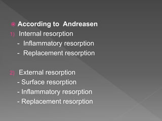
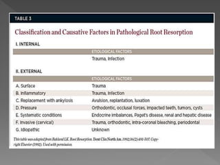
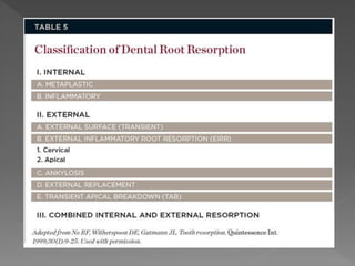
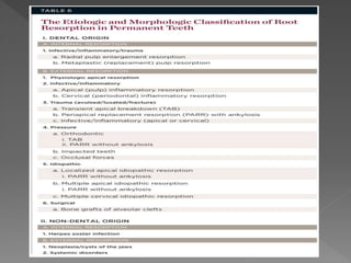
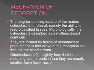
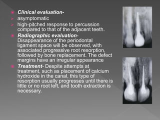
Call Girl Coimbatore Prisha☎️ 8250192130 Independent Escort Service Coimbatore
root resorption.ppt
1.
2. INTRODUCTION
DEFINITION
CLASSIFICATION
MECHANISM OF RESORPTION
TYPES OF RESORPTION
CONCLUSION
REFRENCES
3. Root resorption in deciduous teeth is a normal physiologic
response, resulting in exfoliation of the deciduous teeth with
replacement by the permanent dentition.
However, the process of root resorption in the permanent
dentition has a pathologic basis. Resorption of permanent teeth
has been considered as an unfortunate and unpredictable
phenomenon.
Dental clinicians can be faced with difficult diagnostic and
treatment decisions with respect to tooth resorption.
Tooth resorption in the primary and permanent dentition has
been extensively studied and the complex processes involved in
the removal of the organic and inorganic components of tooth
structure by clastic cells continue to evolve through basic
research.
4. Knowledge gained from experimental studies and
observations of histopathological material has provided
a sound basis for the diagnosis and treatment of many
tooth resorptive processes.
5. RESORPTION
A condition associated with either a physiologic or
pathologic process that results in loss of substance from
a tissue such as dentin , cementum or alveolar bone.
11. The singular defining feature of the mature
osteoclast is functional, namely the ability to
resorb calcified tissues. Morphologically, the
osteoclast is described as a multinucleated
giant cell.
They are formed by fusion of mononuclear
precursor cells that arrive at the resorption site
through the blood stream.
Odontoclasts differ slightly from their bone-
resorbing counterparts in that they are usually
smaller, have fewer nuclei.
12. Their differentiation is under the control of factors
produced by bone marrow stromal cells or found on the
mature osteoblast.
Two such factors are RANK (receptor activator of nuclear
factor kappa B) ligand (RANKL) and osteoprotegerin
(OPG).
The receptor of RANKL is RANK and is localized on the
surface of the progenitor osteoclast .
Therefore, physical contact between the osteoblast or
stromal cells and the progenitor osteoclast is essential for
a direct interaction of RANKL and RANK for osteoclast
formation and activation.
OPG acts as a decoy receptor that can bind to RANKL and
interferes with its ability to bind to RANK receptors, thus
inhibiting osteoclast formation.
Thus, both RANKL and OPG play an important role in
osteoclastinogenesis.
13. The cell organelles of an osteoclast consists of
many nuclei, multiple Golgi complexes,
mitochondria, rough endoplasmic reticulum and
numerous vesicular structures.
The cell membrane of the osteoclast adjacent to the
tissue surface has a series of finger-like projections
called as the ruffled border.
At the periphery of this ruffled border, the plasma
membrane is apposed closely to the bone surface,
and the adjacent cytoplasm, devoid of cell
organelles, is enriched in actin, vinculin, and talin,
fibrillar contractile proteins.
This is called as a clear or sealing zone which
comprises the peripheral non-resorptive area of
adhesion of osteoclasts to bone (i.e. it helps to
attach the cells to the bone).
14. The resorption process occurs in two stages:
Degradation of the inorganic mineral structure
followed by disintegration of the organic matrix.
Degradation of the inorganic crystal structure is
brought about by enzymes like acid phosphatase
and carbonic anhydrase II present in the osteoclasts.
The disintegration of the organic matrix is brought
about by cysteine proteinase, collagenase and
matrix metalloproteinase enzymes.
The inorganic and organic degradation products
then undergo endocytosis at the ruffled border. They
are then translocated in transport vesicles and their
extracellular release occurs along the membrane
opposite the ruffled border (transcytosis).
17. Internal resorption has been associated with
chronic inflammation of the pulp following
caries, trauma or a cracked tooth.
Types of internal resorption
Internal inflammatory resorption
Internal replacement resorption.
Transient apical resorption
Invasive coronal resorption
18. Etiology- It frequently results from chronic
inflammation of the pulp or tooth infarctions.
Pathogenesis- The progression of inflammatory
resorption is dependent on the interaction between
vital pulp tissue and necrotic tissue. Chronic
irritation of pulpal tissues occurs when bacteria and
their components enter root canals via dentinal
tubules that are exposed. Bacteria can also enter
the canals at areas of dilaceration or cracks in the
cervical area of the root.
19. Clinical evaluation-
Generally asymptomatic
The process of resorption is active only if part of the
pulp remains vital; therefore, pulp testing can be
positive. However, usually the coronal pulp is necrotic
while the apical pulp is vital, resulting in a
nonresponsive test.
Pain may be present if perforafion of the crown or root
occurs.
If resorption takes place in the coronal portion of the
tooth, the tooth may exhibit a pinkish or reddish hue
because of the presence of numerous capillaries in
the pulpal granulation tissue undermining the coronal
enamel and this is called as pink tooth of mummery.
Radiographic evaluation – It appears as a
circumscribed, oval enlargment (radiolucency)
continuous with the root canal.
20. Treatment –
Nonsurgical root canal therapy is
recommended. Treatment generally
consists of the preparation of the
canal to the apical foramen with
particular emphasis on irrigation and
ultrasonic activation of iirigants so that
the resorbed area is cleansed as
thoroughly as feasible.
The obturation of the canal can be
achieved by a variety of techniques
including hot vertically condensed
gutta-percha, thermoplasticized gutta-
percha.
Failure to treat internal inflammatory
resorption can lead to its eventual
extension to the periodontal ligament,
via a crown or root perforation. In
these cases, a periodontal procedure,
such as crown lengthening or root
extrusion, may be implemented to
gain access for repair.
21. No episode of pain was reported by the
patient in tooth #8.
The patient gave a history of trauma to
her anterior teeth about 15 years back.
On clinical examination, tooth #8 was
discoloured and displayed an Ellis class
IV fracture.
It failed to respond to thermal and
electric pulp testing while the adjacent
teeth responded within normal limits.
Ca(OH)2 paste was placed as an
intracanal medicament for 1 week.
At the next appointment, the Ca(OH)2
paste was flushed out; the canal was
dried and obturated with warm vertical
compaction technique using gutta-percha.
22. Maxillary lateral incisor with advanced
perforating internal root resorption in the
middle third of the root and the presence
of a sinus tract.
Mineral Trioxide Aggregate (MTA) was
used with the aid of a surgical
microscope in order to fill the resorption
area after conventional root canal therapy
of the apical segment.
At the follow-up after 11 years and 8
months, the patient was clinically
asymptomatic and the sinus tract had
disappeared.
The radiographic examination and
computerized tomography indicated
periodontal bone repair.
23. Etiology. This disease process appears to result from a
low-grade irritation of pulpal tissue, such as chronic
irreversible pulpitis or partial necrosis or trauma.
Pathology- Root canal replacement resorption involves
resorption of the dentin and a subsequent deposition of
hard tissue that resembles bone or cementum, but not
dentin.
Clinical evaluation-
Asymptomatic .It can become painful if the process
perforates the root or crown of the tooth.
Teeth may respond within normal limits to thermal or
electric pulp testing.
24. Radiographic evaluation- Internal replacement
resorption generally appears as enlargement of canal
space, including discontinuity of the normal canal space.
The enlarged canal space appeared radiographically to be
obliterated by a fuzzy-appearing material of mild to
moderate radiodensity
Treatment-
Management consist of pulpectomy, curettage of the
resorptive defect and root filling.
In more extensive cases, the resorptive tissue may
communicate with the periodontal ligament and
pulpectomy should be supplemented by the careful topical
application to the defect of 90% aqueous trichloracetic
acid on a small cotton pellet or a mini-applicator. This will
aid in the inactivation of any communicating resorptive
tissue.
Generally, this treatment can be followed by the insertion
of a conventional root filling although in communicating
lesions there may be occasions where mineral trioxide
aggregate (ProRoot MTA) may be used to advantage in
sealing the defect prior to the placement of a root filling.
25. Transient apical internal resorption is another
form of trauma induced non-infective root
resorption which was identified by
Andreasen in 1986.
This resorptive process can follow injuries
recognized by a confined periapical
radiolucency which resolves within a few
months.
26. Often there is an associated
colour change due to intra-
pulpal haemorrhage and this
may resolve spontaneously if
revasularization to the coronal
pulp chamber occurs.
In the longer term, as this is a
transient process, the
internally resorbed apex will
close uneventfully.
27. INVASIVE CORONAL RESORPTION:
This rare condition generally develops in
erupting teeth where a localized coronal
enamel defect allows the invasion of
aggressive hyperplastic resorptive tissue.
In this case a pink resorptive defect can be
observed in an area of hypomineralization on
the labial surface of the crown of the erupting
central incisor tooth.
28. Invasive coronal resorption has also been
observed in teeth which have been injured
by the intrusion of a primary tooth.
Radiographically the image of the
resorptive defect is generally irregular in
outline and, depending on its extent, the
radiolucency may extend both coronally and
into the radicular tooth structure.
29.
30. Treatment
Treatment is directed towards the total removal or
inactivation of all resorptive tissue and the
restoration of the coronal defect.
This may be achieved by physical curettage of the
defect with round burs and hand instruments, but is
more conveniently and effectively treated by the
topical application of 90% aqueous trichloracetic
acid, curettage, endodontic therapy if there is pulp
involvement, and restoration of the defect with a
glass ionomer cement.
Orthodontic extrusion to render the defect into a
supragingival position may supplement treatment if
the resorption extends deeply .
31. Herpes zoster is a secondary, reactivation infection
caused by the varicella-zoster virus.
Herpes zoster may impact the trigeminal nerve,
and, consequently, dental structures may be
adversely affected.
Rarely, prodromal odontalgia with pulpitis and
devitalization of the teeth has been reported
together with both internal and external root
resorption.
Herpes zoster infection, therefore, should be
considered when unexplained root resorption occur
in multiple adjacent teeth on one side of the dental
arch.
32. External resorption can be classified as
External surface resorption
External inflammatory root resorption
External trauma related resorption
External Replacement resorption
External cervical resorption
Transient apical breakdown
Systemic related resorption
Idiopathic
Neoplasia and cysts of the jaws
33. Etiology- Surface resorption usually
occurs as the response to mild localized
injury of the periodontal ligament and
cementum or some orthodontic
treatment.
Pathophysiology – After 2-4 weeks
repair is seen by cells from the
periodontal ligament . New cementum is
formed with insertion of PDL.
Radiographically- Localized widening
of PDL can be seen. Most repair related
root resorptions have such limited size
that they cannot be recognized
radiographically.
Treatment- This type of resorption is
self-limiting.
34. Etiology: This type of root resorption is a complication that can follow
dental trauma.
Pathogenesis: It begins as a surface resorption due to damage to the
periodontal ligament in conjunction with the traumatic injury. The pulp is
also damaged and becomes necrotic. As the surface resorption
approaches the dentine, the osteoclasts will carry on their resorptive
activity, as necrotic and possible infected pulp matter is released from
the exposed dentine tubules. The pulp products will then maintain an
inflammatory process in the adjacent periodontal tissues that in turn will
trigger the continuance of the resorption.
Clinical Findings:
•Increased mobility
•Dull percussion tone
•Tooth may be extruded
•No response on sensitivity tests
35. Radiographic findings: Progressive
cavitations are seen involving root
and bone after 2-4 weeks. Total
loss of tooth structure after few
months.
Treatment-
Treatment involves the thorough
debridement and preparation of
the root canal system.
Irrigation is a most important
component of this debridement
process.
Calcium hydroxide has been
widely used in the treatment of
external inflammatory root
resorption.
36. Prevention of inflammatory root resorption:
A tooth with complete root development which has been subjected
to avulsion, intrusion or a severe luxation injury should, after
replantation or repositioning, have the pulp removed as soon as
possible and the canal dressed with Ledermix paste.
In a tooth with an immature apex, with a diameter greater than
2mm, there is a chance of revascularization in all the above
injuries, provided the case of a replanted tooth has short extra-oral
period (30 minutes) or the tooth has been stored in a medium
which maintains the viability of the periodontal ligament.
Teeth with wide apices subjected to such trauma should be
monitored carefully at monthly intervals for three months and at
longer intervals thereafter.
If radiographic signs of inflammatory root resorption become
evident immediate endodontic intervention is required.
37. Effect of immediate intracanal placement of Ledermix Paste(R)
on healing of replanted dog teeth after extended dry times.
Bryson EC et al Dental Traumatology 2002
A total of 29 premolar roots were used. Fifteen of these roots were
then filled with a calcium hydroxide (Ca(OH)2) slurry and 14 roots
were filled with Ledermix Paste.
The roots prepared for histological evaluation. Five-micrometer thick
cross-sections of the root and surrounding tissue were evaluated for
healing.
In addition, residual root mass was also measured to determine the
extent of root structure loss for each treatment method.
The Ledermix Paste-treated roots had statistically significantly more
healing and less resorption than the roots treated with Ca(OH)2.
Root filling with Ledermix Paste also resulted in significantly less loss
in root mass due to resorption compared to those roots filled with
Ca(OH)2.
Immediate intracanal placement of Ledermix Paste at the emergency
visit after an avulsion injury appears to decrease resorption and
increase favorable healing.
38. Treatment of inflammatory external root resorption resulting from dental
avulsion and pulp necrosis: Clinical case report
Rodrigo Sanches Cunha, Flavia Casale Abe, Roberta Aranha Araujo,
Eduardo Rodrigues Fregnani, Carlos Eduardo da Silveira Bueno General
Dentistry May/June 2011
39. Etiology-Trauma, resorptions may have resulted from
pressure from unerupted or erupting teeth or some
neoplasms, from biomechanical forces involved in
orthodontics, mechanical , and surgical, thermal or
chemical trauma.
Pathogenesis-In all trauma induced (non-infective) tooth
resorption some damage to the cementum/cementoid-
periodontal membrane complex has occurred which
stimulates clastic activity.
Radiographic Evaluation- It is difflcult to identify
ankylosis on radiographs because of overlapping
structures and bone marrow spaces. However, a complete
disappearance of the periodontal space and an uneven
root surface contour is common
40. Treatment- With the removal of the initiating trauma,
these non-infective resorptions will become inactive and
uncomplicated repair will occur.
Ultimately the crown of the tooth will fracture off at the
gingival crest as ankylosis progresses, resulting in a
complete replacement of the root by bone.
41. Etiology- The primary cause of replacement
resorption is luxation injuries.
Pathogenesis- It differs from ankylosis
because of the presence of intervening inflamed
connective tissue. During tooth avulsion, large
areas of the periodontal ligament are lost or
damaged. Healing then occurs from the alveolar
side of the socket and leads to a union between
the tooth and alveolar bone.
42. Clinical evaluation-
asymptomatic
high-pitched response to percussion
compared to that of the adjacent teeth.
Radiographic evaluation-
Disappearance of the periodontal
ligament space will be observed, with
associated progressive root resorption,
followed by bone replacement. The defect
margins have an irregular appearance
Treatment- Despite attempts at
treatment, such as placement of calcium
hydroxide in the canal, this type of
resorption usually progresses until there is
little or no root left, and tooth extraction is
necessary.
43. This form of external resorption has been
described at length by Heithersay who preferred
the term invasive cervical resorption, which
describes its invasive and aggressive nature.
Other terms used to describe ECR include
odontoclastoma, peripheral cervical resorption ,
extracanal invasive resorption ,supraosseous
extracanal invasive resorption , peripheral
inflammatory root resorption, and subepithelial
external root resorption .
44. ECR usually occurs immediately below the
epithelial attachment of the tooth at the
cervical region.
ECR has been described as an ‘‘aseptic
resorptive process, which may on occasions
become secondarily invaded with
microorganisms’’.
46. Clinical signs:
Located in cervical region of tooth.
Pink spot might be present.
Tooth usually responds positively to vitality
tests unless there is pulpal involvement (in
very advanced cases)
47. Spontaneous and profuse bleeding on
probing.
Sharp, thinned out edges around the
resorptive cavity.
48. Radiologic signs:
Detected on radiologic finding because
tooth is usually asymptomatic.
Varies from asymmetrically located
radiolucency with irregular margins in
cervical/proximal region of tooth to
uniformly round radiolucency centered
over the root.
Early lesions are usually radiolucent in
appearance.
Advanced lesions might have mottled
appearance because of fibro-osseous
nature of the lesion.
Root canal should be visible and intact
(indicating lesion is external).
54. Heithersay concluded that classes 1–3 were treatable, but class
4 lesions were not amenable to treatment, and these cases
would have benefited from alternative treatment such as
extraction and replacement with an implant retained crown
restoration.
In the instance of classes 1–3, the root canal should be
nonsurgically accessed and temporarily occluded with a finger
spreader or gutta-percha point.
Then the surgical repair to the resorptive defect might be carried
out without blocking the root canal with filling material.
Heithersay recommended topical application of a 90%
aqueous solution of trichloroacetic acid, curettage, and
restoration with glass ionomer cement.
55. Topical application of trichloroaceticacid results in
coagulation necrosis of the ECR resorptive tissue, with no
damage to adjacent periodontal tissues.
It also infiltrates the small channels and recesses of ECR
that would otherwise be unreachable by mechanical
instrumentation.
Once the ECR cavity has been restored, endodontic
treatment might be completed confidently without risk of
expelling irrigants and debris via the ECR cavity into the
adjacent periodontium.
Alternatively, the defect could be filled with the mineral
trioxide aggregrate material, Pro-Root MTA which would
appear to possess ideal properties for this type of repair.
56. Kitchens et al in 2007 found that iontophoresis
greatly increased transdentinal delivery of
calcitonin.
They suggested a new method of treatment of ECR
that involves chemomechanical debridement of the
root canal, obturation of the apical part of the canal,
removal of the smear layer, and subsequent
iontophoretic delivery of calcitonin via the dentinal
tubules directly to the ECR lesion.
Some other potential medicaments that could also
be used with this method are osteoprotegerin and
bisphosphonates.
Surgical treatment of varying degrees of invasive
cervical resorption has generally involved
periodontal flap reflection, curettage, restoration of
the defect with MTA.
57. Transient apical breakdown is a temporary
phenomenon in which the apex of the tooth
displays the radiographic appearance of
resorption that is linked to the repair processes
of a traumatically injured pulp and/or
periodontium of luxated mature teeth. This
process is invariably followed by surface
resorption and/or obliteration of the pulp canal.
The injured periradicular tissue generally
returns to normal following repair, which usually
takes place 1 year after trauma.
58. Etiology- Typically, it is caused by moderate
injuries to the pulp, such as subluxation,
extrusion, and lateral luxation, or a moderate
combined injury to the periodontai ligament
and the pulp in mature teeth.
Clinical evaluation- The tooth wiil often
respond within normal limits to pulp tests.
Clinicaliy, in some instances, the tooth may
undergo a color change and/or have varying
results from electrical pulp testing. However,
following repair, clinical and radiographic
findings will return to normal.
Radiographic evaluation- Transient apical
breakdown can only be found in teeth with fully
formed roots and closed or half-closed apices.
A transient localized change in the size of the
apical periodontal ligament space, ranging
from 2-times normal width to a semicircular
radiolucency, combined with a blunting of the
apex from surface resorption, can be observed.
Treatment- No treatment is recommended.
59. Systemic-related resorption may be seen in patients with
hyperparathyroidism, Pagets disease, renal and liver disease.
Occasionally teeth, typically first permanent molars, develop apical
root resorption for which no cause can be elicited.
These teeth have vital pulps and endodontic treatment does not
arrest the condition and so is contraindicated.
NEOPLASIA AND CYSTS OF THE JAWS
Various odontogenic and non-odontogenic cysts and tumors of the
jaws may produce different patterns of external root resorption that
are not of dental origins. Differentiating these types of lesions from
those originating from dental sources is important for avoiding
unnecessary dental therapy or delays in appropriate medical
treatments.
68. Where resorption has extended
from an internal inflammatory
resorption to involve the external
surface a communicating lesion is
created.
This can be recognized
radiographically by a radiolucency
within the tooth structure extending
to the exterior surface and the
surrounding bone.
69. Treatment
Calcium hydroxide has been used.
Another approach involves the topical application
of 90% aqueous trichloracetic acid to the
resorptive tissue following endodontic preparation
to the level of the resorptive defect.
Trichloracetic acid is applied for 1–2 minutes on a
mini-applicator or a small cotton pellet attached to
an endodontic file.
This will induce a sterile necrosis of the resorptive
tissue. This will aid in the inactivation of any
communicating resorptive tissue.
70. Treatment of perforating internal root resorption with MTA: a case
reportEduardo Nunes et al, Journal of Oral Science, Vol. 54, No. 1,
127-131, 2012
Treatment of inflammatory external root resorption resulting from
dental avulsion and pulp necrosis: Clinical case report Rodrigo
Sanches Cunha, Flavia Casale Abe, Roberta Aranha Araujo,
Eduardo Rodrigues Fregnani, Carlos Eduardo da Silveira Bueno
General Dentistry May/June 2011
Tooth resorption part II - external resorption: Case series Marina
Fernandes, Ida de Ataide, and Rahul WagleJ Conserv Dent. 2013
Mar-Apr; 16(2): 180–185.
Tooth resorption part I - pathogenesis and case series of internal
resorption Marina Fernandes, Ida de Ataide, and Rahul WagleJ
Conserv Dent. 2013 Jan-Feb; 16(1): 4–8.
71. Effect of immediate intracanal placement of Ledermix Paste(R) on
healing of replanted dog teeth after extended dry times. Bryson EC
et al Dental Traumatology 2002
Kitchens JA, Schwartz SA, Schindler WG, Hargreaves KM.
Iontophoresis significantly increases the trans-dentinal delivery of
osteoprotegerin, alendroate and calcitonin. J Endod 2007;33:1208–
11.
Internal Resorption in Human Teeth - A Histological, Scanning
Electron Microscopic, and Enzyme Histochemical Study Cecilia
Wedenberg and Lars Zetterqvist. JOE VOL. 13. NO 6. JUNE 1987
Dental root resorption and repair: histology and histometry during
physiological drift of rat molars. Ryusei Kimura, Hisashi Anan, Akiko
Matsumoto, Daisuke Noda, Katsumasa MaedaJ Periodont Res
2003; 38; 525–532
72. Congenitally absence of successor teeth
Impaction of successor teeth
Translation or transmigration of successor
teeth
Existence of pathology, such as cysts,
tumours, under the primary tooth that results
in the impaction of successor teeth
73.
74. The acetazolamide is a potent inhibitor of
carbonic anhydrase. Therefore, with the
inhibition of carbonic anhydrase, there will be
consequently no resorption. Several studies
confirm the efficacy of acetazolamide in
inhibiting the resorption. The idea of using
AZ as a substance to inhibit root resorption
was presented by Mori and Garcia in 2002.
75. If extraoral dry time exceeds 60 min then the treatment
of the root surface should be done with fluoride prior to
replantation by soaking the tooth in a sodium fluoride
2.4% with PH of 4.3 for 20 minutes to slow down
osseous replacement of the tooth.
Fluoride acts on cementum and dentin by converting
hydroxyapatite to fluorapatite which renders the
cementum and dentin more able to resist dissolution.
Fluoride loosely bound to the organic constituents of
cementum is readily released and may inhibit
odontoclastic cell activity, probably by interferring with
"clast" cell enzymes.
76. When an orthodontist identifies root resorption in a patient, the
severity of the condition is decreased with a pause in active
orthodontic movement for two to three months with a use of a
passive archwire or by use of lower forces and by decreasing
the treatment duration .
However, if the resorption is severe, the orthodontist should
reassess the treatment plan.
Alternative options include prosthetic solutions to close spaces,
releasing teeth from active archwires when possible.
If root resorption is diagnosed on the final radiographs after
treatment, follow-up radiographic examinations are
recommended until the resorption has stabilized .
However, if it continues, sequential root canal therapy with
calcium hydroxide may be considered .
77. The high pH of Calcium hydroxide has beneficial effects
which include the
neutralization of acid products
antimicrobial property
activation of alkaline phosphatase.
The hydroxyl group is considered to be the most important
component of calcium hydroxide as it provides an alkaline
environment which encourages repair and active
calcification.
The alkaline pH induced not only neutralises lactic acid
from the osteoclasts, thus preventing a dissolution of the
mineral components of dentine, but could also activate
alkaline phosphatases which play an important role in hard
tissue formation
78. Identification of the stimulation factor of root
resorption is useful in order to render proper
treatment by removing the etiological factors.
Successful management of potential or
established resorption in patient is of
paramount importance, as it has been
recognized that poorly treated injuries can have
not only physical consequences but also a
significant psychological impact on the patient.
79. Pathways of pulp- 10th edition
Ingle- 6th edition
Grossman- 12th edition
Tooth resorption, Rita F. Ne, DDSVDavid E. Witherspooti,
BDSc, BEcon MSVJames L. Gutmafin(Quintessence Int
1999;30:9-25
External Cervical Resorption: A Review Shanon Patel, Shalini
Kanagasingam, and Thomas Pitt Ford, JOE — Volume 35,
Number 5, May 2009
Internal Root Resorption: A Review Shanon Patel, Domenico
Ricucci, Conor Durak, and Franklin Tay,JOE— Volume 36,
Number 7, July 2010
Invasive cervical resorption: an analysis of potential
predisposing factors.Heithersay GS, Quintessence Int.1999
Feb;30(2):83-95.
a molecule, as an antibody, hormone, or drug, that binds to a receptor.- ligand
Decoy - lure or entice or pulling away from their intended course
Endocytosis is an energy-using process by which cells absorb molecules (such as proteins) by engulfing them. It is used by all cells of the body because most substances important to them are large polar molecules that cannot pass through the hydrophobic plasma or cell membrane. The opposite process is exocytosis.
Exocytosis (/ˌɛksoʊsaɪˈtoʊsɪs/; from Greek ἔξω "out" and English cyto- "cell" from Gk. κύτος "receptacle") is the durable, energy-consuming process by which a cell directs the contents of secretory vesicles out of the cell membrane and into the extracellular space.
Tooth infraction can be defined as an incomplete tooth fracture extending partially through a tooth.
Vertical root fractures are longitudinal fractures that originates in the root of the teeth in contrast to tooth infractions that originates in the crown.
NaOCl an effective antimicrobial irrigating
solution, aids in dissolving necrotic tissue.[15] Ultrasonic activation of irrigants has also proved to be an
effective method to remove debris lodged in inaccessible resorptive sites.
Metaplasia refers to
a reversible change in which one adult cell type (epithelial or
mesenchymal) is replaced by another cell type (73). In the present
context, the metaplastic tissue appears lamella-like, with entrapped
osteocyte-like cells that resemble osteons of compact bone
Herpes zoster (or simply zoster), commonly known as shingles and also known as zona, is a viral disease characterized by a painful skin rash with blisters in a limited area on one side of the body (left or right), often in a stripeThe earliest symptoms of herpes zoster, which include headache, fever, and malaise, are nonspecific, and may result in an incorrect diagnosis.[5][11]These symptoms are commonly followed by sensations of burning pain, itching, hyperesthesia (oversensitivity), or paresthesia ("pins and needles": tingling, pricking, or numbness). acyclovirhas been the standard treatment, but the new drugs valaciclovir and famciclovir .
Herpes simplex virus infection causes recurring episodes of small, painful, fluid-filled blisters on the skin, mouth, lips (cold sores), eyes, or genitals.* This very contagious infection is spread by direct contact with sores or sometimes with the affected area when no sores are present.* Herpes causes blisters or sores in the mouth or on the genitals and, often with the first infection, a fever and general feeling of illness.* The virus sometimes infects other parts of the body, including the eyes and brain.
In medicine, a prodrome is an early symptom (or set of symptoms) that might indicate the start of a disease before specific symptoms occur
Ledermix Paste is a paste containing triamcinolone and demeclocycline with demonstrated anti-inflammatory activity that may slow down resorptive processes after severe traumatic injuries to the dentition
After 4 months dogs were killed
Resorption had arrested and a return of the normal lamina dura was observed on the radiograph, indicating that the therapy was successful.
Ankylosis is a dental situation in which the roots of primary teeth lose their normal attachment to the bone (small ligaments) and become fused directly to the bone
Ankylosis is a dental situation in which the roots of primary teeth lose their normal attachment to the bone (small ligaments) and become fused directly to the bone.
Spotted or blotched with different shades or colors
Class 1 – Denotes a small invasive resorptive lesion near the cervical area with shallow penetration into dentine.
Class 2 – Denotes a well-defined invasive resorptive lesion that has penetrated close to the coronal pulp chamber but shows little or no extension into the
radicular dentine.
Class 3 – Denotes a deeper invasion of dentine by resorbing tissue, not only involving the coronal dentine but also extending into the coronal third of the root.
Class 4 – Denotes a large invasive resorptive process that has extended beyond the coronal third of the root.
75-icr
Calcitonin decreases osteoclast cytoplasmic motility, stimulates pseudopodial
retraction, causes a loss of the ruffled border, and reduces tartrate-resistant acid phosphatase
secretionIontophoresis is a physical process in which ions flow diffusively in a medium driven by an electric field
Paget's disease of bone or Paget disease of bone (/ˈpædʒɨt/, rhymes with "gadget") is a chronic disorder that can result in enlarged and misshapen bones. Paget's is caused by the excessive breakdown and formation of bone, followed by disorganized bone remodeling. This causes affected bone to weaken, resulting in pain, misshapen bones, fractures, and arthritis in the joints near the affected bones. have elevated levels of serum alkaline phosphatase (ALP) in their blood; steoclasts in a localised area of bone become out of control is not known. Facial disfigurement may be consequence of enlargement of the maxilla and/or mandible.[3] Radiograph shows characteristic cotton wool appearance and other radiographic finding include well-circumscribed radiolucency, loss of lamina dura, pulpal radio-opacity, root resorption, and hypercementosis.
Primary hyperparathyroidism results from a hyperfunction of the parathyroid glands themselves. There is oversecretion of PTH due to adenoma, hyperplasia or, rarely, carcinoma of the parathyroid glands. weakness and fatigue, depression, bone pain, muscle soreness (myalgias), decreased appetite, feelings of nausea and vomiting, constipation, polyuria,polydipsia, cognitive impairment, kidney stones and osteoporosis pulp calcified max and man demineralized
When there is irritation of some kind, the tissue responds by some kind of inflammation iwith an infiltrate consisting predominantly of lymphocytes, macrophages, and some neutrophils. The blood vessels become dilated. Odontoblasts could not be observed in any of the teeth and predentin was rarely seen outlining the mineralized dentin. characterized by large, multinucleated dentinoclasts in resorption lacunae on the pulpal dentin surface (Fig. 3). These cells showed intense tartrate-resistant acid phosphatase activity
In the early stages, the résorption cavity contains a mass of fibrous tissue, numerous blood vessels and clastic resorbing cells adjacent to the dentin surface [Fig 13). Clastic cells lining the resorptive cavities are generally mononuclear, but some multinucleated cells can also be identified. A layer of dentin and predentin separates the resorbing tissue from the dental pulp, which remains free of inflammation until late in the process. The resorbing tissue is usually devoid of acute or chronic inflammatory cells unless the lesion has been invaded by oral microorganisms. These early lesions contain fibrovascular
tissue, but they appear to progress to fibro-osseous lesions by the deposition of ectopic bonelike calcifications both within the resorbing tissue and directly
onto the resorbed dentin surface.
The odontoclast in the lacuna exhibits a developed ruffled border
(small arrows), a clear zone (CZ), abundant vacuoles (V) and mitochondria (small arrowheads), and scattered rER (open arrowheads), which
indicate that this odontoclast is activated for root resorption. Mononuclear cells (arrows) close to the odontoclast-detached surface are
extending cytoplasmic processes (arrowheads) toward the root surface.
Resorption channels are created; these burrow
deeply into the dentin surrounding the root canal and
later interconnect more apically with the periodontal
ligamentAs a result, a delta of channels may
be created, which has the effect of increasing the
potential for further résorption. The ectopic bonelike
tissue that is deposited within the fibrovaseuiar tissue
and directly onto dentin has a eanalicular structure
with cellular inclusions and resembles cancellous
bone. As lesions become more extensive and there is cavitation
into the oral cavity, secondary invasion of the
tissue by microorganisms attracts a normal inflammatory
response cbaracterized by tbe presence of acute
and cbronic inflammatory cells. Despite this, the pulp
will often remain free of inflammation (Fig 15) unless
tbere is penetration into the pulp space by tissue
accompanied by microorganisms. In some instanceswhen the pulp space is invaded by resorbirtg tissue
a place in which something is formed or deposited; a site of origin- nidus