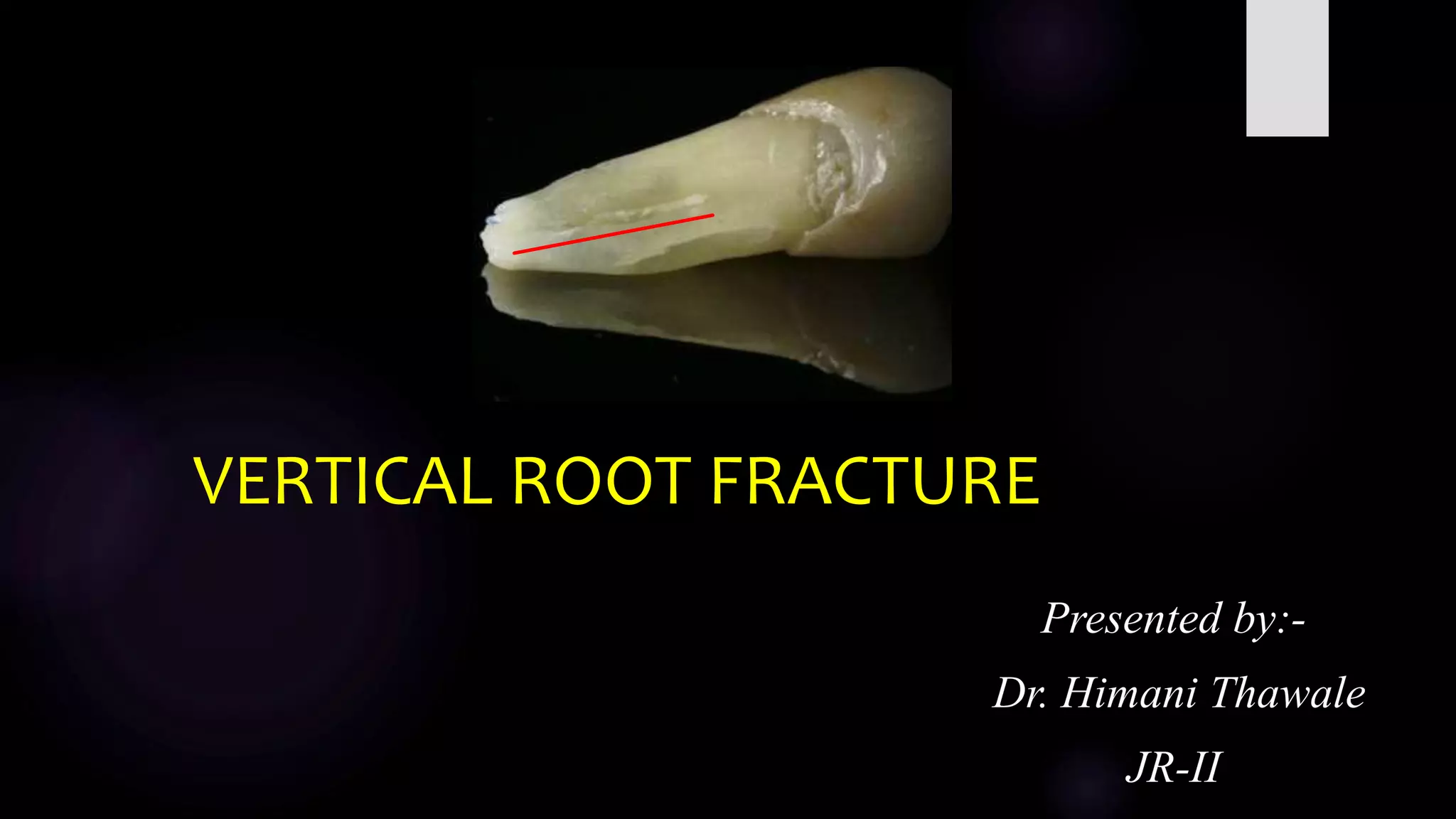
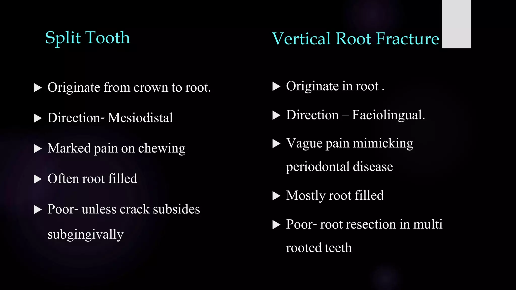
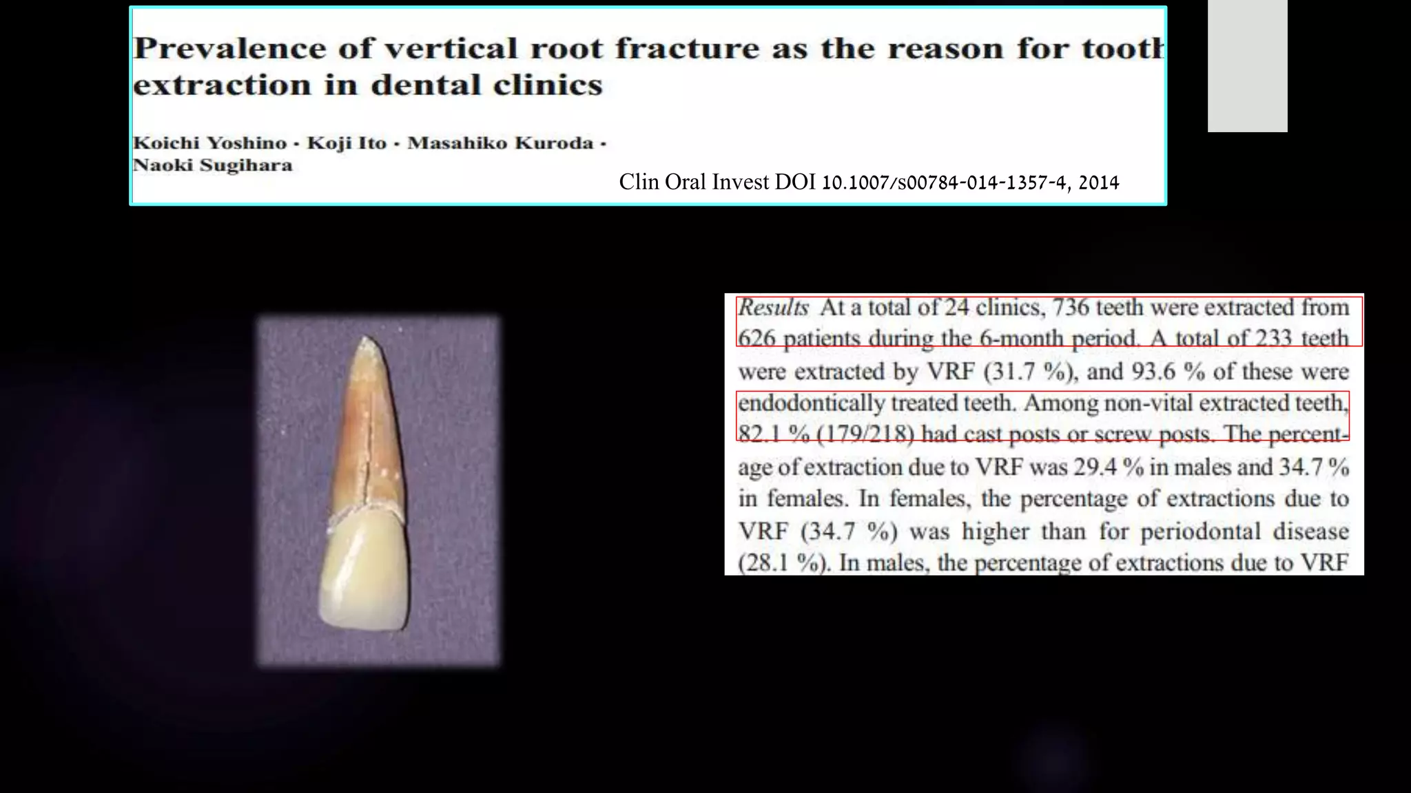
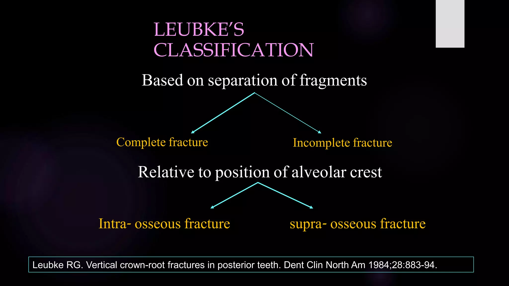
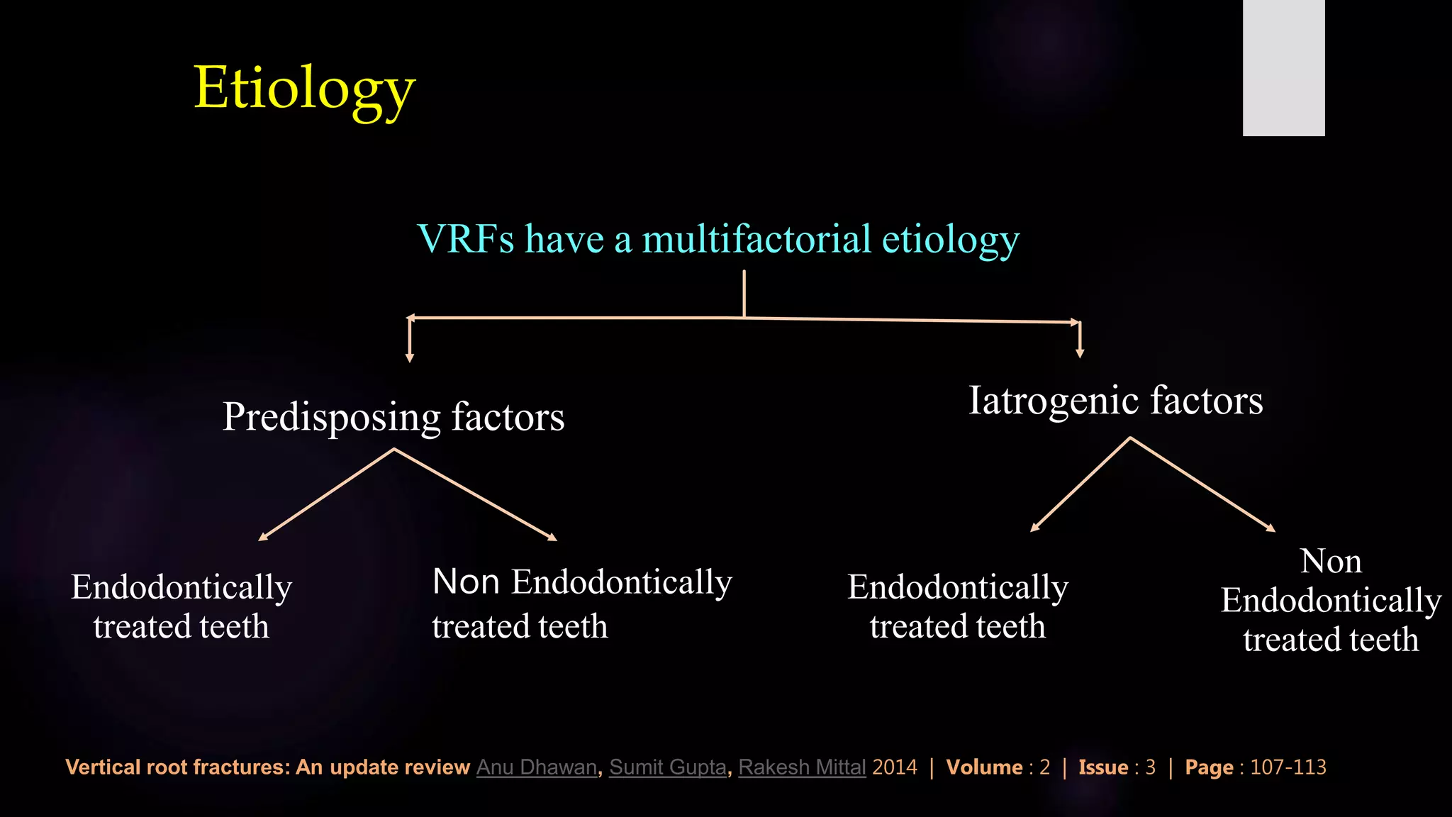
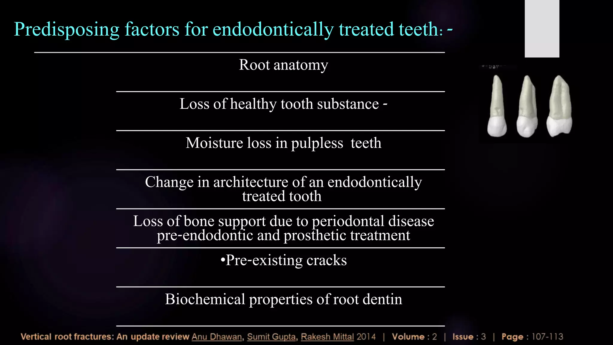
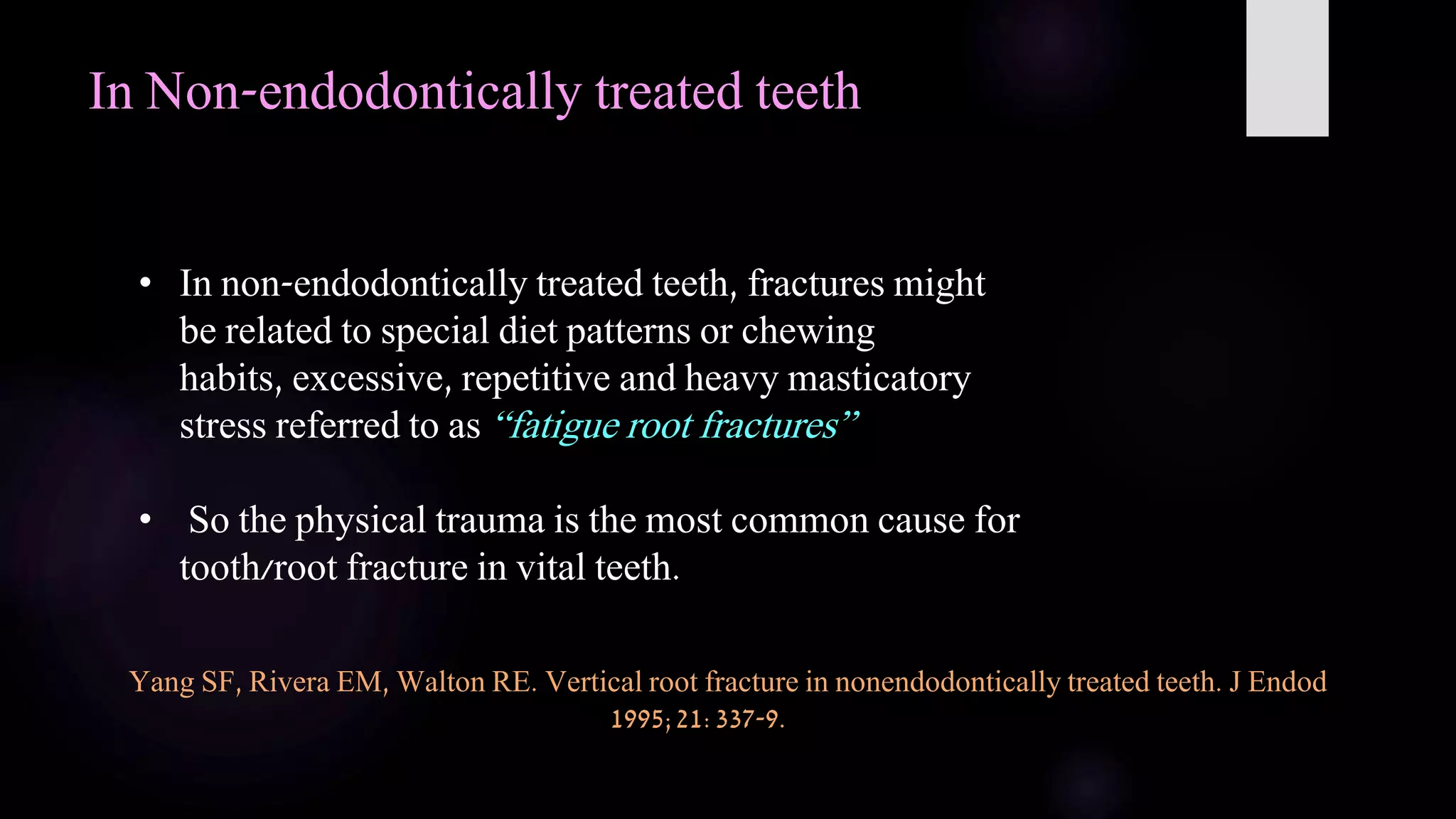
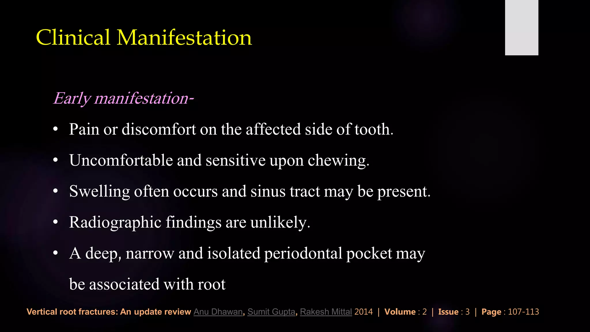
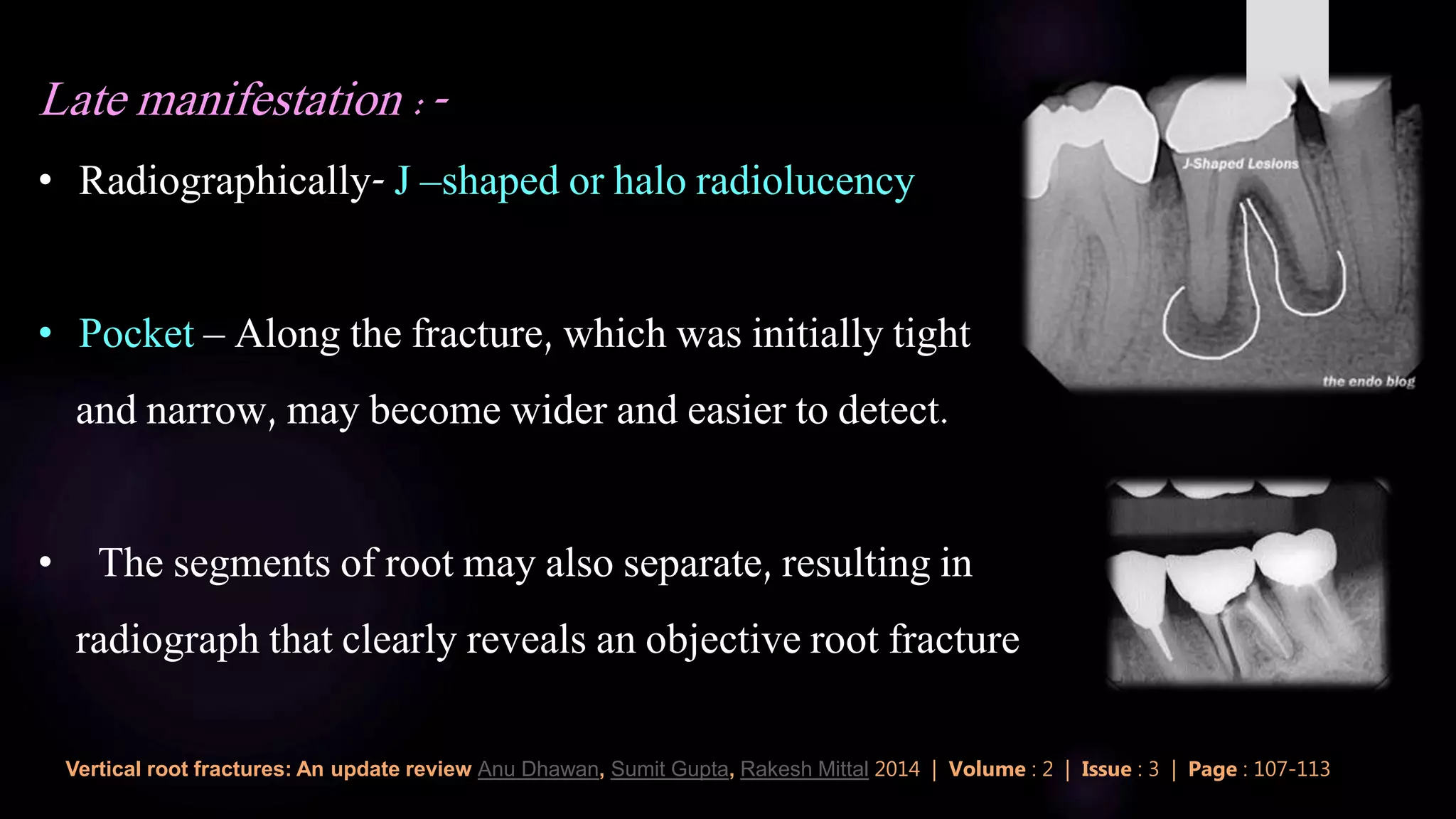
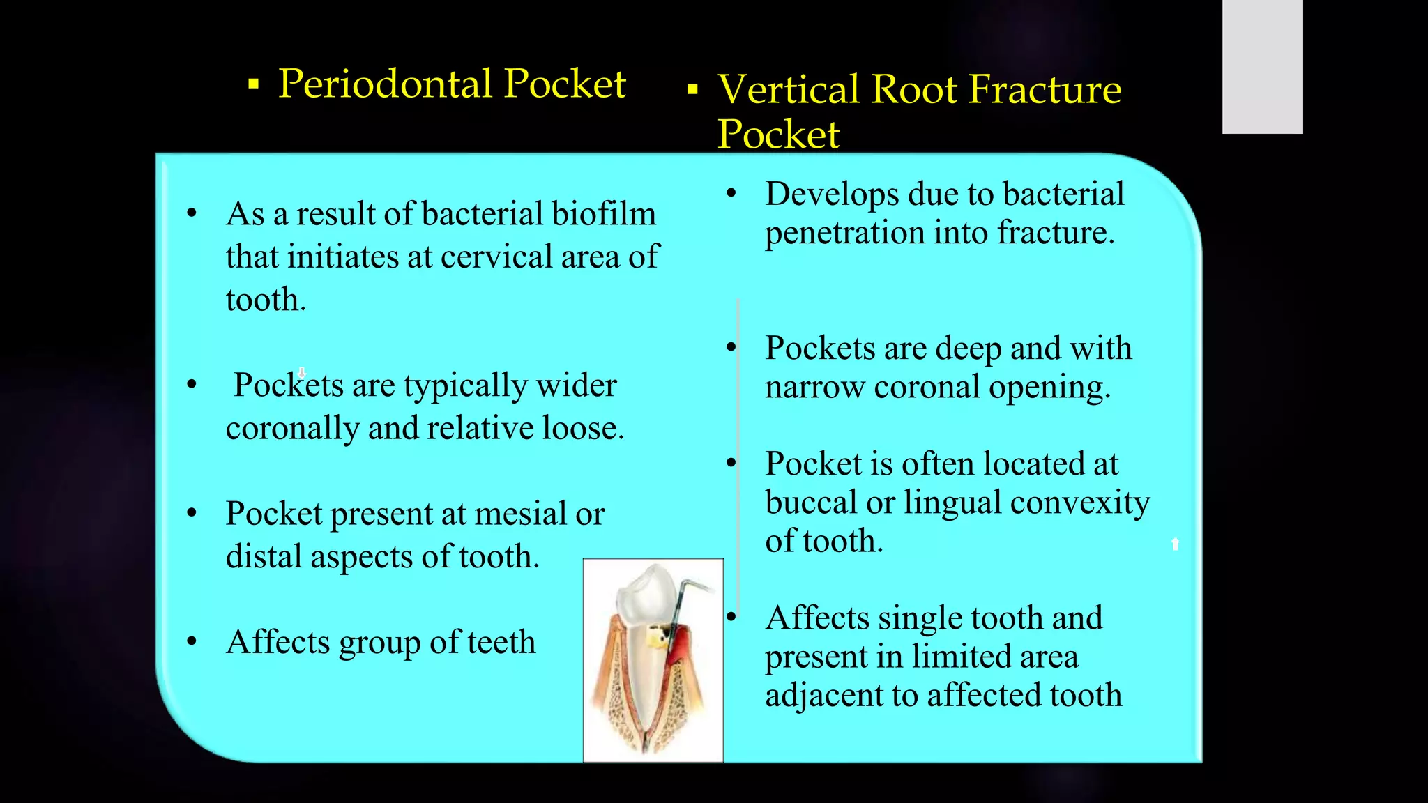
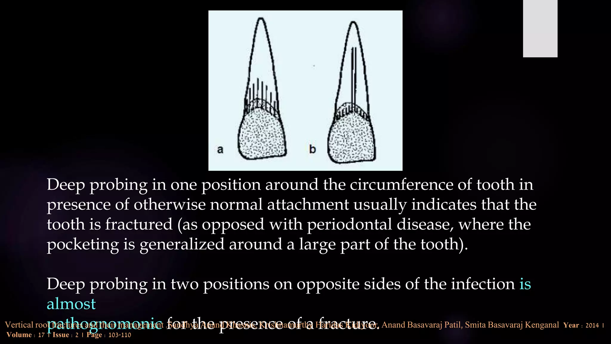
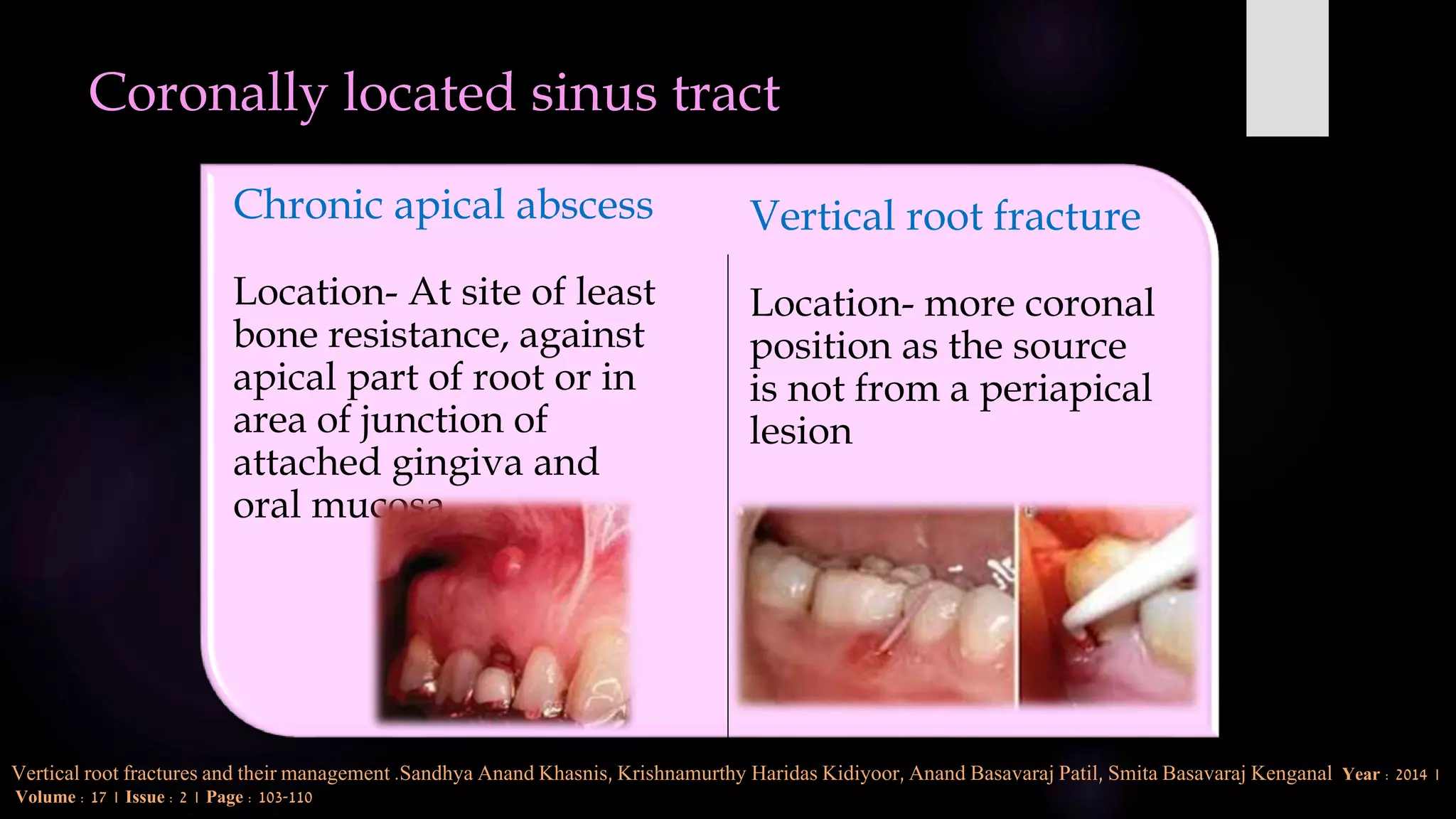
VERTICAL ROOT FRACTURE
Definition
 A ‘‘true’’ VRF is defined as a longitudinally oriented
complete or incomplete fracture initiated in the root at
any level, usually directed buccolingually
American association of endodontics
Split Tooth
 Originate from crown to root.
 Direction- Mesiodistal
 Marked pain on chewing
 Often root filled
 Poor- unless crack subsides
subgingivally
Vertical Root Fracture
 Originate in root .
 Direction – Faciolingual.
 Vague pain mimicking
periodontal disease
 Mostly root filled
 Poor- root resection in multi
rooted teeth
Incidence
 Gher et al. have reported a low incidence of 2.3%.
Gher ME Jr, Dunlap RM, AndersoLV.Clnical survey
of fractured teeth. J Am Dent Assoc 1987;114:174-7.
 Highest incidence has been observed in endodontically treated teeth and in patients older than
40 years of age.
 Premolars are the most susceptible teeth for vertical root fracture followed by molars, incisors
and cuspids in descending order.
 The most susceptible roots to fracture are maxillary and mandibular premolars, mesial roots of
mandibular molars and mandibular incisors.
Cohen . Pathways of pulp , 11th edition , chp-21, pg no- 800.
JOE 2009
• VRF is rare in vital anterior teeth.
• VRF in vital teeth occurs more frequently in males due to factors such as
stronger masticatory force, increased attrition, habitual chewing of hard
food and less pliable supporting bone.
• Fracture most commonly occurs in bucco-lingual direction in individual
roots of molar teeth. Mesio-distal fractures are less common.
• In anterior teeth, the fractures are most commonly in a bucco-lingual
direction.
Holcomb JQ, Pitts DL, Nicholls JI. Further investigation of spreader loads required to
cause vertical root fracture during lateral condensation. J Endod 1987;13:277-84.
Chan CP, Lin CP, Tseng SC, Jeng JH. Vertical root fracture in endodontically
versus nonendodontically treated teeth: A survey of 315 cases in Chinese
patients. Oral Surg Oral Med Oral Pathol Oral Radio Endod 1999;87:504-7
Testori et al. reported premolars to have the highest incidence of VRF in
endodontically treated teeth; however,
Chan et al. reported first molars to be the teeth most frequently fractured.
On the other hand, canines had the lowest incidence.
Testori T, Badino M, Castagnola M. Vertical root fractures in
endodontically treated teeth: A clinical survey of 36
cases.JEndo1993;19:87-91.
Chan CP, Lin CP, Tseng SC, Jeng JH. Vertical root fracture
in endodontically versus nonendodontically treated teeth: A
survey of 315 cases in Chinese patients. Oral Surg Oral Med
Oral Pathol Oral Radio Endod 1999;87:504-7
Prevalence of vertical root fractures in teeth planned for apical surgery. A
retrospective cohort study. M. Maddalone1 , M. Gagliani , C.L. Citterio , A.
Pellegatta & M. Del Fabbro . Int Endo . Journal ,2018
using a finite elements model, concluded that the lack of bonding between
post and canal walls increased the frequency of VRFs, stating that
“Tensile stress peaks for the non-bonded models were approximately three
times higher than for the bonded or intact models”.
Santos AF, Tanaka CB, Lima RG et al. (2009) Vertical root
fracture in upper premolars with endodontic posts: finite
element analysis. Journal of Endodontics 35, 117-20.
Suggested that the improper selection of intra-canal posts or excessive
pressure in positioning them could cause fractures in roots
Tamse A (1988) Iatrogenic vertical root fractures in
endodontically treated teeth. Endodontics and Dental
Traumatology 4, 190-6.
History
Clinical Diagnostic Test
Radiographic Examination
Endodontic Status After Healing
Has Occured
Surgical Exploration
Lasers
DIAGNOSIS OF VERTICAL ROOT FRACTURE IS
BASED ON:-
HISTORY:-
• History of facial trauma (could result in a VRF if the trauma is directed
accordingly.
• History of pain, swelling, presence of sinus tract, mobility, or any
history of post or restoration dislodgement.
• A thorough clinical examination, including age and gender of patient;
involved tooth, its location, pulp vitality; history of previous dental
treatment (including endodontic and restorative treatments); and type
of restoration (with or without post and crown) using glass ionomer
cement, resin-based composite or amalgam
Vertical root fractures: An update review Anu Dhawan, Sumit Gupta, Rakesh Mittal 2014 | Volume : 2 | Issue : 3 | Page : 107-113
Direct visualization
staining
Pulp testing
Bite test
Transillumination test
Periodontal probing test
Sinus tract tracing
CLINICAL DIAGNOSTIC TESTS:-
Vertical root fractures: An update review Anu Dhawan, Sumit Gupta, Rakesh Mittal 2014 | Volume : 2 | Issue : 3 | Page : 107-113
Prevalence
An overall prevalence of 3% to 5% has been reported in retrospective studies.
However, the percentage of extracted teeth with VRF has been reported to be much
higher - 10-20%.
Bergman B, Lundquist P, Sjögren U, Sundquist G. Restorative and
endodontic results after treatment with cast posts and cores. J Prosthet
Dent 1989;61:10-5.
Fuss Z, Lustig J, Tamse A. Prevalence of vertical root fractures in
extracted endoodontically treated teeth. Int Endod J 1999;32:283-6.
According to Chan et al.,11- 40% of VRFs occurred in nonendodontically treated teeth
of Chinese patients, which may result from excessive, repetitive, and heavy
masticatory stress exerted vertically on attrited occlusal surface
Chan CP, Lin CP, Tseng SC, Jeng JH. Vertical root fracture in
endodontically versus nonendodontically treated teeth: a survey of
315 cases in Chinese patients. Oral Surg Oral Med Oral Pathol Oral
Radiol Endod 1999;87:504e7.
It has been reported that VRFs accounted for 8.8 -20% of all extracted root-filled
teeth
Zadik Y, Sandler V, Bechor R, Salehrabi R. Analysis of
factors related to extraction of endodontically treated
teeth. Oral Surg Oral Med Oral Pathol Oral Radiol Endod
2008;106:
Clin Oral Invest DOI 10.1007/s00784-014-1357-4, 2014
CLASSIFICATION
Leubke RG. Vertical crown-root fractures in posterior teeth. Dent Clin North Am 1984;28:883-94.
Based on separation of fragments
Complete fracture Incomplete fracture
Relative to position of alveolar crest
Intra- osseous fracture supra- osseous fracture
LEUBKE’S
CLASSIFICATION
Pathogenesis
Vertical root fracture results from wedging forces
within the canal
These excessive forces exceed the binding strength of
root dentin, causing fatigue and fracture
Irritants that induce severe inflammation in or extend
the length of the root, that is, from apical to cervical
The fracture likely initiates internally (canal wall) and
grows outward to the root surface
Holcomb JQ, Pitts DL, Nicholls JI. Further investigation of spreader loads required to cause vertical root fracture during lateral condensation. J Endod1987:13: 277–284
Etiology
VRFs have a multifactorial etiology
Predisposing factors Iatrogenic factors
Endodontically
treated teeth
Non Endodontically
treated teeth
Non
Endodontically
treated teeth
Endodontically
treated teeth
Vertical root fractures: An update review Anu Dhawan, Sumit Gupta, Rakesh Mittal 2014 | Volume : 2 | Issue : 3 | Page : 107-113
Predisposing factors for endodontically treated teeth:-
Root anatomy
Loss of healthy tooth substance -
Moisture loss in pulpless teeth
Change in architecture of an endodontically
treated tooth
Loss of bone support due to periodontal disease
pre-endodontic and prosthetic treatment
•Pre-existing cracks
Biochemical properties of root dentin
• Excessive cutting during various phase of root canal treatment
• Increased stress generation with threaded and tapered posts
• Increased wedging forces with lateral compaction of gutta-percha
accounts for 48% to 84% of cases of VRFs. The development of these
stresses initiates crack introduction and propagation, leading to final root
fracture.
Iatrogenic errors for endodontically treated teeth
Yang HS, Lang LA, Molina A, Felton DA. The effects of dowel design and load
direction on dowel and core restorations. J Prosthet Dent 2001;85:558-67.
Akkayan B, Gulmez T. Resistance to fracture of endodontically treated teeth
restored with different post systems. J Prosthet Dent 2002;87:431-7.
Tamse A. Iatrogenic vertical root fractures in endodontically treated
teeth. Endod Dent Traumatol 1988;4:190-6.
In Non-endodontically treated teeth
• In non-endodontically treated teeth, fractures might
be related to special diet patterns or chewing
habits, excessive, repetitive and heavy masticatory
stress referred to as “fatigue root fractures”
• So the physical trauma is the most common cause for
tooth/root fracture in vital teeth.
Yang SF, Rivera EM, Walton RE. Vertical root fracture in nonendodontically treated teeth. J Endod
1995;21:337-9.
Predisposing factors in vital teeth
• Loss of tooth material
• Anatomy of the susceptible teeth
• Previous dental cracks and alveolar bone support
• Habits:- Bruxism and clenching
• Increased stress in compromised teeth.
• Poor cavity preparation design.
• Poorly fitting intracoronal restoration.
• Improper choice of teeth for bridge abutment.
Clinical Manifestation
Early manifestation-
• Pain or discomfort on the affected side of tooth.
• Uncomfortable and sensitive upon chewing.
• Swelling often occurs and sinus tract may be present.
• Radiographic findings are unlikely.
• A deep, narrow and isolated periodontal pocket may
be associated with root
Vertical root fractures: An update review Anu Dhawan, Sumit Gupta, Rakesh Mittal 2014 | Volume : 2 | Issue : 3 | Page : 107-113
Late manifestation :-
• Radiographically- J –shaped or halo radiolucency
• Pocket – Along the fracture, which was initially tight
and narrow, may become wider and easier to detect.
• The segments of root may also separate, resulting in
radiograph that clearly reveals an objective root fracture
Vertical root fractures: An update review Anu Dhawan, Sumit Gupta, Rakesh Mittal 2014 | Volume : 2 | Issue : 3 | Page : 107-113
Pathognomic for presence of VRF
Sinus tract
&
Narrow , Isolated periodontal probing defect associated
with a tooth that has undergone a root canal treatment ,
with or without post placement
According To American Association Of
Endodontists
▪ Periodontal Pocket ▪ Vertical Root Fracture
Pocket
• As a result of bacterial biofilm
that initiates at cervical area of
tooth.
• Pockets are typically wider
coronally and relative loose.
• Pocket present at mesial or
distal aspects of tooth.
• Affects group of teeth
• Develops due to bacterial
penetration into fracture.
• Pockets are deep and with
narrow coronal opening.
• Pocket is often located at
buccal or lingual convexity
of tooth.
• Affects single tooth and
present in limited area
adjacent to affected tooth
Deep probing in one position around the circumference of tooth in
presence of otherwise normal attachment usually indicates that the
tooth is fractured (as opposed with periodontal disease, where the
pocketing is generalized around a large part of the tooth).
Deep probing in two positions on opposite sides of the infection is
almost
pathognomonic for the presence of a fracture.Vertical root fractures and their management .Sandhya Anand Khasnis, Krishnamurthy Haridas Kidiyoor, Anand Basavaraj Patil, Smita Basavaraj Kenganal Year : 2014 |
Volume : 17 | Issue : 2 | Page : 103-110
• Rigid metal periodontal probing may
be ineffective in probing VRF.
• A flexible probe should be used –
probe from Premier dental products.
As reported by tamse & colleagues
typical VRF pocket was observed in 67%
of VRF cases.
Tamse A. Iatrogenic vertical root fractures in endodontically treated
teeth. Endod Dent Traumatol 1988;4:190-6.
Vertical root fractures and their management .Sandhya Anand Khasnis, Krishnamurthy Haridas Kidiyoor, Anand Basavaraj Patil, Smita Basavaraj Kenganal Year : 2014 |
Volume : 17 | Issue : 2 | Page : 103-110
Coronally located sinus tract
Chronic apical abscess
Location- At site of least
bone resistance, against
apical part of root or in
area of junction of
attached gingiva and
oral mucosa.
Vertical root fracture
Location- more coronal
position as the source
is not from a periapical
lesion
Vertical root fractures and their management .Sandhya Anand Khasnis, Krishnamurthy Haridas Kidiyoor, Anand Basavaraj Patil, Smita Basavaraj Kenganal Year : 2014 |
Volume : 17 | Issue : 2 | Page : 103-110
• J-shaped or “halo” radiolucency demonstrated the greatest association with VRF (52.2%),
• Followed by periodontal pocket depth >5 mm( 25.5%)
• Sinus tract alone- 11.9%
• Periodontal swelling or abscess- 7.3%
•
• Approximately 70% of cases manifested themselves as combinations of at least two of these
factors.
• Teeth having two and three or four of these factors had 3.14 times and 11.64 times higher
risks for the presentations of vrfs, respectively (p < 0.001)
Journal of the Formosan Medical Asso
(2018)
Direct visualization
• Direct visual examination (with good illumination and
magnification) of tooth especially the marginal ridges is
important.
• When excess coronal structure is missing, or when a crown has
dislodged, fracture may be directly viewed by examining the
remaining tooth structure.
• Fracture is clearly visible when separation of fragments has
occurred.
• A sharp probe may aid in identifying the fracture line where
separation has not occurred.
Staining
• Disclosing dyes stain the fracture line and aid the clinician to visualize a suspected
crack.
• Also, cleaning the occlusal surface with a cotton pellet moistened with 70%
isopropyl alcohol, washes away the food coloring on the surface, but the food
coloring within the fracture line remains and becomes apparent.
Pulp testing
• Pulp vitality tests can be helpful in diagnosing a VRF (especially in sound teeth) as
fracture line may extend to the pulp causing inflammation and necrosis.
• Diagnostic information may be obtained when the patient complains of a sharp,
sudden pain, especially while chewing.
Bite test:
Rubber wheels, cottonwood sticks or aids such as Tooth Slooth may be used
to reproduce the biting pain described by the patient.
This test is performed tooth-by-tooth or cusp-by-cusp. Usually the patient
feels relaxed on biting and pain starts while releasing the pressure.
Transillumination test:
Fiberoptic light may be used to visualize a crack.
Vertical root fractures: An update review Anu Dhawan, Sumit Gupta, Rakesh Mittal 2014 | Volume : 2 | Issue : 3 | Page : 107-113
Periodontal probing test:
Probing with periodontal probe or a no. 25 silver cone may
reveal a narrow, isolated, periodontal defect in the gingival
attachment.
Tracing the sinus tract:
Gutta percha, endodontic explorer, etc., may be used to trace the
sinus tract back to its origin.
Vertical root fractures: An update review Anu Dhawan, Sumit Gupta, Rakesh Mittal 2014 | Volume : 2 | Issue : 3 | Page : 107-113
Radiographic features
 In early stages of VRF , no radiolucent bone lesions observed
 In a study of pattern of bone resorption in 110 VRF Cases, Lustig and
associates found 72% of patients with either chronic signs and symptoms
or acute exacerbations - as greater bone loss compared to patients for
whom a VRF diagnosis was made at an early stage.
 J shaped or halo appearance are associated with high probability of VRF
 An angular bone resorption of crestal bone along the root on one or both
sides , without involvement of periapical area was found in 14% cases
Tamse & coworkers reported radiographic appearance of halo
and periodontal radiolucencies's in vertically fractured mesial
roots of mandibular molars- 37 & 29% respectively.
Tamse A (1988) Iatrogenic vertical root fractures in
endodontically treated teeth. Endodontics and Dental
Traumatology 4, 190-6.
Radiolucency in bone along root
 Substantial destruction of cortical plate of alveolar bone is seen
 In early stages, bone resorption is limited in buccolingual plane
and is usually obscured by superimposition of roots.
 As the VRF progresses to intermediate stage, radiographs taken
at different angulations may detect bone resorption.
 This feature should be differentiated from split tooth , in which
fracture plane is typically mesiodistal.
Vertical root fractures: An update review Anu Dhawan, Sumit Gupta, Rakesh Mittal 2014 | Volume : 2 | Issue : 3 | Page : 107-113
 A periapical radiograph can detect a fracture line only in
35.7% cases. The reasons for this may be,
i. Superimpositions of root canals on fracture line
ii. X-ray beam not parallel to the plane of fracture
iii. Fracture line present in the fused root superimposed by
radiopaque anatomic structures
iv. Location of fracture line precludes the use radiograph.
Vertical root fractures: An update review Anu Dhawan, Sumit Gupta, Rakesh Mittal 2014 | Volume : 2 | Issue : 3 | Page : 107-113
Cone-Beam computed tomography
 American association of endodontics recommended use of
CBCT for diagnosis of VRF.
 Unique feature of CBCT is its ability to study the suspected
tooth and associated bone in an axial plane.
 At a voxel size of 0.3mm, the detection of early , unseparated
VRFs is not reliable, however small voxel size is used.
 Smallest voxel size of 0.075mm is available for CBCT device, &
CBCT imaging would visualize fracture when the width of
fracture is greater than 0.15mm
 Results showed better sensitivity and specificity of CBCT
scans than PRs in the detection of VRFs in unfilled teeth,
particularly when a voxel size of 0.2 mm was used.
Vertical root fractures: An update review Anu Dhawan, Sumit Gupta, Rakesh Mittal 2014 | Volume : 2 | Issue : 3 | Page : 107-113
• The sensitivity and specificity of VRF diagnosis in assessing gutta-percha
filled canals were 32% and 68%
• The sensitivity and specificity of VRF diagnosis in assessing the empty
canals (without gutta-percha) were 72% and 96% .
• And concluded that intracanal filling materials such as gutta-percha
reduce the diagnostic ability of vertical root fractures. Hence, it is
recommended to remove those materials from root canals before imaging
to improve the diagnostic potential of CBCT.
The Scientific World Journal volume
2018,
Vertical root fractures: An update review Anu Dhawan, Sumit Gupta, Rakesh Mittal 2014 | Volume : 2 | Issue : 3 | Page : 107-113
Endodontic status after healing has
occurred
Rapid deterioration of endodontic status of a tooth after a long time
without symptoms, or reappearance of radiolucencies after
healing has previously taken place, is indicative of fracture.
Vertical root fractures: An update review Anu Dhawan, Sumit Gupta, Rakesh Mittal 2014 | Volume : 2 | Issue : 3 | Page : 107-113
Laser in diagnosis
 Kimura et al. suggested that root fracture could be diagnosed by DIAGNOdent
with methylene blue (MB) dye solution.
 In a further study, they proposed that the use of detergent allows better
penetration of dye thereby enhancing the detection of root fractures.
Kimura Y, Tanabe M, Amano Y, Kinoshita J, Yamada Y, Masuda Y. Basic study of use of
laser on detection of vertical root fracture. J Dent 2009;37:909-12
Kimura Y, Tanabe M, Yamazaki N, Amano Y, Kinoshita JI, Yamada
Y, et al. Basic study on diagnosis of root fracture by DIAGNOdent 1.
Jap J Cons Dent 2009;52:12-20.
Exploratory Surgery
Full
thickness
flap raised
Granulation
tissue
removed
VRF may
often be
directly
visualized.
Vertical root fractures: An update review Anu Dhawan, Sumit Gupta, Rakesh Mittal 2014 | Volume : 2 | Issue : 3 | Page : 107-113
Misdiagnosis of VRF
Fuzz Z, Lusting J, Katz A, Tamse A. An evaluation of
endodontically treated vertical root fractured teeth: Impact
of operative procedures. J Endod 2001;27:46-8.
Chan CP, Lin CP, Tseng SC, Jeng JH. Vertical root fracture in endodontically
versus nonendodontically treated teeth: A survey of 315 cases in Chinese
patients. Oral Surg Oral Med Oral Pathol Oral Radio Endod 1999
• Reported that general practitioners often misdiagnose VRFs.
• The teeth that were extracted in studies had often been diagnosed as
endodontic failures or refractive periodontal pockets, only to realize after
extraction that in some of them actual cause was a VRF
Management:
 Single rooted teeth Multirooted teeth
 Unfavourable prognosis Resecting the root
 Extraction Root Amputation
Hemisection
Vertical root fractures: An update review Anu Dhawan, Sumit Gupta, Rakesh Mittal 2014 | Volume : 2 | Issue : 3 | Page : 107-113
1) Extraction and replantation after bonding
2)Application of a bio-resorbable membrane
3) Other treatment options for bonding can be use of:-
Composite Resin
Mineral Trioxide Aggregate
4) Other alternative attempts
Vertical root fractures: An update review Anu Dhawan, Sumit Gupta, Rakesh Mittal 2014 | Volume : 2 | Issue : 3 | Page : 107-113
various Treatment modalities
• Chief complaint- 52-year-old lady, complaint of pus discharge
and discomfort in her upper anterior teeth.
• History – RCT in upper 21 one year previously.
• Radiographically, it was noted that the obturation was
satisfactory. However, a large periapical lesion was
noted with respect to that tooth .
• Diagnosis - A non healing radicular cyst
• Treatment plan - surgical enucleation of the cyst
Vertical root fracture- A Case report
Hegde MN, Hegde ND, Haldar C. Vertical root fractures: Review and case report. J Interdiscip Dentistry
2011;1:101-4
• . A flap was raised and the periapical area of the tooth was
examined
• It was noted that the tooth exhibited a vertical root
fracture.
• The treatment plan modified and the tooth was
atraumatically extracted.
• The two segments were thoroughly cleaned, irrigated and
dried.
• They were then bonded with a cyanoacrylate based
adhesive material, held under pressure for three minutes
and any excess adhesive was cleaned from the surface.
Cohen S, Burns RC. Pathways of the pulp. 8 th ed. St Louis: Mosby; 2002.
• The tooth was re-implanted into the
socket and splinting was done on the
lingual surface for a period of two weeks.
• The patient reported that her symptoms
were relieved and the tooth was noted to
be firm in the socket.
• Post operative evaluation after two years
showed healed periapical lesion and
functional tooth in the anterior segment.
2) Application of a bio-resorbable membrane :-
Reinforce periodontal healing, by preventing any gingival
connective tissue from making contact with the
curetted root surfaces during healing and allowing
for regeneration of periodontal ligament cells around
the teeth has been suggested in few reports.This
membrane also prevents the ankylosis after replantation.
3) Other treatment options like use of:-
Composite Resin
Mineral Trioxide Aggregate
Silver Glass Ionomer Cement
for bonding the fracture line have also been tried.
• Calcium hydroxide to promote tissue repair and resolve
osseous defects before the roots were restored has also
been used.
• Poor long-term prognosis has been reported with teeth
cemented extra-orally with cyanoacrylate.
Other alternative attempts at treating VRF include:
• Bonding the fractured segments with glass ionomer cement and
replanting the tooth in conjunction with an e-PTFE membrane.
Trope M, Rosenberg ES. Multidisciplinary approach to the repair of
vertically fractured teeth. J Endod 1992;18:460-3
• Two-stage surgical procedure of bonding with silver glass ionomer
cement, placement of a bone graft material and GTR therapy.
Seiden HS. Repair of incomplete vertical root fractures in
endodontically treated teeth in vivo trials. J Endod 1996;22:426-9.
Replantation with intentional rotation of a complete
vertically fractured root using adhesive resin cement
Fidel SR, Sassone L, Alvares GR, Guimarães RP, Fidel RA. Use of glass fiber post
and composite resin in restoration of a vertical fractured tooth. Dent Traumatol
2006;22:337-9.
KudouY, Kubota M. Replantation with intentional rotation of a complete verticallyfractured
root using adhesive resin cement. Dent Traumatol 2003;19:115^117
Combined technique of glass fiber-post and composite for
aesthetic and functional results
Use of dual-cured adhesive resin cement is preferred for bonding the fractured
fragments, as it has a controlled polymerization and is easy to apply
Oztürk M, Unal GC. A successful treatment of vertical root fracture:
A case report and 4 year follow-up. Dent Traumatol 2008;24:e56-60.
Use of CO2 and Nd.YAG laser to fuse fractured tooth roots
Arakawa S, Cobb CM, Rapley JW, Killoy WJ, Spencer P. Treatment of root
fracture by C02 and ND: YAG lasers: An in vitro study. J Endod 2012;22:662-7.
• Four cases were presented in which 1 endodontically treated maxillary or
mandibular molar had an incomplete vertical root fracture involving 1 of the
roots.
• The tooth underwent a flap elevation procedure to visualize the pattern of
bone loss and assess the extent of root fracture.
• The fracture line was eliminated by resecting the root in a beveled manner,
after which root-end preparation and root-end filling were performed by using
mineral trioxide aggregate.
JOE 2012
The osteotomy was covered with an absorbable collagen
membrane. Cases were followed up for 8–24 months after
surgery.
Results: The procedure was shown to be predictable and
successful in this series. Root length was preserved, and
tooth extraction was avoided.
Conclusions: The microsurgical treatment option for
multirooted teeth with incomplete vertical root fracture
resulted in long-term clinical success
Chief Complaint:- Five patients referred to a periodontal practice for management of severe
vertical bone loss and suspected VRF were evaluated and found to have vertical fractures
located subgingivally, on the root surface and only on one side of the tooth (unilateral).
History:- All teeth had previously been treated endodontically, but without resolution of the
chronic infection and periodontal pocket associated with the tooth. In all cases, the bony
defect was 10 mm or greater and demonstrated bleeding on probing and associated
inflammation.
Radiographic examination failed to demonstrate the fracture in any of the cases.
The area root surface was exposed using a full-thickness
mucogingival flap as part of a diagnostic and therapeutic approach
toward resolution.
Periodontal debridement, consisting of ultrasonic and hand
instrumentation, was completed
The VRFs were repaired by enlarging the fracture with an
inverted cone bur and filling the defect with a resin-ionomer
The flap was closed using interrupted 4-0 gut sutures and
the occlusion was adjusted to eliminate heavy interferences
Standard postoperative instructions were given, and pain
medications and chlorhexidine prescribed
The teeth were examined at 1-week (for suture removal) and
reprobed at the 1-, 3- and 6-month recall
All teeth failed during this period and required
extraction due to recurrent periodontal abscess,
increased probing depth, inflammation and patient
discomfort
Attempts to repair a fracture by filling the
crevice with a variety of restorative material
have been reported ; however none of these
repair is considered as reliable long term
solution.
Cohen . Pathways of pulp , 11th edition , chp-21, pg no- 800.
Prevention
 Avoiding or correcting all the etiological factors provides the best prevention.
This may include
Extensive cutting
of dentin during
preparation of
canal
Over-preparation
of the canal for a
dowel, selection of
an improper dowel
and traumatic
seating of
intra-canal
restorations
Nightguards may
be used in patients
with bruxism to
minimize the risk
of VRFs
Kishen A. Mechanisms and risk factors for fracture predilection in endodontically treated teeth. Endodontic topics 2006;13:57-83.
Conclusion:-
Cracked tooth syndrome and Vertical root fracture is a
common and well-documented entity in the clinical
practice. Patients usually present with a wide variety of
signs and symptoms, thus making the diagnosis difficult
and complicated. Detailed history and thorough clinical
examination may help in establishing a correct diagnosis
and hence that an appropriate treatment plan can be
instituted.
Thank you

Vertical root fracture

  • 1.
  • 2.
    Definition  A ‘‘true’’VRF is defined as a longitudinally oriented complete or incomplete fracture initiated in the root at any level, usually directed buccolingually American association of endodontics
  • 3.
    Split Tooth  Originatefrom crown to root.  Direction- Mesiodistal  Marked pain on chewing  Often root filled  Poor- unless crack subsides subgingivally Vertical Root Fracture  Originate in root .  Direction – Faciolingual.  Vague pain mimicking periodontal disease  Mostly root filled  Poor- root resection in multi rooted teeth
  • 4.
    Incidence  Gher etal. have reported a low incidence of 2.3%. Gher ME Jr, Dunlap RM, AndersoLV.Clnical survey of fractured teeth. J Am Dent Assoc 1987;114:174-7.  Highest incidence has been observed in endodontically treated teeth and in patients older than 40 years of age.  Premolars are the most susceptible teeth for vertical root fracture followed by molars, incisors and cuspids in descending order.  The most susceptible roots to fracture are maxillary and mandibular premolars, mesial roots of mandibular molars and mandibular incisors. Cohen . Pathways of pulp , 11th edition , chp-21, pg no- 800.
  • 5.
  • 6.
    • VRF israre in vital anterior teeth. • VRF in vital teeth occurs more frequently in males due to factors such as stronger masticatory force, increased attrition, habitual chewing of hard food and less pliable supporting bone. • Fracture most commonly occurs in bucco-lingual direction in individual roots of molar teeth. Mesio-distal fractures are less common. • In anterior teeth, the fractures are most commonly in a bucco-lingual direction. Holcomb JQ, Pitts DL, Nicholls JI. Further investigation of spreader loads required to cause vertical root fracture during lateral condensation. J Endod 1987;13:277-84. Chan CP, Lin CP, Tseng SC, Jeng JH. Vertical root fracture in endodontically versus nonendodontically treated teeth: A survey of 315 cases in Chinese patients. Oral Surg Oral Med Oral Pathol Oral Radio Endod 1999;87:504-7
  • 7.
    Testori et al.reported premolars to have the highest incidence of VRF in endodontically treated teeth; however, Chan et al. reported first molars to be the teeth most frequently fractured. On the other hand, canines had the lowest incidence. Testori T, Badino M, Castagnola M. Vertical root fractures in endodontically treated teeth: A clinical survey of 36 cases.JEndo1993;19:87-91. Chan CP, Lin CP, Tseng SC, Jeng JH. Vertical root fracture in endodontically versus nonendodontically treated teeth: A survey of 315 cases in Chinese patients. Oral Surg Oral Med Oral Pathol Oral Radio Endod 1999;87:504-7
  • 8.
    Prevalence of verticalroot fractures in teeth planned for apical surgery. A retrospective cohort study. M. Maddalone1 , M. Gagliani , C.L. Citterio , A. Pellegatta & M. Del Fabbro . Int Endo . Journal ,2018
  • 9.
    using a finiteelements model, concluded that the lack of bonding between post and canal walls increased the frequency of VRFs, stating that “Tensile stress peaks for the non-bonded models were approximately three times higher than for the bonded or intact models”. Santos AF, Tanaka CB, Lima RG et al. (2009) Vertical root fracture in upper premolars with endodontic posts: finite element analysis. Journal of Endodontics 35, 117-20. Suggested that the improper selection of intra-canal posts or excessive pressure in positioning them could cause fractures in roots Tamse A (1988) Iatrogenic vertical root fractures in endodontically treated teeth. Endodontics and Dental Traumatology 4, 190-6.
  • 10.
    History Clinical Diagnostic Test RadiographicExamination Endodontic Status After Healing Has Occured Surgical Exploration Lasers DIAGNOSIS OF VERTICAL ROOT FRACTURE IS BASED ON:-
  • 11.
    HISTORY:- • History offacial trauma (could result in a VRF if the trauma is directed accordingly. • History of pain, swelling, presence of sinus tract, mobility, or any history of post or restoration dislodgement. • A thorough clinical examination, including age and gender of patient; involved tooth, its location, pulp vitality; history of previous dental treatment (including endodontic and restorative treatments); and type of restoration (with or without post and crown) using glass ionomer cement, resin-based composite or amalgam Vertical root fractures: An update review Anu Dhawan, Sumit Gupta, Rakesh Mittal 2014 | Volume : 2 | Issue : 3 | Page : 107-113
  • 12.
    Direct visualization staining Pulp testing Bitetest Transillumination test Periodontal probing test Sinus tract tracing CLINICAL DIAGNOSTIC TESTS:- Vertical root fractures: An update review Anu Dhawan, Sumit Gupta, Rakesh Mittal 2014 | Volume : 2 | Issue : 3 | Page : 107-113
  • 13.
    Prevalence An overall prevalenceof 3% to 5% has been reported in retrospective studies. However, the percentage of extracted teeth with VRF has been reported to be much higher - 10-20%. Bergman B, Lundquist P, Sjögren U, Sundquist G. Restorative and endodontic results after treatment with cast posts and cores. J Prosthet Dent 1989;61:10-5. Fuss Z, Lustig J, Tamse A. Prevalence of vertical root fractures in extracted endoodontically treated teeth. Int Endod J 1999;32:283-6.
  • 14.
    According to Chanet al.,11- 40% of VRFs occurred in nonendodontically treated teeth of Chinese patients, which may result from excessive, repetitive, and heavy masticatory stress exerted vertically on attrited occlusal surface Chan CP, Lin CP, Tseng SC, Jeng JH. Vertical root fracture in endodontically versus nonendodontically treated teeth: a survey of 315 cases in Chinese patients. Oral Surg Oral Med Oral Pathol Oral Radiol Endod 1999;87:504e7. It has been reported that VRFs accounted for 8.8 -20% of all extracted root-filled teeth Zadik Y, Sandler V, Bechor R, Salehrabi R. Analysis of factors related to extraction of endodontically treated teeth. Oral Surg Oral Med Oral Pathol Oral Radiol Endod 2008;106:
  • 15.
    Clin Oral InvestDOI 10.1007/s00784-014-1357-4, 2014
  • 16.
  • 17.
    Leubke RG. Verticalcrown-root fractures in posterior teeth. Dent Clin North Am 1984;28:883-94. Based on separation of fragments Complete fracture Incomplete fracture Relative to position of alveolar crest Intra- osseous fracture supra- osseous fracture LEUBKE’S CLASSIFICATION
  • 18.
    Pathogenesis Vertical root fractureresults from wedging forces within the canal These excessive forces exceed the binding strength of root dentin, causing fatigue and fracture Irritants that induce severe inflammation in or extend the length of the root, that is, from apical to cervical The fracture likely initiates internally (canal wall) and grows outward to the root surface Holcomb JQ, Pitts DL, Nicholls JI. Further investigation of spreader loads required to cause vertical root fracture during lateral condensation. J Endod1987:13: 277–284
  • 19.
    Etiology VRFs have amultifactorial etiology Predisposing factors Iatrogenic factors Endodontically treated teeth Non Endodontically treated teeth Non Endodontically treated teeth Endodontically treated teeth Vertical root fractures: An update review Anu Dhawan, Sumit Gupta, Rakesh Mittal 2014 | Volume : 2 | Issue : 3 | Page : 107-113
  • 20.
    Predisposing factors forendodontically treated teeth:- Root anatomy Loss of healthy tooth substance - Moisture loss in pulpless teeth Change in architecture of an endodontically treated tooth Loss of bone support due to periodontal disease pre-endodontic and prosthetic treatment •Pre-existing cracks Biochemical properties of root dentin
  • 21.
    • Excessive cuttingduring various phase of root canal treatment • Increased stress generation with threaded and tapered posts • Increased wedging forces with lateral compaction of gutta-percha accounts for 48% to 84% of cases of VRFs. The development of these stresses initiates crack introduction and propagation, leading to final root fracture. Iatrogenic errors for endodontically treated teeth Yang HS, Lang LA, Molina A, Felton DA. The effects of dowel design and load direction on dowel and core restorations. J Prosthet Dent 2001;85:558-67. Akkayan B, Gulmez T. Resistance to fracture of endodontically treated teeth restored with different post systems. J Prosthet Dent 2002;87:431-7. Tamse A. Iatrogenic vertical root fractures in endodontically treated teeth. Endod Dent Traumatol 1988;4:190-6.
  • 22.
    In Non-endodontically treatedteeth • In non-endodontically treated teeth, fractures might be related to special diet patterns or chewing habits, excessive, repetitive and heavy masticatory stress referred to as “fatigue root fractures” • So the physical trauma is the most common cause for tooth/root fracture in vital teeth. Yang SF, Rivera EM, Walton RE. Vertical root fracture in nonendodontically treated teeth. J Endod 1995;21:337-9.
  • 23.
    Predisposing factors invital teeth • Loss of tooth material • Anatomy of the susceptible teeth • Previous dental cracks and alveolar bone support • Habits:- Bruxism and clenching • Increased stress in compromised teeth. • Poor cavity preparation design. • Poorly fitting intracoronal restoration. • Improper choice of teeth for bridge abutment.
  • 24.
    Clinical Manifestation Early manifestation- •Pain or discomfort on the affected side of tooth. • Uncomfortable and sensitive upon chewing. • Swelling often occurs and sinus tract may be present. • Radiographic findings are unlikely. • A deep, narrow and isolated periodontal pocket may be associated with root Vertical root fractures: An update review Anu Dhawan, Sumit Gupta, Rakesh Mittal 2014 | Volume : 2 | Issue : 3 | Page : 107-113
  • 25.
    Late manifestation :- •Radiographically- J –shaped or halo radiolucency • Pocket – Along the fracture, which was initially tight and narrow, may become wider and easier to detect. • The segments of root may also separate, resulting in radiograph that clearly reveals an objective root fracture Vertical root fractures: An update review Anu Dhawan, Sumit Gupta, Rakesh Mittal 2014 | Volume : 2 | Issue : 3 | Page : 107-113
  • 26.
    Pathognomic for presenceof VRF Sinus tract & Narrow , Isolated periodontal probing defect associated with a tooth that has undergone a root canal treatment , with or without post placement According To American Association Of Endodontists
  • 27.
    ▪ Periodontal Pocket▪ Vertical Root Fracture Pocket • As a result of bacterial biofilm that initiates at cervical area of tooth. • Pockets are typically wider coronally and relative loose. • Pocket present at mesial or distal aspects of tooth. • Affects group of teeth • Develops due to bacterial penetration into fracture. • Pockets are deep and with narrow coronal opening. • Pocket is often located at buccal or lingual convexity of tooth. • Affects single tooth and present in limited area adjacent to affected tooth
  • 28.
    Deep probing inone position around the circumference of tooth in presence of otherwise normal attachment usually indicates that the tooth is fractured (as opposed with periodontal disease, where the pocketing is generalized around a large part of the tooth). Deep probing in two positions on opposite sides of the infection is almost pathognomonic for the presence of a fracture.Vertical root fractures and their management .Sandhya Anand Khasnis, Krishnamurthy Haridas Kidiyoor, Anand Basavaraj Patil, Smita Basavaraj Kenganal Year : 2014 | Volume : 17 | Issue : 2 | Page : 103-110
  • 29.
    • Rigid metalperiodontal probing may be ineffective in probing VRF. • A flexible probe should be used – probe from Premier dental products. As reported by tamse & colleagues typical VRF pocket was observed in 67% of VRF cases. Tamse A. Iatrogenic vertical root fractures in endodontically treated teeth. Endod Dent Traumatol 1988;4:190-6. Vertical root fractures and their management .Sandhya Anand Khasnis, Krishnamurthy Haridas Kidiyoor, Anand Basavaraj Patil, Smita Basavaraj Kenganal Year : 2014 | Volume : 17 | Issue : 2 | Page : 103-110
  • 30.
    Coronally located sinustract Chronic apical abscess Location- At site of least bone resistance, against apical part of root or in area of junction of attached gingiva and oral mucosa. Vertical root fracture Location- more coronal position as the source is not from a periapical lesion Vertical root fractures and their management .Sandhya Anand Khasnis, Krishnamurthy Haridas Kidiyoor, Anand Basavaraj Patil, Smita Basavaraj Kenganal Year : 2014 | Volume : 17 | Issue : 2 | Page : 103-110
  • 31.
    • J-shaped or“halo” radiolucency demonstrated the greatest association with VRF (52.2%), • Followed by periodontal pocket depth >5 mm( 25.5%) • Sinus tract alone- 11.9% • Periodontal swelling or abscess- 7.3% • • Approximately 70% of cases manifested themselves as combinations of at least two of these factors. • Teeth having two and three or four of these factors had 3.14 times and 11.64 times higher risks for the presentations of vrfs, respectively (p < 0.001) Journal of the Formosan Medical Asso (2018)
  • 32.
    Direct visualization • Directvisual examination (with good illumination and magnification) of tooth especially the marginal ridges is important. • When excess coronal structure is missing, or when a crown has dislodged, fracture may be directly viewed by examining the remaining tooth structure. • Fracture is clearly visible when separation of fragments has occurred. • A sharp probe may aid in identifying the fracture line where separation has not occurred.
  • 33.
    Staining • Disclosing dyesstain the fracture line and aid the clinician to visualize a suspected crack. • Also, cleaning the occlusal surface with a cotton pellet moistened with 70% isopropyl alcohol, washes away the food coloring on the surface, but the food coloring within the fracture line remains and becomes apparent. Pulp testing • Pulp vitality tests can be helpful in diagnosing a VRF (especially in sound teeth) as fracture line may extend to the pulp causing inflammation and necrosis. • Diagnostic information may be obtained when the patient complains of a sharp, sudden pain, especially while chewing.
  • 34.
    Bite test: Rubber wheels,cottonwood sticks or aids such as Tooth Slooth may be used to reproduce the biting pain described by the patient. This test is performed tooth-by-tooth or cusp-by-cusp. Usually the patient feels relaxed on biting and pain starts while releasing the pressure. Transillumination test: Fiberoptic light may be used to visualize a crack. Vertical root fractures: An update review Anu Dhawan, Sumit Gupta, Rakesh Mittal 2014 | Volume : 2 | Issue : 3 | Page : 107-113
  • 35.
    Periodontal probing test: Probingwith periodontal probe or a no. 25 silver cone may reveal a narrow, isolated, periodontal defect in the gingival attachment. Tracing the sinus tract: Gutta percha, endodontic explorer, etc., may be used to trace the sinus tract back to its origin. Vertical root fractures: An update review Anu Dhawan, Sumit Gupta, Rakesh Mittal 2014 | Volume : 2 | Issue : 3 | Page : 107-113
  • 36.
    Radiographic features  Inearly stages of VRF , no radiolucent bone lesions observed  In a study of pattern of bone resorption in 110 VRF Cases, Lustig and associates found 72% of patients with either chronic signs and symptoms or acute exacerbations - as greater bone loss compared to patients for whom a VRF diagnosis was made at an early stage.  J shaped or halo appearance are associated with high probability of VRF  An angular bone resorption of crestal bone along the root on one or both sides , without involvement of periapical area was found in 14% cases
  • 37.
    Tamse & coworkersreported radiographic appearance of halo and periodontal radiolucencies's in vertically fractured mesial roots of mandibular molars- 37 & 29% respectively. Tamse A (1988) Iatrogenic vertical root fractures in endodontically treated teeth. Endodontics and Dental Traumatology 4, 190-6.
  • 38.
    Radiolucency in bonealong root  Substantial destruction of cortical plate of alveolar bone is seen  In early stages, bone resorption is limited in buccolingual plane and is usually obscured by superimposition of roots.  As the VRF progresses to intermediate stage, radiographs taken at different angulations may detect bone resorption.  This feature should be differentiated from split tooth , in which fracture plane is typically mesiodistal. Vertical root fractures: An update review Anu Dhawan, Sumit Gupta, Rakesh Mittal 2014 | Volume : 2 | Issue : 3 | Page : 107-113
  • 39.
     A periapicalradiograph can detect a fracture line only in 35.7% cases. The reasons for this may be, i. Superimpositions of root canals on fracture line ii. X-ray beam not parallel to the plane of fracture iii. Fracture line present in the fused root superimposed by radiopaque anatomic structures iv. Location of fracture line precludes the use radiograph. Vertical root fractures: An update review Anu Dhawan, Sumit Gupta, Rakesh Mittal 2014 | Volume : 2 | Issue : 3 | Page : 107-113
  • 40.
    Cone-Beam computed tomography American association of endodontics recommended use of CBCT for diagnosis of VRF.  Unique feature of CBCT is its ability to study the suspected tooth and associated bone in an axial plane.  At a voxel size of 0.3mm, the detection of early , unseparated VRFs is not reliable, however small voxel size is used.  Smallest voxel size of 0.075mm is available for CBCT device, & CBCT imaging would visualize fracture when the width of fracture is greater than 0.15mm
  • 41.
     Results showedbetter sensitivity and specificity of CBCT scans than PRs in the detection of VRFs in unfilled teeth, particularly when a voxel size of 0.2 mm was used. Vertical root fractures: An update review Anu Dhawan, Sumit Gupta, Rakesh Mittal 2014 | Volume : 2 | Issue : 3 | Page : 107-113
  • 42.
    • The sensitivityand specificity of VRF diagnosis in assessing gutta-percha filled canals were 32% and 68% • The sensitivity and specificity of VRF diagnosis in assessing the empty canals (without gutta-percha) were 72% and 96% . • And concluded that intracanal filling materials such as gutta-percha reduce the diagnostic ability of vertical root fractures. Hence, it is recommended to remove those materials from root canals before imaging to improve the diagnostic potential of CBCT. The Scientific World Journal volume 2018, Vertical root fractures: An update review Anu Dhawan, Sumit Gupta, Rakesh Mittal 2014 | Volume : 2 | Issue : 3 | Page : 107-113
  • 43.
    Endodontic status afterhealing has occurred Rapid deterioration of endodontic status of a tooth after a long time without symptoms, or reappearance of radiolucencies after healing has previously taken place, is indicative of fracture. Vertical root fractures: An update review Anu Dhawan, Sumit Gupta, Rakesh Mittal 2014 | Volume : 2 | Issue : 3 | Page : 107-113
  • 44.
    Laser in diagnosis Kimura et al. suggested that root fracture could be diagnosed by DIAGNOdent with methylene blue (MB) dye solution.  In a further study, they proposed that the use of detergent allows better penetration of dye thereby enhancing the detection of root fractures. Kimura Y, Tanabe M, Amano Y, Kinoshita J, Yamada Y, Masuda Y. Basic study of use of laser on detection of vertical root fracture. J Dent 2009;37:909-12 Kimura Y, Tanabe M, Yamazaki N, Amano Y, Kinoshita JI, Yamada Y, et al. Basic study on diagnosis of root fracture by DIAGNOdent 1. Jap J Cons Dent 2009;52:12-20.
  • 45.
    Exploratory Surgery Full thickness flap raised Granulation tissue removed VRFmay often be directly visualized. Vertical root fractures: An update review Anu Dhawan, Sumit Gupta, Rakesh Mittal 2014 | Volume : 2 | Issue : 3 | Page : 107-113
  • 46.
    Misdiagnosis of VRF FuzzZ, Lusting J, Katz A, Tamse A. An evaluation of endodontically treated vertical root fractured teeth: Impact of operative procedures. J Endod 2001;27:46-8. Chan CP, Lin CP, Tseng SC, Jeng JH. Vertical root fracture in endodontically versus nonendodontically treated teeth: A survey of 315 cases in Chinese patients. Oral Surg Oral Med Oral Pathol Oral Radio Endod 1999 • Reported that general practitioners often misdiagnose VRFs. • The teeth that were extracted in studies had often been diagnosed as endodontic failures or refractive periodontal pockets, only to realize after extraction that in some of them actual cause was a VRF
  • 47.
    Management:  Single rootedteeth Multirooted teeth  Unfavourable prognosis Resecting the root  Extraction Root Amputation Hemisection Vertical root fractures: An update review Anu Dhawan, Sumit Gupta, Rakesh Mittal 2014 | Volume : 2 | Issue : 3 | Page : 107-113
  • 48.
    1) Extraction andreplantation after bonding 2)Application of a bio-resorbable membrane 3) Other treatment options for bonding can be use of:- Composite Resin Mineral Trioxide Aggregate 4) Other alternative attempts Vertical root fractures: An update review Anu Dhawan, Sumit Gupta, Rakesh Mittal 2014 | Volume : 2 | Issue : 3 | Page : 107-113 various Treatment modalities
  • 49.
    • Chief complaint-52-year-old lady, complaint of pus discharge and discomfort in her upper anterior teeth. • History – RCT in upper 21 one year previously. • Radiographically, it was noted that the obturation was satisfactory. However, a large periapical lesion was noted with respect to that tooth . • Diagnosis - A non healing radicular cyst • Treatment plan - surgical enucleation of the cyst Vertical root fracture- A Case report Hegde MN, Hegde ND, Haldar C. Vertical root fractures: Review and case report. J Interdiscip Dentistry 2011;1:101-4
  • 50.
    • . Aflap was raised and the periapical area of the tooth was examined • It was noted that the tooth exhibited a vertical root fracture. • The treatment plan modified and the tooth was atraumatically extracted. • The two segments were thoroughly cleaned, irrigated and dried. • They were then bonded with a cyanoacrylate based adhesive material, held under pressure for three minutes and any excess adhesive was cleaned from the surface. Cohen S, Burns RC. Pathways of the pulp. 8 th ed. St Louis: Mosby; 2002.
  • 51.
    • The toothwas re-implanted into the socket and splinting was done on the lingual surface for a period of two weeks. • The patient reported that her symptoms were relieved and the tooth was noted to be firm in the socket. • Post operative evaluation after two years showed healed periapical lesion and functional tooth in the anterior segment.
  • 52.
    2) Application ofa bio-resorbable membrane :- Reinforce periodontal healing, by preventing any gingival connective tissue from making contact with the curetted root surfaces during healing and allowing for regeneration of periodontal ligament cells around the teeth has been suggested in few reports.This membrane also prevents the ankylosis after replantation.
  • 53.
    3) Other treatmentoptions like use of:- Composite Resin Mineral Trioxide Aggregate Silver Glass Ionomer Cement for bonding the fracture line have also been tried. • Calcium hydroxide to promote tissue repair and resolve osseous defects before the roots were restored has also been used. • Poor long-term prognosis has been reported with teeth cemented extra-orally with cyanoacrylate.
  • 54.
    Other alternative attemptsat treating VRF include: • Bonding the fractured segments with glass ionomer cement and replanting the tooth in conjunction with an e-PTFE membrane. Trope M, Rosenberg ES. Multidisciplinary approach to the repair of vertically fractured teeth. J Endod 1992;18:460-3 • Two-stage surgical procedure of bonding with silver glass ionomer cement, placement of a bone graft material and GTR therapy. Seiden HS. Repair of incomplete vertical root fractures in endodontically treated teeth in vivo trials. J Endod 1996;22:426-9.
  • 55.
    Replantation with intentionalrotation of a complete vertically fractured root using adhesive resin cement Fidel SR, Sassone L, Alvares GR, Guimarães RP, Fidel RA. Use of glass fiber post and composite resin in restoration of a vertical fractured tooth. Dent Traumatol 2006;22:337-9. KudouY, Kubota M. Replantation with intentional rotation of a complete verticallyfractured root using adhesive resin cement. Dent Traumatol 2003;19:115^117 Combined technique of glass fiber-post and composite for aesthetic and functional results
  • 56.
    Use of dual-curedadhesive resin cement is preferred for bonding the fractured fragments, as it has a controlled polymerization and is easy to apply Oztürk M, Unal GC. A successful treatment of vertical root fracture: A case report and 4 year follow-up. Dent Traumatol 2008;24:e56-60. Use of CO2 and Nd.YAG laser to fuse fractured tooth roots Arakawa S, Cobb CM, Rapley JW, Killoy WJ, Spencer P. Treatment of root fracture by C02 and ND: YAG lasers: An in vitro study. J Endod 2012;22:662-7.
  • 57.
    • Four caseswere presented in which 1 endodontically treated maxillary or mandibular molar had an incomplete vertical root fracture involving 1 of the roots. • The tooth underwent a flap elevation procedure to visualize the pattern of bone loss and assess the extent of root fracture. • The fracture line was eliminated by resecting the root in a beveled manner, after which root-end preparation and root-end filling were performed by using mineral trioxide aggregate. JOE 2012
  • 58.
    The osteotomy wascovered with an absorbable collagen membrane. Cases were followed up for 8–24 months after surgery. Results: The procedure was shown to be predictable and successful in this series. Root length was preserved, and tooth extraction was avoided. Conclusions: The microsurgical treatment option for multirooted teeth with incomplete vertical root fracture resulted in long-term clinical success
  • 59.
    Chief Complaint:- Fivepatients referred to a periodontal practice for management of severe vertical bone loss and suspected VRF were evaluated and found to have vertical fractures located subgingivally, on the root surface and only on one side of the tooth (unilateral). History:- All teeth had previously been treated endodontically, but without resolution of the chronic infection and periodontal pocket associated with the tooth. In all cases, the bony defect was 10 mm or greater and demonstrated bleeding on probing and associated inflammation. Radiographic examination failed to demonstrate the fracture in any of the cases.
  • 60.
    The area rootsurface was exposed using a full-thickness mucogingival flap as part of a diagnostic and therapeutic approach toward resolution. Periodontal debridement, consisting of ultrasonic and hand instrumentation, was completed The VRFs were repaired by enlarging the fracture with an inverted cone bur and filling the defect with a resin-ionomer The flap was closed using interrupted 4-0 gut sutures and the occlusion was adjusted to eliminate heavy interferences Standard postoperative instructions were given, and pain medications and chlorhexidine prescribed The teeth were examined at 1-week (for suture removal) and reprobed at the 1-, 3- and 6-month recall
  • 61.
    All teeth failedduring this period and required extraction due to recurrent periodontal abscess, increased probing depth, inflammation and patient discomfort
  • 62.
    Attempts to repaira fracture by filling the crevice with a variety of restorative material have been reported ; however none of these repair is considered as reliable long term solution. Cohen . Pathways of pulp , 11th edition , chp-21, pg no- 800.
  • 63.
    Prevention  Avoiding orcorrecting all the etiological factors provides the best prevention. This may include Extensive cutting of dentin during preparation of canal Over-preparation of the canal for a dowel, selection of an improper dowel and traumatic seating of intra-canal restorations Nightguards may be used in patients with bruxism to minimize the risk of VRFs Kishen A. Mechanisms and risk factors for fracture predilection in endodontically treated teeth. Endodontic topics 2006;13:57-83.
  • 64.
    Conclusion:- Cracked tooth syndromeand Vertical root fracture is a common and well-documented entity in the clinical practice. Patients usually present with a wide variety of signs and symptoms, thus making the diagnosis difficult and complicated. Detailed history and thorough clinical examination may help in establishing a correct diagnosis and hence that an appropriate treatment plan can be instituted.
  • 65.

Editor's Notes

  • #3 It starts from an internal dentinal crack, and develops over time,. 2-. However, the primary pathogenic cause is not always clear as it develops over a long period. Fractures or splits represent the third most common reason for tooth loss Average time between root filling and the appearance of a vertical root fracture (VRF) has been estimated to be between 39 months  and 52.5 months with a range of three days to 14 years.
  • #4 Facio- in those teeth and roots that are are typically narrow mesiodistally and wide buccolingually.
  • #5 Root depressions in the mesial roots of mandibular molars and buccal roots of bifurcated maxillary premolars are the anatomical entities that can predispose the roots to fracture.
  • #7 , possibly because the direction of masticatory force is usually more lateral than vertical
  • #8 Teeth with flat or thin roots of smaller mesio-distal diameter and an oval diameter in a bucco-oral direction, such as those in maxillary and mandibular premolars; mesio-buccal roots of maxillary and mesial roots of mandibular molars; and mandibular incisors, are more susceptible to fracture.
  • #12 1-for example, in patients with seizure disorders, stroke, heart attack or any other ailment that might have resulted in lack of consciousness)
  • #18 Total separation is visible, absence of visible seperation ,
  • #19 In addition, the fracture may beginat the apex or at mid-root . Although vertical root fractures usually show onlymild clinical signs and symptoms, the effects on theperiodontium are eventually devastating andirresolvable (by current therapeutic means)
  • #21 1-. roots with narrow mesio-distal diameter, root curvature [13],[14] and root depressions in mesial root of mandibular molars and the buccal root of bifurcated maxillary premolars, [15] predisposes these roots to fracture, 2- Combined with tooth loss due to caries, the result of intraradicular procedures, the remaining tooth structure is directly related to the ability of endodontically treated tooth to resist fracture 3-was reported to make the endodontically treated teeth more brittle. However, it was not supported 4-makes the tooth more prone to fracture and require a restoration (full cuspal coverage) that will protect the tooth during function 5- result in reduced ability of the tooth to withstand functional stresses 7-In a study on stress-strain response in human dentin, it was found that the dentin adaptation to functional stress-strain distribution results in greater mineralization in the bucco-lingual areas. This may increase the likelihood for a fracture to propagate in this direction, compared with less mineralization and more collagen in the mesio-distal areas
  • #22 Shemesh and colleagues observed that root canal preparation using nicket titanium rotary and reciprocating files often result in microcracks in remaining dentin
  • #25 2- pain is often a dull nature , as opposed to sharp pain typical of cracked cusp or tooth with vital pulp. 3- as the fracture and subsequent infection progreses. st- may be present at a location more coronal than a sinus tract associated in case of chronic apical abscess. 3- root canal filing may obstruct the detection of fracture. 2- bone destruction (which still has limited mesiodistal dimension) may obstructed by superimposed root structures. Pock- this specific type of periodontal defect occurs secondary to bony dehiscence caused by VRF.
  • #26 A long standing vRF is easy to detect. The major destruction of alveolar bone adjacent to root has already occurred, allowing VRF to be more likely revealed in a periapical radio. J- combination of periapical and periradicular radiolucency ( bone loss apically and along the side of root , extending coronally.
  • #28 Vrf- triggering a destructive host response that occurs into pdl along the entire length of fracture. Bavcteria may also leak from infected root canal
  • #30 As pocket is deep, narrow and tight , the bulge of tooth crown may prevent the insertion of metal probe pocket.
  • #31 Prevelance of sinus tract was found in 13-35% cases The presence of two sinus tracts (at both buccal and lingual aspects) or multiple sinus tracts is almost pathognomonic for a VRF.
  • #32 With regard to logistic regression analysis,
  • #33 1-(areas most predisposed to crack)
  • #35 Light is deflected at the crack, reducing its transmission through the tooth, making the fractured segment appear darker.
  • #37 1- which may be one of the reson y vrf remain undetected , delaying diagnosis and treatment.3- combtn of periapical and periradicular radiolucencies
  • #39 As the bone loss increases , radiolucency becomes greater allowing it to be detected clearly.4- with the bone resorption occurig on mesial or distal aspect of root.
  • #46 When clinical and radiographic evaluations are equivocal in detecting a suspected VRF , exploratory surgery may be indicated.
  • #48 However, many innovative attempts to treat and retain anterior teeth have been described in various case reports
  • #50 The patient gave a history of bruxism.
  • #51 2-The fracture line propagated from the cementoenamel junction to the apex of the tooth 3-using Emdent Upper Anterior Forceps
  • #55 2-The two-stage surgical procedure incorporated ultrasonic fracture cleaning, bonding of the fracture repair with silver glass-ionomer cement, placement of a bone graft material, and application of guided-tissue regeneration. Of the six roots in the study, five failed within 2 to 11 months. One root continued to be symptom-free, without periodontal pocket formation for 1 yr, but then failed because of extension of the incomplete root fracture to the lingual of the root.
  • #56 The case reported showed that a combined technique of glass fiber post and composite could be a simple and efficient procedure for the treatment of anterior traumatized teeth with excellent esthetic and functional results. 4 meta/mma- 4-Acryloyloxyethylene trimelliate anhydride/ methyl methacrylate tri-n-butylborane.
  • #64 So over‑instrumentation should be avoided especially in the teeth and roots most susceptible to fracture, i.e. the maxillary and mandibular premolars[17] and the mesial roots of the mandibular molars 2-An ill‑fitting post may exert intra‑radicular stresses leading to fracture. Use of either prefabricated, parallel‑sided posts with round edges and passive insertion, or the fiber‑reinforced resin based composite posts that have the same modulus of elasticity as dentin is recommended