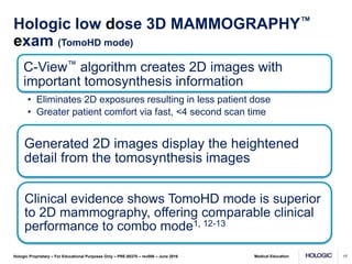
Medical Education
Hologic Proprietary – For Educational Purposes Only – PRE-00370 – rev006 – June 2016
style
Breast Tomosynthesis and Hologic
Genius™ 3D MAMMOGRAPHY™ exams
1
Medical Education
Hologic Proprietary – For Educational Purposes Only – PRE-00370 – rev006 – June 2016
style
Terminology
Selenia Dimensions system
Hologic low dose 3D MAMMOGRAPHY™ exam
• Hologic’s unique low dose 2-view exam made possible
with the Hologic Selenia® Dimensions® system and the
C-View™ software option
• also known as TomoHD mode
Combo mode
• Hologic exclusive mode that takes co-registered 2D
and tomosynthesis images under one compression
2
Medical Education
Hologic Proprietary – For Educational Purposes Only – PRE-00370 – rev006 – June 2016
style
Overview
2D mammography challenges & limitations
Why perform Genius™ 3D MAMMOGRAPHY™ exams?
The technology behind Genius™ 3D MAMMOGRAPHY™
exam
3D MAMMOGRAPHY™ exam clinical performance?
Generating a 2D image with C-View™ software
Clinical examples
3
Medical Education
Hologic Proprietary – For Educational Purposes Only – PRE-00370 – rev006 – June 2016
style
Challenges of conventional mammography
Prompt annual
mammography has
shown the ability to
reduce the mortality
rate from breast
cancer in a
population by 15%
to 50% 1-3
As many as 20% of
breast cancers will
be missed by
conventional
mammography 4
In the U.S. ~10% of
women are recalled
for additional
diagnostic work-up
A significant portion
prove to have no
abnormality,
resulting in
unnecessary
anxiety and cost 5
4
1. Smith RA, Duffy SW, Gabe R et al. The randomized trials of breast cancer screening: what have we learned? Radiol Clin N Am
42 (2004) 793 – 806.
2. Hendrick RE, Smith RA, Rutledge JH, Smart CR. Benefit of screening mammography in women ages 40-49: a new meta-
analysis of randomized controlled trials. Monogr Natl Cancer Inst 1997;22:87-92.
3. Tabar L, Vitak B, Tony HH, Yen MF, Duffy SW, Smith RA. Beyond randomized controlled trials: organized mammographic
screening substantially reduces breast carcinoma mortality. Cancer 2001;91:1724-31.
4. http://www.cancer.gov/cancertopics/factsheet/detection/mammograms
5. http://radiology.rsna.org/content/244/2/381.full#T3
Medical Education
Hologic Proprietary – For Educational Purposes Only – PRE-00370 – rev006 – June 2016
style
2D FFDM imaging
Tissue superimposition hides
or mimics pathologies in 2D
5
Medical Education
Hologic Proprietary – For Educational Purposes Only – PRE-00370 – rev006 – June 2016
style
Breast Tomosynthesis Improves Visibility
by Reducing Tissue Superimposition
6
Medical Education
Hologic Proprietary – For Educational Purposes Only – PRE-00370 – rev006 – June 2016
style
3D MAMMOGRAPHY™ exam
Principle of Operation
• X-ray tube moves in an
arc across the breast
• A series of low dose
images are acquired
from different angles
• Total dose approximately
the same as one 2D
mammogram
• Projection images are
reconstructed into
1 mm slices Compression Paddle
Compressed Breast
Detector Housing
Reconstructed
Slices {
X-ray tube arc of motion
7
Medical Education
Hologic Proprietary – For Educational Purposes Only – PRE-00370 – rev006 – June 2016
style
Why combine tomosynthesis and 2D
images?
Comparison of current images with prior images is
standard mammography practice and critical to
perceive subtle changes that may be associated with a
cancer
Obtaining a 2D exam with the tomosynthesis exam will
allow direct comparison of current 2D images with prior
2D images
Segmental and clustered calcifications are more easily
and quickly appreciated with 2D because they can
traverse multiple slices in the tomosynthesis image set
By minimizing structure overlap, tomosynthesis image
slices optimally demonstrates masses and
architectural distortion
8
Medical Education
Hologic Proprietary – For Educational Purposes Only – PRE-00370 – rev006 – June 2016
style
Genius™ 3D MAMMOGRAPHY™ exams
improve clinical outcomes
Over 40% more
invasive
cancer
detection
versus 2D2-7
Earlier detection
15-40% fewer
recalls
depending on
practices2,5,8
PPV increase
of 49% for
recalls and
21% biopsies2
Reduced recalls Improved efficiencies
Study exclusively used the Hologic Selenia Dimensions system.
Results are not applicable to other breast tomosynthesis systems.
9
Medical Education
Hologic Proprietary – For Educational Purposes Only – PRE-00370 – rev006 – June 2016
style
A landmark study in JAMA – June 2014
10
454,850 examinations from 13 centers interpreted by 139
radiologists over two time periods were retrospectively analyzed2
4.2 2.9
107
5.4 4.1
91
0
20
40
60
80
100
120
Overall Cancer Detection Rate
(per 1000)
Invasive Cancer Detection Rate
(per 1000)
Recall Rate
(per 1000)
2D 3D™
29% 41%
15%
Medical Education
Hologic Proprietary – For Educational Purposes Only – PRE-00370 – rev006 – June 2016
style
A landmark study in JAMA – June 2014
11
2D 2D + 3D™ exams Relative
Change
P-Value
PPV for
Recall
4.3% 6.4% +49% P<.001
PPV for
Biopsy
24.2% 29.2% +21% P<.001
Medical Education
Hologic Proprietary – For Educational Purposes Only – PRE-00370 – rev006 – June 2016
style
Benefits of a Genius™ 3D MAMMOGRAPHY™
Exam*
Most accurate mammogram available today2-3
Superior to 2D mammography alone2-3,11
Significant increase in efficiencies5
Fewer false positives may result in fewer biopsies2,9
Significant increase in sensitivity and specificity2-3
Reduced healthcare costs14
Reduction in patient anxiety and burden15
12
*Studies exclusively used the Hologic Selenia® Dimensions® system.
Results are not applicable to other tomosynthesis mammography exams or systems.
Medical Education
Hologic Proprietary – For Educational Purposes Only – PRE-00370 – rev006 – June 2016
style
LCC
2D 3D™
Breast cancer screening with the 3D™
exam in two views finds more cancers than
in one-view 15,16
13
Medical Education
Hologic Proprietary – For Educational Purposes Only – PRE-00370 – rev006 – June 2016
style
Image Acquisition: Two methods
TomoHD mode
• Single exposure to acquire 3D™ data set
• Utilizes C-View™ software to create the 2D image
Combo mode exam
• Combination of 3D™ and 2D exposures
14
Medical Education
Hologic Proprietary – For Educational Purposes Only – PRE-00370 – rev006 – June 2016
style
A short video demonstrating a patient examination
15
Medical Education
Hologic Proprietary – For Educational Purposes Only – PRE-00370 – rev006 – June 2016 16
Medical Education
Hologic Proprietary – For Educational Purposes Only – PRE-00370 – rev006 – June 2016
style
C-View™ algorithm creates 2D images with
important tomosynthesis information
• Eliminates 2D exposures resulting in less patient dose
• Greater patient comfort via fast, <4 second scan time
Generated 2D images display the heightened
detail from the tomosynthesis images
Clinical evidence shows TomoHD mode is superior
to 2D mammography, offering comparable clinical
performance to combo mode1, 12-13
Hologic low dose 3D MAMMOGRAPHY™
exam (TomoHD mode)
17
Medical Education
Hologic Proprietary – For Educational Purposes Only – PRE-00370 – rev006 – June 2016
style
Hologic C-View™ software:
generated 2D image
• Perform a standard
tomosynthesis scan*
• 3.7 seconds
18
*Scan time is reduced from ~10 seconds (combo mode)
regardless of breast size. A short scan time lessens patient
motion risk.
Medical Education
Hologic Proprietary – For Educational Purposes Only – PRE-00370 – rev006 – June 2016
style
Generating 2D images
• Perform a standard
tomosynthesis scan
• Reconstruct
tomosynthesis slices
15 Projection Images
Tomosynthesis
Reconstruction
Algorithm
Tomosynthesis Slices
19
Medical Education
Hologic Proprietary – For Educational Purposes Only – PRE-00370 – rev006 – June 2016
style
Generating 2D Images
• Perform a standard
tomosynthesis scan
• Reconstruct tomosynthesis
slices
• Generate 2D image
• Tomo + generated 2D = a
low dose examination
• Available in any
tomosynthesis view
Generated 2D Images
C-View
Software
Algorithm
Tomosynthesis Slices
20
Medical Education
Hologic Proprietary – For Educational Purposes Only – PRE-00370 – rev006 – June 2016
style
C-View™ 2D Image Attributes
Resolution
• Contrast: More value/weight on linear structures and bright
spots (when present)
• Spatial: Reflects tomosynthesis 100µm
Normal tissue density
• Less value/weight on normal tissue; less overlap while
retaining density assessment ability
Skin line appearance
• More akin to tomosynthesis versus FFDM
Total patient radiation dose
• TomoHD mode: 1.45 mGy vs. combo mode: 2.65 mGy*
*Based on 4.5 cm compressed breast.
21
Medical Education
Hologic Proprietary – For Educational Purposes Only – PRE-00370 – rev006 – June 2016
style
Clinical Performance:
Low dose Genius™ 3D MAMMOGRAPHY™
exam vs. 2D alone1, 12-13
Superior diagnostic accuracy for
all breast types
Fewer non-cancer recalls
Recall rate
reduction
Increase in
cancer detection
22
Medical Education
Hologic Proprietary – For Educational Purposes Only – PRE-00370 – rev006 – June 2016
style
C-View™ 2D Image Uses
• Calcification detection and distribution
• Side by side symmetry
• Interval comparison
Diagnostic image replacement to FFDM
within the tomosynthesis screening exam
Navigation aid to the tomosynthesis image
review
Must always review the tomosynthesis
slices
23
Image Comparison
C-View™ image
FFDM 2D Tomosynthesis Slice
Medical Education
Hologic Proprietary – For Educational Purposes Only – PRE-00370 – rev006 – June 2016
style
Image Comparison
C-View™ image
FFDM 2D Tomosynthesis Slice
Medical Education
Hologic Proprietary – For Educational Purposes Only – PRE-00370 – rev006 – June 2016
style
Summary: Low dose Genius™ 3D
MAMMOGRAPHY™ exam1
• Lower patient dose
• Shorter compression time
• Increased patient comfort
• Lowered risk of patient motion
• Superior performance
compared to traditional 2D
alone1, 12-13
26
Medical Education
Hologic Proprietary – For Educational Purposes Only – PRE-00370 – rev006 – June 2016
style
Combo Mode examination
27
Medical Education
Hologic Proprietary – For Educational Purposes Only – PRE-00370 – rev006 – June 2016
style
Combo Mode
• A series of projection images are acquired across 15 degrees (3.7 seconds)
• Tube head returns to center; a 2D image is acquired under the same
compression, giving perfectly co-registered 2D & breast tomosynthesis
images (~6 seconds)
• The Genius™ 3D MAMMOGRAPHY™ exam is ~10 seconds total
• Total dose for both 2D and the 3D™ scan is below the MQSA/EUREF
limits
• Two exposures under a single compression
28
Medical Education
Hologic Proprietary – For Educational Purposes Only – PRE-00370 – rev006 – June 2016
style
Positioning & Compression for
Combo Mode examination
Standard
positioning &
compression
3D™ = 15 low
dose images
are acquired
from different
angles -
no grid
2D = grid is
automatically
brought back
into place &
the 2D image
is acquired
Medical Education
Hologic Proprietary – For Educational Purposes Only – PRE-00370 – rev006 – June 2016
style
Clinical image review
Genius™ 3D MAMMOGRAPHY™ exams are
revolutionizing breast imaging
30
Medical Education
Hologic Proprietary – For Educational Purposes Only – PRE-00370 – rev006 – June 2016
style
Clinical Study: Cancer Case 1
• Patient presented with area of thickening
• Several suspicious areas
• Multifocality clearly seen with tomosynthesis
• Pathology: Invasive ductal carcinoma and
infiltrating lobular carcinoma
31
RMLO
2D
Cancer Case 1
2D
Cancer Case 1
Tomosynthesis
slice
2D
Cancer Case 1
Medical Education
Hologic Proprietary – For Educational Purposes Only – PRE-00370 – rev006 – June 2016
style
Clinical Study: Cancer Case 2
• Routine screening with combo mode
• 2D mammogram essentially negative
• Tomosynthesis RMLO reveals an area of
architectural distortion in the superior aspect
• 2D diagnostic views added no useful information
and did not reveal the lesion
• Pathology: Invasive ductal carcinoma
35
2D Tomosynthesis
Slice 16
Cancer Case 2
2D Tomosynthesis
Slice 28
Cancer Case 2
Breast Tomosynthesis
Slice 16
Breast Tomosynthesis
Slice 28
RCC RML
O
Cancer Case 2
Diagnostic Work-up
Images 12/09/11
Diagnostic Work-up Views
Cancer Case 2
Medical Education
Hologic Proprietary – For Educational Purposes Only – PRE-00370 – rev006 – June 2016
style
Clinical Study: Cancer Case 3
• Routine screening mammogram
• 2D mammogram shows calcifications in the Left
upper inner quadrant (UIQ) in both projections -
one view only finding
• Tomo slices show calcifications well along with a
subtle spiculated mass in the Left CC
• No definitive correlate on MLO
• Pathology: Invasive Ductal Carcinoma
40
2D Breast Tomosynthesis
Slice 37
Screening Combo
06/20/2012
Cancer Case 3
2D Breast Tomosynthesis
Slice 27
Screening Combo
06/20/2012
Cancer Case 3
LCC LCC
Tomosynthesis Reconstructed Slices
Cancer Case 3
2D Tomosynthesis
Slice 27
LCC LCC
Cancer Case 3
Medical Education
Hologic Proprietary – For Educational Purposes Only – PRE-00370 – rev006 – June 2016
style
Clinical Study:
Superimposition of Tissues – Case 1
• Combo mode exam completed
• 2D right CC displays density in the subareolar
region of the breast
• Tomosynthesis displays overlapping tissue
45
RCC
2D
SIP Case 1
RCC
Tomosynthesis
slice
Medical Education
Hologic Proprietary – For Educational Purposes Only – PRE-00370 – rev006 – June 2016
style
Slice 14 18 22 26 30
SIP Case 1
47
Medical Education
Hologic Proprietary – For Educational Purposes Only – PRE-00370 – rev006 – June 2016
style
The Future of Breast Imaging
Hologic, Dimensions, Selenia, and other associated logos are trademarks and/or registered trademarks of Hologic, Inc. and/or its subsidiaries in the United States and /or other countries.
48
Medical Education
Hologic Proprietary – For Educational Purposes Only – PRE-00370 – rev006 – June 2016
style
Additional publication information
1. FDA PMA submission P080003/S001.
2. Friedewald SM, Rafferty EA, Rose SL, Durand MA, Plecha DM, Greenberg JS, Hayes MK, Copit DS, Carlson KL, Cink TM, Barke LD, Greer LN, Miller DP, Conant EF.
Breast cancer screening using tomosynthesis in combination with digital mammography. JAMA. 2014 Jun 25;311(24):2499-507. doi: 10.1001/jama.2014.6095.
3. Skaane P, Bandos AI, Gullien R, Eben EB, Ekseth U, Haakenaasen U, Izadi M, Jebsen IN, Jahr G, Krager M, Niklason LT, Hofvind S, Gur D. Comparison of digital
mammography alone and digital mammography plus tomosynthesis in a population-based screening program. Radiology. 2013 Apr;267(1):47-56.
doi:10.1148/radiol.12121373. Epub 2013 Jan 7.
4. Ciatto S, Houssami N, Bernardi D, Caumo F, Pellegrini M, Brunelli S, Tuttobene P, Bricolo P, Fantò C, Valentini M, Montemezzi S, Macaskill P. Integration of 3D digital
mammography with tomosynthesis for population breast-cancer screening (STORM): a prospective comparison study. Lancet Oncol. 2013 Jun;14(7):583-9.
doi:10.1016/S1470-2045(13)70134-7. Epub 2013 Apr 25.
5. Rose SL, Tidwell AL, Bujnoch LJ, Kushwaha AC, Nordmann AS, Sexton R Jr. Implementation of breast tomosynthesis in a routine screening practice: an observational
study. AJR Am J Roentgenol. 2013 Jun;200(6):1401-8. doi:10.2214/AJR.12.9672.
6. Conant E, Mitra N, McCarthy A, et al. “Implementing Digital Breast Tomosynthesis (DBT) in a Screening Population: PPV1 as a Measure of Outcome” (paper presented
at the annual meeting of the Radiological Society of North America, Chicago, Il, December 2013).
7. Greenberg J, Javitt M, Katzen J, et al. “Clinical Performance Metrics of 3D Digital Breast Tomosynthesis Compared With 2D Digital Mammography for Breast Cancer
Screening in Community Practice.” AJR Am J Roentgenol. 2014 Sept; 203:687-693. Epub 2014 Jun 11.
8. Haas B, Kalra V, Geisel J et al. “Comparison of Tomosynthesis Plus Digital Mammography and Digital Mammography Alone for Breast Cancer Screening” Radiology.
2013 Dec;269(3):694-700. Epub 2013 Jul 30.
9. Zuley ML, Bandos AI, Ganott MA, Sumkin JH, Kelly AE, Catullo VJ, Rathfon GY, Lu AH, Gur D. Digital breast tomosynthesis versus supplemental diagnostic
mammographic views for evaluation of noncalcified breast lesions. Radiology. 2013 Jan;266(1):89-95. doi: 10.1148/radiol.12120552. Epub 2012 Nov 9.
10. Dang P, Humphrey K, Freer P, Halpern E, Saksena M, Rafferty E. “Comparison of Lesion Detection and Characterization in Invasive Cancers Using Breast
Tomosynthesis versus Conventional Mammography” (paper presented at the annual meeting of the Radiological Society of North America, Chicago, Il, December 2013).
11. Rafferty EA, Park JM, Philpotts LE, Poplack SP, Sumkin JH, Halpern EF, Niklason LT. Assessing radiologist performance using combined digital mammography and
breast tomosynthesis compared with digital mammography alone: results of a multicenter, multireader trial. Radiology. 2013 Jan;266(1):104-13. doi:
10.1148/radiol.12120674. Epub 2012 Nov 20
12. Skaane P, Bandos AI, Eben EB, Jebsen IN, Krager M, Haakenaasen U, Ekseth U, Izadi M, Hofvind S, Gullien R. Two-View Digital Breast Tomosynthesis Screening with
Synthetically Reconstructed Projection Images: Comparison with Digital Breast Tomosynthesis with Full-Field Digital Mammographic Images. Radiology. 2014 Jan
24:131391.
13. Zuley ML, Guo B, Catullo VJ, Chough DM, Kelly AE, Lu AH, Rathfon GY, Lee Spangler M, Sumkin JH, Wallace LP, Bandos AI. Comparison of Two-dimensional
Synthesized Mammograms versus Original Digital Mammograms Alone and in Combination with Tomosynthesis Images. Radiology. 2014 Jan 21:131530.
14. Kalra V, Haas B, Forman H et al. “Cost-Effectiveness of Digital Breast Tomosynthesis.” (paper presented at the annual meeting of the Radiological Society of North
America, Chicago, Il, November 2012).
15. Rafferty EA, Park JM, Philpotts LE, Poplack SP, Sumkin JH, Halpern EF, Niklason LT. Diagnostic accuracy and recall rates for digital mammography and digital
mammography combined with one-view and two-view tomosynthesis: results of an enriched reader study. AJR Am J Roentgenol. 2014 Feb;202(2):273-81. doi:
10.2214/AJR.13.11240.
16. Beck, N, Butler, R.; Durand, M.; Andrejeva, L.; Hooley, R.; Horvath, L.; Raghu, M.; Philpotts, L. “One-View Versus Two-View Tomosynthesis: A Comparison of Breast
Cancer Visibility in the Mediolateral Oblique and Craniocaudal Views.” Paper presented at the annual meeting for the American Roentgen Ray Society, Washington, D.C.
April 2013.
17. Brodersen J, Siersma V. “Long-Term Psychosocial Consequences of False-Positive Screening Mammography.” The Annals of Family Medicine 2013 Mar;11(2):106-15.
49

Breast-Tomosynthesis-for-MDs-PPT.pptx

  • 1.
    Medical Education Hologic Proprietary– For Educational Purposes Only – PRE-00370 – rev006 – June 2016 style Breast Tomosynthesis and Hologic Genius™ 3D MAMMOGRAPHY™ exams 1
  • 2.
    Medical Education Hologic Proprietary– For Educational Purposes Only – PRE-00370 – rev006 – June 2016 style Terminology Selenia Dimensions system Hologic low dose 3D MAMMOGRAPHY™ exam • Hologic’s unique low dose 2-view exam made possible with the Hologic Selenia® Dimensions® system and the C-View™ software option • also known as TomoHD mode Combo mode • Hologic exclusive mode that takes co-registered 2D and tomosynthesis images under one compression 2
  • 3.
    Medical Education Hologic Proprietary– For Educational Purposes Only – PRE-00370 – rev006 – June 2016 style Overview 2D mammography challenges & limitations Why perform Genius™ 3D MAMMOGRAPHY™ exams? The technology behind Genius™ 3D MAMMOGRAPHY™ exam 3D MAMMOGRAPHY™ exam clinical performance? Generating a 2D image with C-View™ software Clinical examples 3
  • 4.
    Medical Education Hologic Proprietary– For Educational Purposes Only – PRE-00370 – rev006 – June 2016 style Challenges of conventional mammography Prompt annual mammography has shown the ability to reduce the mortality rate from breast cancer in a population by 15% to 50% 1-3 As many as 20% of breast cancers will be missed by conventional mammography 4 In the U.S. ~10% of women are recalled for additional diagnostic work-up A significant portion prove to have no abnormality, resulting in unnecessary anxiety and cost 5 4 1. Smith RA, Duffy SW, Gabe R et al. The randomized trials of breast cancer screening: what have we learned? Radiol Clin N Am 42 (2004) 793 – 806. 2. Hendrick RE, Smith RA, Rutledge JH, Smart CR. Benefit of screening mammography in women ages 40-49: a new meta- analysis of randomized controlled trials. Monogr Natl Cancer Inst 1997;22:87-92. 3. Tabar L, Vitak B, Tony HH, Yen MF, Duffy SW, Smith RA. Beyond randomized controlled trials: organized mammographic screening substantially reduces breast carcinoma mortality. Cancer 2001;91:1724-31. 4. http://www.cancer.gov/cancertopics/factsheet/detection/mammograms 5. http://radiology.rsna.org/content/244/2/381.full#T3
  • 5.
    Medical Education Hologic Proprietary– For Educational Purposes Only – PRE-00370 – rev006 – June 2016 style 2D FFDM imaging Tissue superimposition hides or mimics pathologies in 2D 5
  • 6.
    Medical Education Hologic Proprietary– For Educational Purposes Only – PRE-00370 – rev006 – June 2016 style Breast Tomosynthesis Improves Visibility by Reducing Tissue Superimposition 6
  • 7.
    Medical Education Hologic Proprietary– For Educational Purposes Only – PRE-00370 – rev006 – June 2016 style 3D MAMMOGRAPHY™ exam Principle of Operation • X-ray tube moves in an arc across the breast • A series of low dose images are acquired from different angles • Total dose approximately the same as one 2D mammogram • Projection images are reconstructed into 1 mm slices Compression Paddle Compressed Breast Detector Housing Reconstructed Slices { X-ray tube arc of motion 7
  • 8.
    Medical Education Hologic Proprietary– For Educational Purposes Only – PRE-00370 – rev006 – June 2016 style Why combine tomosynthesis and 2D images? Comparison of current images with prior images is standard mammography practice and critical to perceive subtle changes that may be associated with a cancer Obtaining a 2D exam with the tomosynthesis exam will allow direct comparison of current 2D images with prior 2D images Segmental and clustered calcifications are more easily and quickly appreciated with 2D because they can traverse multiple slices in the tomosynthesis image set By minimizing structure overlap, tomosynthesis image slices optimally demonstrates masses and architectural distortion 8
  • 9.
    Medical Education Hologic Proprietary– For Educational Purposes Only – PRE-00370 – rev006 – June 2016 style Genius™ 3D MAMMOGRAPHY™ exams improve clinical outcomes Over 40% more invasive cancer detection versus 2D2-7 Earlier detection 15-40% fewer recalls depending on practices2,5,8 PPV increase of 49% for recalls and 21% biopsies2 Reduced recalls Improved efficiencies Study exclusively used the Hologic Selenia Dimensions system. Results are not applicable to other breast tomosynthesis systems. 9
  • 10.
    Medical Education Hologic Proprietary– For Educational Purposes Only – PRE-00370 – rev006 – June 2016 style A landmark study in JAMA – June 2014 10 454,850 examinations from 13 centers interpreted by 139 radiologists over two time periods were retrospectively analyzed2 4.2 2.9 107 5.4 4.1 91 0 20 40 60 80 100 120 Overall Cancer Detection Rate (per 1000) Invasive Cancer Detection Rate (per 1000) Recall Rate (per 1000) 2D 3D™ 29% 41% 15%
  • 11.
    Medical Education Hologic Proprietary– For Educational Purposes Only – PRE-00370 – rev006 – June 2016 style A landmark study in JAMA – June 2014 11 2D 2D + 3D™ exams Relative Change P-Value PPV for Recall 4.3% 6.4% +49% P<.001 PPV for Biopsy 24.2% 29.2% +21% P<.001
  • 12.
    Medical Education Hologic Proprietary– For Educational Purposes Only – PRE-00370 – rev006 – June 2016 style Benefits of a Genius™ 3D MAMMOGRAPHY™ Exam* Most accurate mammogram available today2-3 Superior to 2D mammography alone2-3,11 Significant increase in efficiencies5 Fewer false positives may result in fewer biopsies2,9 Significant increase in sensitivity and specificity2-3 Reduced healthcare costs14 Reduction in patient anxiety and burden15 12 *Studies exclusively used the Hologic Selenia® Dimensions® system. Results are not applicable to other tomosynthesis mammography exams or systems.
  • 13.
    Medical Education Hologic Proprietary– For Educational Purposes Only – PRE-00370 – rev006 – June 2016 style LCC 2D 3D™ Breast cancer screening with the 3D™ exam in two views finds more cancers than in one-view 15,16 13
  • 14.
    Medical Education Hologic Proprietary– For Educational Purposes Only – PRE-00370 – rev006 – June 2016 style Image Acquisition: Two methods TomoHD mode • Single exposure to acquire 3D™ data set • Utilizes C-View™ software to create the 2D image Combo mode exam • Combination of 3D™ and 2D exposures 14
  • 15.
    Medical Education Hologic Proprietary– For Educational Purposes Only – PRE-00370 – rev006 – June 2016 style A short video demonstrating a patient examination 15
  • 16.
    Medical Education Hologic Proprietary– For Educational Purposes Only – PRE-00370 – rev006 – June 2016 16
  • 17.
    Medical Education Hologic Proprietary– For Educational Purposes Only – PRE-00370 – rev006 – June 2016 style C-View™ algorithm creates 2D images with important tomosynthesis information • Eliminates 2D exposures resulting in less patient dose • Greater patient comfort via fast, <4 second scan time Generated 2D images display the heightened detail from the tomosynthesis images Clinical evidence shows TomoHD mode is superior to 2D mammography, offering comparable clinical performance to combo mode1, 12-13 Hologic low dose 3D MAMMOGRAPHY™ exam (TomoHD mode) 17
  • 18.
    Medical Education Hologic Proprietary– For Educational Purposes Only – PRE-00370 – rev006 – June 2016 style Hologic C-View™ software: generated 2D image • Perform a standard tomosynthesis scan* • 3.7 seconds 18 *Scan time is reduced from ~10 seconds (combo mode) regardless of breast size. A short scan time lessens patient motion risk.
  • 19.
    Medical Education Hologic Proprietary– For Educational Purposes Only – PRE-00370 – rev006 – June 2016 style Generating 2D images • Perform a standard tomosynthesis scan • Reconstruct tomosynthesis slices 15 Projection Images Tomosynthesis Reconstruction Algorithm Tomosynthesis Slices 19
  • 20.
    Medical Education Hologic Proprietary– For Educational Purposes Only – PRE-00370 – rev006 – June 2016 style Generating 2D Images • Perform a standard tomosynthesis scan • Reconstruct tomosynthesis slices • Generate 2D image • Tomo + generated 2D = a low dose examination • Available in any tomosynthesis view Generated 2D Images C-View Software Algorithm Tomosynthesis Slices 20
  • 21.
    Medical Education Hologic Proprietary– For Educational Purposes Only – PRE-00370 – rev006 – June 2016 style C-View™ 2D Image Attributes Resolution • Contrast: More value/weight on linear structures and bright spots (when present) • Spatial: Reflects tomosynthesis 100µm Normal tissue density • Less value/weight on normal tissue; less overlap while retaining density assessment ability Skin line appearance • More akin to tomosynthesis versus FFDM Total patient radiation dose • TomoHD mode: 1.45 mGy vs. combo mode: 2.65 mGy* *Based on 4.5 cm compressed breast. 21
  • 22.
    Medical Education Hologic Proprietary– For Educational Purposes Only – PRE-00370 – rev006 – June 2016 style Clinical Performance: Low dose Genius™ 3D MAMMOGRAPHY™ exam vs. 2D alone1, 12-13 Superior diagnostic accuracy for all breast types Fewer non-cancer recalls Recall rate reduction Increase in cancer detection 22
  • 23.
    Medical Education Hologic Proprietary– For Educational Purposes Only – PRE-00370 – rev006 – June 2016 style C-View™ 2D Image Uses • Calcification detection and distribution • Side by side symmetry • Interval comparison Diagnostic image replacement to FFDM within the tomosynthesis screening exam Navigation aid to the tomosynthesis image review Must always review the tomosynthesis slices 23
  • 24.
  • 25.
    Medical Education Hologic Proprietary– For Educational Purposes Only – PRE-00370 – rev006 – June 2016 style Image Comparison C-View™ image FFDM 2D Tomosynthesis Slice
  • 26.
    Medical Education Hologic Proprietary– For Educational Purposes Only – PRE-00370 – rev006 – June 2016 style Summary: Low dose Genius™ 3D MAMMOGRAPHY™ exam1 • Lower patient dose • Shorter compression time • Increased patient comfort • Lowered risk of patient motion • Superior performance compared to traditional 2D alone1, 12-13 26
  • 27.
    Medical Education Hologic Proprietary– For Educational Purposes Only – PRE-00370 – rev006 – June 2016 style Combo Mode examination 27
  • 28.
    Medical Education Hologic Proprietary– For Educational Purposes Only – PRE-00370 – rev006 – June 2016 style Combo Mode • A series of projection images are acquired across 15 degrees (3.7 seconds) • Tube head returns to center; a 2D image is acquired under the same compression, giving perfectly co-registered 2D & breast tomosynthesis images (~6 seconds) • The Genius™ 3D MAMMOGRAPHY™ exam is ~10 seconds total • Total dose for both 2D and the 3D™ scan is below the MQSA/EUREF limits • Two exposures under a single compression 28
  • 29.
    Medical Education Hologic Proprietary– For Educational Purposes Only – PRE-00370 – rev006 – June 2016 style Positioning & Compression for Combo Mode examination Standard positioning & compression 3D™ = 15 low dose images are acquired from different angles - no grid 2D = grid is automatically brought back into place & the 2D image is acquired
  • 30.
    Medical Education Hologic Proprietary– For Educational Purposes Only – PRE-00370 – rev006 – June 2016 style Clinical image review Genius™ 3D MAMMOGRAPHY™ exams are revolutionizing breast imaging 30
  • 31.
    Medical Education Hologic Proprietary– For Educational Purposes Only – PRE-00370 – rev006 – June 2016 style Clinical Study: Cancer Case 1 • Patient presented with area of thickening • Several suspicious areas • Multifocality clearly seen with tomosynthesis • Pathology: Invasive ductal carcinoma and infiltrating lobular carcinoma 31
  • 32.
  • 33.
  • 34.
  • 35.
    Medical Education Hologic Proprietary– For Educational Purposes Only – PRE-00370 – rev006 – June 2016 style Clinical Study: Cancer Case 2 • Routine screening with combo mode • 2D mammogram essentially negative • Tomosynthesis RMLO reveals an area of architectural distortion in the superior aspect • 2D diagnostic views added no useful information and did not reveal the lesion • Pathology: Invasive ductal carcinoma 35
  • 36.
  • 37.
  • 38.
    Breast Tomosynthesis Slice 16 BreastTomosynthesis Slice 28 RCC RML O Cancer Case 2
  • 39.
  • 40.
    Medical Education Hologic Proprietary– For Educational Purposes Only – PRE-00370 – rev006 – June 2016 style Clinical Study: Cancer Case 3 • Routine screening mammogram • 2D mammogram shows calcifications in the Left upper inner quadrant (UIQ) in both projections - one view only finding • Tomo slices show calcifications well along with a subtle spiculated mass in the Left CC • No definitive correlate on MLO • Pathology: Invasive Ductal Carcinoma 40
  • 41.
    2D Breast Tomosynthesis Slice37 Screening Combo 06/20/2012 Cancer Case 3
  • 42.
    2D Breast Tomosynthesis Slice27 Screening Combo 06/20/2012 Cancer Case 3
  • 43.
  • 44.
  • 45.
    Medical Education Hologic Proprietary– For Educational Purposes Only – PRE-00370 – rev006 – June 2016 style Clinical Study: Superimposition of Tissues – Case 1 • Combo mode exam completed • 2D right CC displays density in the subareolar region of the breast • Tomosynthesis displays overlapping tissue 45
  • 46.
  • 47.
    Medical Education Hologic Proprietary– For Educational Purposes Only – PRE-00370 – rev006 – June 2016 style Slice 14 18 22 26 30 SIP Case 1 47
  • 48.
    Medical Education Hologic Proprietary– For Educational Purposes Only – PRE-00370 – rev006 – June 2016 style The Future of Breast Imaging Hologic, Dimensions, Selenia, and other associated logos are trademarks and/or registered trademarks of Hologic, Inc. and/or its subsidiaries in the United States and /or other countries. 48
  • 49.
    Medical Education Hologic Proprietary– For Educational Purposes Only – PRE-00370 – rev006 – June 2016 style Additional publication information 1. FDA PMA submission P080003/S001. 2. Friedewald SM, Rafferty EA, Rose SL, Durand MA, Plecha DM, Greenberg JS, Hayes MK, Copit DS, Carlson KL, Cink TM, Barke LD, Greer LN, Miller DP, Conant EF. Breast cancer screening using tomosynthesis in combination with digital mammography. JAMA. 2014 Jun 25;311(24):2499-507. doi: 10.1001/jama.2014.6095. 3. Skaane P, Bandos AI, Gullien R, Eben EB, Ekseth U, Haakenaasen U, Izadi M, Jebsen IN, Jahr G, Krager M, Niklason LT, Hofvind S, Gur D. Comparison of digital mammography alone and digital mammography plus tomosynthesis in a population-based screening program. Radiology. 2013 Apr;267(1):47-56. doi:10.1148/radiol.12121373. Epub 2013 Jan 7. 4. Ciatto S, Houssami N, Bernardi D, Caumo F, Pellegrini M, Brunelli S, Tuttobene P, Bricolo P, Fantò C, Valentini M, Montemezzi S, Macaskill P. Integration of 3D digital mammography with tomosynthesis for population breast-cancer screening (STORM): a prospective comparison study. Lancet Oncol. 2013 Jun;14(7):583-9. doi:10.1016/S1470-2045(13)70134-7. Epub 2013 Apr 25. 5. Rose SL, Tidwell AL, Bujnoch LJ, Kushwaha AC, Nordmann AS, Sexton R Jr. Implementation of breast tomosynthesis in a routine screening practice: an observational study. AJR Am J Roentgenol. 2013 Jun;200(6):1401-8. doi:10.2214/AJR.12.9672. 6. Conant E, Mitra N, McCarthy A, et al. “Implementing Digital Breast Tomosynthesis (DBT) in a Screening Population: PPV1 as a Measure of Outcome” (paper presented at the annual meeting of the Radiological Society of North America, Chicago, Il, December 2013). 7. Greenberg J, Javitt M, Katzen J, et al. “Clinical Performance Metrics of 3D Digital Breast Tomosynthesis Compared With 2D Digital Mammography for Breast Cancer Screening in Community Practice.” AJR Am J Roentgenol. 2014 Sept; 203:687-693. Epub 2014 Jun 11. 8. Haas B, Kalra V, Geisel J et al. “Comparison of Tomosynthesis Plus Digital Mammography and Digital Mammography Alone for Breast Cancer Screening” Radiology. 2013 Dec;269(3):694-700. Epub 2013 Jul 30. 9. Zuley ML, Bandos AI, Ganott MA, Sumkin JH, Kelly AE, Catullo VJ, Rathfon GY, Lu AH, Gur D. Digital breast tomosynthesis versus supplemental diagnostic mammographic views for evaluation of noncalcified breast lesions. Radiology. 2013 Jan;266(1):89-95. doi: 10.1148/radiol.12120552. Epub 2012 Nov 9. 10. Dang P, Humphrey K, Freer P, Halpern E, Saksena M, Rafferty E. “Comparison of Lesion Detection and Characterization in Invasive Cancers Using Breast Tomosynthesis versus Conventional Mammography” (paper presented at the annual meeting of the Radiological Society of North America, Chicago, Il, December 2013). 11. Rafferty EA, Park JM, Philpotts LE, Poplack SP, Sumkin JH, Halpern EF, Niklason LT. Assessing radiologist performance using combined digital mammography and breast tomosynthesis compared with digital mammography alone: results of a multicenter, multireader trial. Radiology. 2013 Jan;266(1):104-13. doi: 10.1148/radiol.12120674. Epub 2012 Nov 20 12. Skaane P, Bandos AI, Eben EB, Jebsen IN, Krager M, Haakenaasen U, Ekseth U, Izadi M, Hofvind S, Gullien R. Two-View Digital Breast Tomosynthesis Screening with Synthetically Reconstructed Projection Images: Comparison with Digital Breast Tomosynthesis with Full-Field Digital Mammographic Images. Radiology. 2014 Jan 24:131391. 13. Zuley ML, Guo B, Catullo VJ, Chough DM, Kelly AE, Lu AH, Rathfon GY, Lee Spangler M, Sumkin JH, Wallace LP, Bandos AI. Comparison of Two-dimensional Synthesized Mammograms versus Original Digital Mammograms Alone and in Combination with Tomosynthesis Images. Radiology. 2014 Jan 21:131530. 14. Kalra V, Haas B, Forman H et al. “Cost-Effectiveness of Digital Breast Tomosynthesis.” (paper presented at the annual meeting of the Radiological Society of North America, Chicago, Il, November 2012). 15. Rafferty EA, Park JM, Philpotts LE, Poplack SP, Sumkin JH, Halpern EF, Niklason LT. Diagnostic accuracy and recall rates for digital mammography and digital mammography combined with one-view and two-view tomosynthesis: results of an enriched reader study. AJR Am J Roentgenol. 2014 Feb;202(2):273-81. doi: 10.2214/AJR.13.11240. 16. Beck, N, Butler, R.; Durand, M.; Andrejeva, L.; Hooley, R.; Horvath, L.; Raghu, M.; Philpotts, L. “One-View Versus Two-View Tomosynthesis: A Comparison of Breast Cancer Visibility in the Mediolateral Oblique and Craniocaudal Views.” Paper presented at the annual meeting for the American Roentgen Ray Society, Washington, D.C. April 2013. 17. Brodersen J, Siersma V. “Long-Term Psychosocial Consequences of False-Positive Screening Mammography.” The Annals of Family Medicine 2013 Mar;11(2):106-15. 49