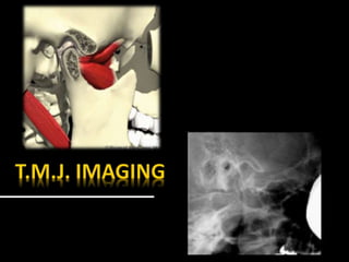
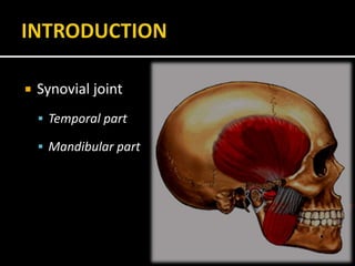
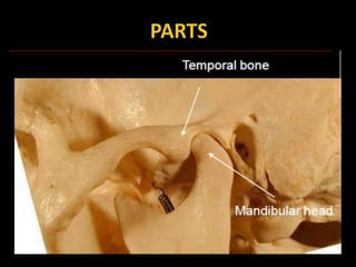
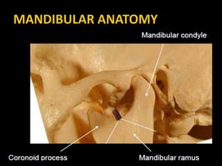
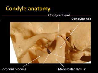
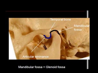
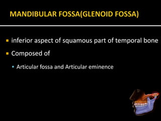
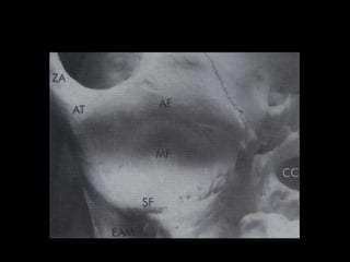
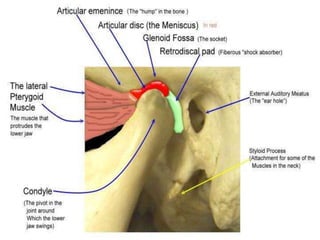
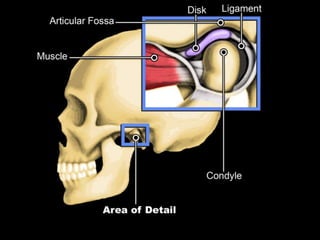
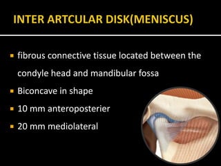
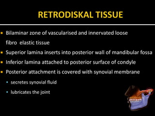
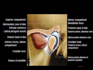
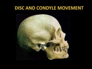
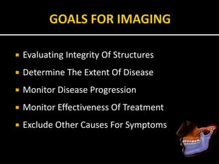
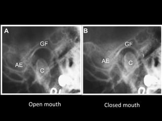
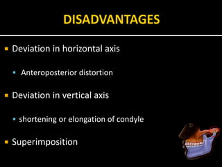
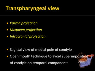
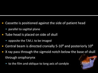
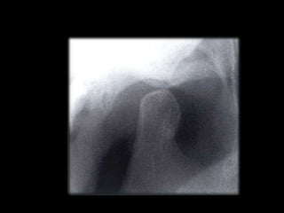
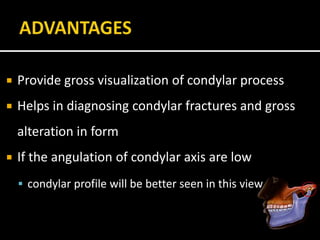
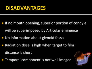
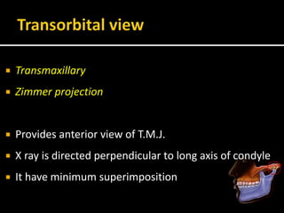
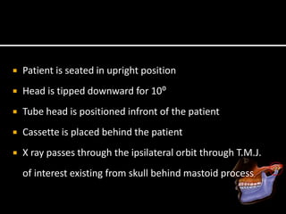
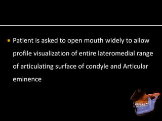
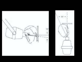
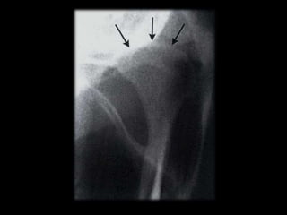
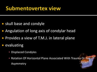
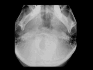
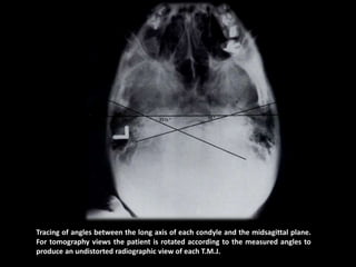
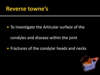
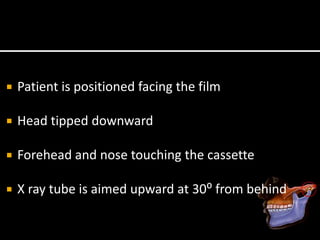
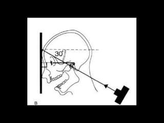
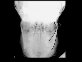
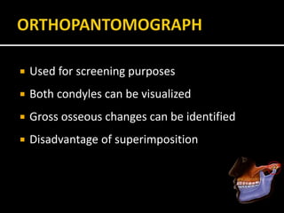
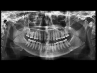
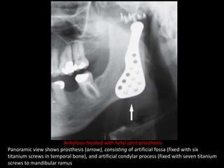
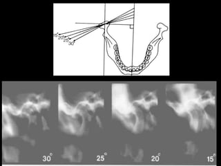
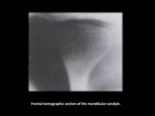
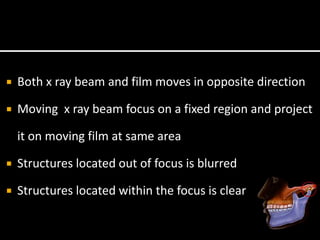
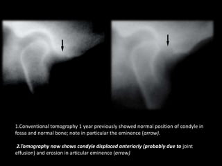
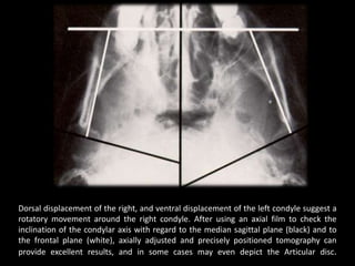
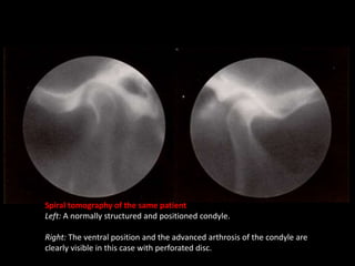
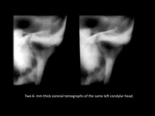
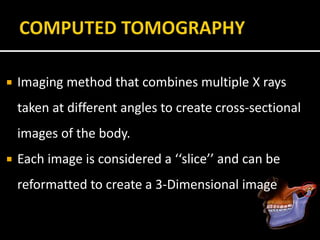
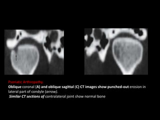
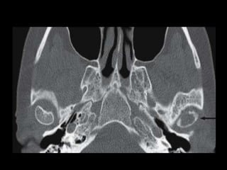
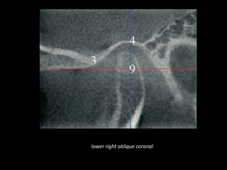
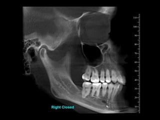
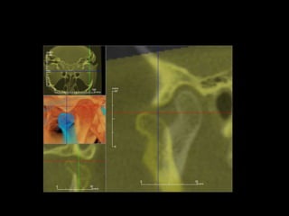
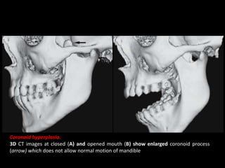
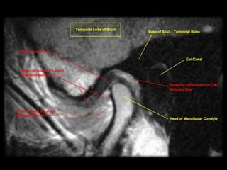
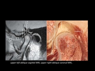
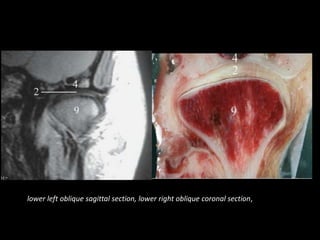
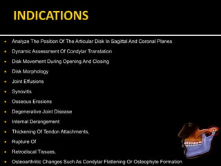
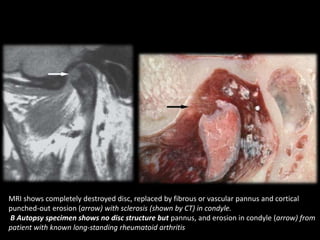
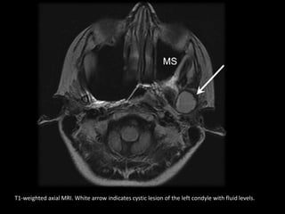
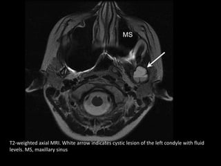
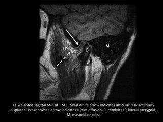
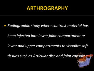
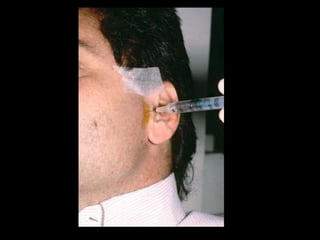
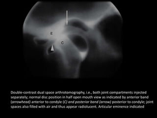
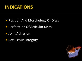
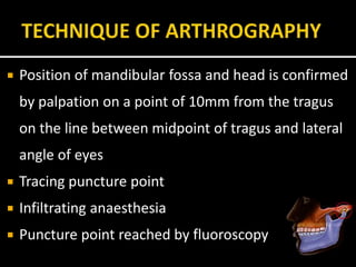
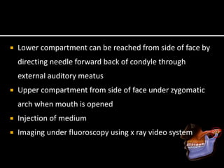
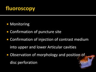
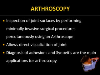
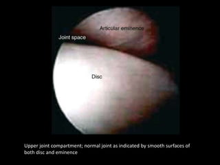
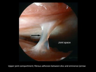
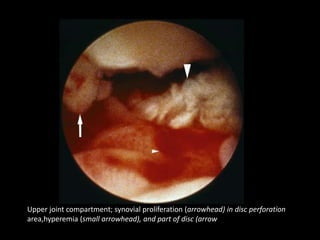
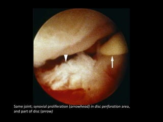
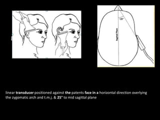
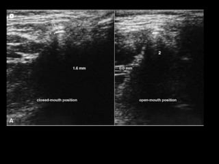
1. The document describes the anatomy and imaging techniques of the temporomandibular joint (TMJ).
2. It details the components of the TMJ including the articular disc, condyle, and fossa.
3. Various radiographic and advanced imaging modalities for evaluating the TMJ are discussed such as panoramic radiography, tomography, CT, MRI, and arthrography.
4. Each imaging technique has advantages and limitations for assessing abnormalities, injuries, and diseases affecting the TMJ structures.
This is a presentation describing in brief regarding the physics behind MRI and it's application from dental point of view. It contains few videos as well.
This presentation covers routinely used intraoral & extraoral plain radiographs used in assessment of maxillofacial trauma patients with extended coverage on occlusal radiographs. This PPT is echanced with addition of images for all radiographs
This presentation will give you a detailed knowledge about the various techniques that can be performed for imaging various aspects and diseases of TM Joint.
Temporomandibular joint imaging 2 /certified fixed orthodontic courses by Ind...Indian dental academy
The Indian Dental Academy is the Leader in continuing dental education , training dentists in all aspects of dentistry and offering a wide range of dental certified courses in different formats.
Indian dental academy provides dental crown & Bridge,rotary endodontics,fixed orthodontics,
Dental implants courses.for details pls visit www.indiandentalacademy.com ,or call
0091-9248678078
Report Back from SGO 2024: What’s the Latest in Cervical Cancer?bkling
Are you curious about what’s new in cervical cancer research or unsure what the findings mean? Join Dr. Emily Ko, a gynecologic oncologist at Penn Medicine, to learn about the latest updates from the Society of Gynecologic Oncology (SGO) 2024 Annual Meeting on Women’s Cancer. Dr. Ko will discuss what the research presented at the conference means for you and answer your questions about the new developments.
Title: Sense of Taste
Presenter: Dr. Faiza, Assistant Professor of Physiology
Qualifications:
MBBS (Best Graduate, AIMC Lahore)
FCPS Physiology
ICMT, CHPE, DHPE (STMU)
MPH (GC University, Faisalabad)
MBA (Virtual University of Pakistan)
Learning Objectives:
Describe the structure and function of taste buds.
Describe the relationship between the taste threshold and taste index of common substances.
Explain the chemical basis and signal transduction of taste perception for each type of primary taste sensation.
Recognize different abnormalities of taste perception and their causes.
Key Topics:
Significance of Taste Sensation:
Differentiation between pleasant and harmful food
Influence on behavior
Selection of food based on metabolic needs
Receptors of Taste:
Taste buds on the tongue
Influence of sense of smell, texture of food, and pain stimulation (e.g., by pepper)
Primary and Secondary Taste Sensations:
Primary taste sensations: Sweet, Sour, Salty, Bitter, Umami
Chemical basis and signal transduction mechanisms for each taste
Taste Threshold and Index:
Taste threshold values for Sweet (sucrose), Salty (NaCl), Sour (HCl), and Bitter (Quinine)
Taste index relationship: Inversely proportional to taste threshold
Taste Blindness:
Inability to taste certain substances, particularly thiourea compounds
Example: Phenylthiocarbamide
Structure and Function of Taste Buds:
Composition: Epithelial cells, Sustentacular/Supporting cells, Taste cells, Basal cells
Features: Taste pores, Taste hairs/microvilli, and Taste nerve fibers
Location of Taste Buds:
Found in papillae of the tongue (Fungiform, Circumvallate, Foliate)
Also present on the palate, tonsillar pillars, epiglottis, and proximal esophagus
Mechanism of Taste Stimulation:
Interaction of taste substances with receptors on microvilli
Signal transduction pathways for Umami, Sweet, Bitter, Sour, and Salty tastes
Taste Sensitivity and Adaptation:
Decrease in sensitivity with age
Rapid adaptation of taste sensation
Role of Saliva in Taste:
Dissolution of tastants to reach receptors
Washing away the stimulus
Taste Preferences and Aversions:
Mechanisms behind taste preference and aversion
Influence of receptors and neural pathways
Impact of Sensory Nerve Damage:
Degeneration of taste buds if the sensory nerve fiber is cut
Abnormalities of Taste Detection:
Conditions: Ageusia, Hypogeusia, Dysgeusia (parageusia)
Causes: Nerve damage, neurological disorders, infections, poor oral hygiene, adverse drug effects, deficiencies, aging, tobacco use, altered neurotransmitter levels
Neurotransmitters and Taste Threshold:
Effects of serotonin (5-HT) and norepinephrine (NE) on taste sensitivity
Supertasters:
25% of the population with heightened sensitivity to taste, especially bitterness
Increased number of fungiform papillae
TEST BANK for Operations Management, 14th Edition by William J. Stevenson, Ve...kevinkariuki227
TEST BANK for Operations Management, 14th Edition by William J. Stevenson, Verified Chapters 1 - 19, Complete Newest Version.pdf
Ethanol (CH3CH2OH), or beverage alcohol, is a two-carbon alcohol
that is rapidly distributed in the body and brain. Ethanol alters many
neurochemical systems and has rewarding and addictive properties. It
is the oldest recreational drug and likely contributes to more morbidity,
mortality, and public health costs than all illicit drugs combined. The
5th edition of the Diagnostic and Statistical Manual of Mental Disorders
(DSM-5) integrates alcohol abuse and alcohol dependence into a single
disorder called alcohol use disorder (AUD), with mild, moderate,
and severe subclassifications (American Psychiatric Association, 2013).
In the DSM-5, all types of substance abuse and dependence have been
combined into a single substance use disorder (SUD) on a continuum
from mild to severe. A diagnosis of AUD requires that at least two of
the 11 DSM-5 behaviors be present within a 12-month period (mild
AUD: 2–3 criteria; moderate AUD: 4–5 criteria; severe AUD: 6–11 criteria).
The four main behavioral effects of AUD are impaired control over
drinking, negative social consequences, risky use, and altered physiological
effects (tolerance, withdrawal). This chapter presents an overview
of the prevalence and harmful consequences of AUD in the U.S.,
the systemic nature of the disease, neurocircuitry and stages of AUD,
comorbidities, fetal alcohol spectrum disorders, genetic risk factors, and
pharmacotherapies for AUD.
Flu Vaccine Alert in Bangalore Karnatakaaddon Scans
As flu season approaches, health officials in Bangalore, Karnataka, are urging residents to get their flu vaccinations. The seasonal flu, while common, can lead to severe health complications, particularly for vulnerable populations such as young children, the elderly, and those with underlying health conditions.
Dr. Vidisha Kumari, a leading epidemiologist in Bangalore, emphasizes the importance of getting vaccinated. "The flu vaccine is our best defense against the influenza virus. It not only protects individuals but also helps prevent the spread of the virus in our communities," he says.
This year, the flu season is expected to coincide with a potential increase in other respiratory illnesses. The Karnataka Health Department has launched an awareness campaign highlighting the significance of flu vaccinations. They have set up multiple vaccination centers across Bangalore, making it convenient for residents to receive their shots.
To encourage widespread vaccination, the government is also collaborating with local schools, workplaces, and community centers to facilitate vaccination drives. Special attention is being given to ensuring that the vaccine is accessible to all, including marginalized communities who may have limited access to healthcare.
Residents are reminded that the flu vaccine is safe and effective. Common side effects are mild and may include soreness at the injection site, mild fever, or muscle aches. These side effects are generally short-lived and far less severe than the flu itself.
Healthcare providers are also stressing the importance of continuing COVID-19 precautions. Wearing masks, practicing good hand hygiene, and maintaining social distancing are still crucial, especially in crowded places.
Protect yourself and your loved ones by getting vaccinated. Together, we can help keep Bangalore healthy and safe this flu season. For more information on vaccination centers and schedules, residents can visit the Karnataka Health Department’s official website or follow their social media pages.
Stay informed, stay safe, and get your flu shot today!
HOT NEW PRODUCT! BIG SALES FAST SHIPPING NOW FROM CHINA!! EU KU DB BK substit...GL Anaacs
Contact us if you are interested:
Email / Skype : kefaya1771@gmail.com
Threema: PXHY5PDH
New BATCH Ku !!! MUCH IN DEMAND FAST SALE EVERY BATCH HAPPY GOOD EFFECT BIG BATCH !
Contact me on Threema or skype to start big business!!
Hot-sale products:
NEW HOT EUTYLONE WHITE CRYSTAL!!
5cl-adba precursor (semi finished )
5cl-adba raw materials
ADBB precursor (semi finished )
ADBB raw materials
APVP powder
5fadb/4f-adb
Jwh018 / Jwh210
Eutylone crystal
Protonitazene (hydrochloride) CAS: 119276-01-6
Flubrotizolam CAS: 57801-95-3
Metonitazene CAS: 14680-51-4
Payment terms: Western Union,MoneyGram,Bitcoin or USDT.
Deliver Time: Usually 7-15days
Shipping method: FedEx, TNT, DHL,UPS etc.Our deliveries are 100% safe, fast, reliable and discreet.
Samples will be sent for your evaluation!If you are interested in, please contact me, let's talk details.
We specializes in exporting high quality Research chemical, medical intermediate, Pharmaceutical chemicals and so on. Products are exported to USA, Canada, France, Korea, Japan,Russia, Southeast Asia and other countries.
New Directions in Targeted Therapeutic Approaches for Older Adults With Mantl...i3 Health
i3 Health is pleased to make the speaker slides from this activity available for use as a non-accredited self-study or teaching resource.
This slide deck presented by Dr. Kami Maddocks, Professor-Clinical in the Division of Hematology and
Associate Division Director for Ambulatory Operations
The Ohio State University Comprehensive Cancer Center, will provide insight into new directions in targeted therapeutic approaches for older adults with mantle cell lymphoma.
STATEMENT OF NEED
Mantle cell lymphoma (MCL) is a rare, aggressive B-cell non-Hodgkin lymphoma (NHL) accounting for 5% to 7% of all lymphomas. Its prognosis ranges from indolent disease that does not require treatment for years to very aggressive disease, which is associated with poor survival (Silkenstedt et al, 2021). Typically, MCL is diagnosed at advanced stage and in older patients who cannot tolerate intensive therapy (NCCN, 2022). Although recent advances have slightly increased remission rates, recurrence and relapse remain very common, leading to a median overall survival between 3 and 6 years (LLS, 2021). Though there are several effective options, progress is still needed towards establishing an accepted frontline approach for MCL (Castellino et al, 2022). Treatment selection and management of MCL are complicated by the heterogeneity of prognosis, advanced age and comorbidities of patients, and lack of an established standard approach for treatment, making it vital that clinicians be familiar with the latest research and advances in this area. In this activity chaired by Michael Wang, MD, Professor in the Department of Lymphoma & Myeloma at MD Anderson Cancer Center, expert faculty will discuss prognostic factors informing treatment, the promising results of recent trials in new therapeutic approaches, and the implications of treatment resistance in therapeutic selection for MCL.
Target Audience
Hematology/oncology fellows, attending faculty, and other health care professionals involved in the treatment of patients with mantle cell lymphoma (MCL).
Learning Objectives
1.) Identify clinical and biological prognostic factors that can guide treatment decision making for older adults with MCL
2.) Evaluate emerging data on targeted therapeutic approaches for treatment-naive and relapsed/refractory MCL and their applicability to older adults
3.) Assess mechanisms of resistance to targeted therapies for MCL and their implications for treatment selection
Factory Supply Best Quality Pmk Oil CAS 28578–16–7 PMK Powder in Stockrebeccabio
Factory Supply Best Quality Pmk Oil CAS 28578–16–7 PMK Powder in Stock
Telegram: bmksupplier
signal: +85264872720
threema: TUD4A6YC
You can contact me on Telegram or Threema
Communicate promptly and reply
Free of customs clearance, Double Clearance 100% pass delivery to USA, Canada, Spain, Germany, Netherland, Poland, Italy, Sweden, UK, Czech Republic, Australia, Mexico, Russia, Ukraine, Kazakhstan.Door to door service
Hot Selling Organic intermediates
Title: Sense of Smell
Presenter: Dr. Faiza, Assistant Professor of Physiology
Qualifications:
MBBS (Best Graduate, AIMC Lahore)
FCPS Physiology
ICMT, CHPE, DHPE (STMU)
MPH (GC University, Faisalabad)
MBA (Virtual University of Pakistan)
Learning Objectives:
Describe the primary categories of smells and the concept of odor blindness.
Explain the structure and location of the olfactory membrane and mucosa, including the types and roles of cells involved in olfaction.
Describe the pathway and mechanisms of olfactory signal transmission from the olfactory receptors to the brain.
Illustrate the biochemical cascade triggered by odorant binding to olfactory receptors, including the role of G-proteins and second messengers in generating an action potential.
Identify different types of olfactory disorders such as anosmia, hyposmia, hyperosmia, and dysosmia, including their potential causes.
Key Topics:
Olfactory Genes:
3% of the human genome accounts for olfactory genes.
400 genes for odorant receptors.
Olfactory Membrane:
Located in the superior part of the nasal cavity.
Medially: Folds downward along the superior septum.
Laterally: Folds over the superior turbinate and upper surface of the middle turbinate.
Total surface area: 5-10 square centimeters.
Olfactory Mucosa:
Olfactory Cells: Bipolar nerve cells derived from the CNS (100 million), with 4-25 olfactory cilia per cell.
Sustentacular Cells: Produce mucus and maintain ionic and molecular environment.
Basal Cells: Replace worn-out olfactory cells with an average lifespan of 1-2 months.
Bowman’s Gland: Secretes mucus.
Stimulation of Olfactory Cells:
Odorant dissolves in mucus and attaches to receptors on olfactory cilia.
Involves a cascade effect through G-proteins and second messengers, leading to depolarization and action potential generation in the olfactory nerve.
Quality of a Good Odorant:
Small (3-20 Carbon atoms), volatile, water-soluble, and lipid-soluble.
Facilitated by odorant-binding proteins in mucus.
Membrane Potential and Action Potential:
Resting membrane potential: -55mV.
Action potential frequency in the olfactory nerve increases with odorant strength.
Adaptation Towards the Sense of Smell:
Rapid adaptation within the first second, with further slow adaptation.
Psychological adaptation greater than receptor adaptation, involving feedback inhibition from the central nervous system.
Primary Sensations of Smell:
Camphoraceous, Musky, Floral, Pepperminty, Ethereal, Pungent, Putrid.
Odor Detection Threshold:
Examples: Hydrogen sulfide (0.0005 ppm), Methyl-mercaptan (0.002 ppm).
Some toxic substances are odorless at lethal concentrations.
Characteristics of Smell:
Odor blindness for single substances due to lack of appropriate receptor protein.
Behavioral and emotional influences of smell.
Transmission of Olfactory Signals:
From olfactory cells to glomeruli in the olfactory bulb, involving lateral inhibition.
Primitive, less old, and new olfactory systems with different path
Ozempic: Preoperative Management of Patients on GLP-1 Receptor Agonists Saeid Safari
Preoperative Management of Patients on GLP-1 Receptor Agonists like Ozempic and Semiglutide
ASA GUIDELINE
NYSORA Guideline
2 Case Reports of Gastric Ultrasound
- Video recording of this lecture in English language: https://youtu.be/lK81BzxMqdo
- Video recording of this lecture in Arabic language: https://youtu.be/Ve4P0COk9OI
- Link to download the book free: https://nephrotube.blogspot.com/p/nephrotube-nephrology-books.html
- Link to NephroTube website: www.NephroTube.com
- Link to NephroTube social media accounts: https://nephrotube.blogspot.com/p/join-nephrotube-on-social-media.html
7. inferior aspect of squamous part of temporal bone
Composed of
Articular fossa and Articular eminence
8.
9.
10.
11. fibrous connective tissue located between the
condyle head and mandibular fossa
Biconcave in shape
10 mm anteroposterier
20 mm mediolateral
12.
13. Superior
Inferior
Superior compartment
3 times larger than the inferior compartment
Anterior recess and posterior recess
Inferior compartment
Encloses the entire neck of the mandible
Divided into anterior and posterior recesses.
14.
15. THICK ANTERIOR BAND
Attached to superior head of lateral pterygoid muscle
THIN CENTRAL BAND
serve as articulating cushion between condyle and
articulating eminence
THICKER POSTERIOR BAND
Attached to retrodiskal tissue
16.
17. Bilaminar zone of vascularised and innervated loose
fibro elastic tissue
Superior lamina inserts into posterior wall of mandibular fossa
Inferior lamina attached to posterior surface of condyle
Posterior attachment is covered with synovial membrane
secretes synovial fluid
lubricates the joint
18.
19.
20. Evaluating Integrity Of Structures
Determine The Extent Of Disease
Monitor Disease Progression
Monitor Effectiveness Of Treatment
Exclude Other Causes For Symptoms
21. Specific Clinical Problem
Diagnostic Information Available
Cost Of Examination
Contraindications
Availability Of Equipment
25. Schüllers Technique
Sagittal view of Lateral part of the condyle
and temporal components
X-ray directed parallel to long axis of condyle
26. Film is positioned against facial skin on side of
interest parallel to sagittal plane
X ray tube head is projected from opposite side
Central beam is projected downwards 25⁰ and
centered on T.M.J.
27.
28. Central beam is projected through cranium above
petrous ridge of temporal bone on film side
through T.M.J. in line with long axis of condyle
Horizontal angle not more than 10⁰
Vertical angle not more than 25⁰
32. Deviation in horizontal axis
Anteroposterior distortion
Deviation in vertical axis
shortening or elongation of condyle
Superimposition
33. Parma projection
Mcqueen projection
Infracranial projection
Sagittal view of medial pole of condyle
Open mouth technique to avoid superimposition
of condyle on temporal components
34. Cassette is positioned against the side of patient head
parallel to sagittal plane
Tube head is placed on side of skull
opposite the T.M.J. to be imaged
Central beam is directed cranially 5-10⁰ and posteriorly 10⁰
X ray pass through the sigmoid notch below the base of skull
through oropharynx
to the film and oblique to long axis of condyle
35. patient is holding the film against the left T.M.J., the mouth is open
and the X-ray beam is aimed across the pharynx.
36. The side of the face with various anatomical structures zygomatic arch, condyle, sigmoid
notch and coronoid process drawn in to clarify the centering point of the X-ray beam
37. positioning from the front showing the film parallel to the sagittal plane and the
X-ray beam aimed across the pharynx
38. positioning from above showing the X-ray beam aimed slightly posteriorly
across the pharynx
39.
40. Provide gross visualization of condylar process
Helps in diagnosing condylar fractures and gross
alteration in form
If the angulation of condylar axis are low
condylar profile will be better seen in this view
41. If no mouth opening, superior portion of condyle
will be superimposed by Articular eminence
No information about glenoid fossa
Radiation dose is high when target to film
distance is short
Temporal component is not well imaged
42. Transmaxillary
Zimmer projection
Provides anterior view of T.M.J.
X ray is directed perpendicular to long axis of condyle
It have minimum superimposition
43. Patient is seated in upright position
Head is tipped downward for 10⁰
Tube head is positioned infront of the patient
Cassette is placed behind the patient
X ray passes through the ipsilateral orbit through T.M.J.
of interest existing from skull behind mastoid process
44. Patient is asked to open mouth widely to allow
profile visualization of entire lateromedial range
of articulating surface of condyle and Articular
eminence
45.
46.
47. Simpler technique
Less superimposition
Erosive condylar changes are seen
Open mouth position will show major portion of
condyle
48. Glenoid fossa is not clear
Superimposition in compromised mouth opening
Less accurate
Reproducibility is difficult
49. skull base and condyle
Angulation of long axis of condylar head
Provides a view of T.M.J. in lateral plane
evaluating
▪ Displaced Condyles
▪ Rotation Of Horizontal Plane Associated With Trauma Or Facial
Asymmetry
50.
51. Tracing of angles between the long axis of each condyle and the midsagittal plane.
For tomography views the patient is rotated according to the measured angles to
produce an undistorted radiographic view of each T.M.J.
52. To investigate the Articular surface of the
condyles and disease within the joint
Fractures of the condylar heads and necks
53. Patient is positioned facing the film
Head tipped downward
Forehead and nose touching the cassette
X ray tube is aimed upward at 30⁰ from behind
54.
55.
56.
57. Used for screening purposes
Both condyles can be visualized
Gross osseous changes can be identified
Disadvantage of superimposition
58.
59. Ankylosis treated with total joint prosthesis
Panoramic view shows prosthesis (arrow), consisting of artificial fossa (fixed with six
titanium screws in temporal bone), and artificial condylar process (fixed with seven titanium
screws to mandibular ramus
60.
61.
62. Multiple thin image slices, permitting visualization
of anatomic structures
Exposed in sagittal plane
Condylar long axis position is determined with
respect to mid sagittal plane using S.M.V.
projection
65. Both x ray beam and film moves in opposite direction
Moving x ray beam focus on a fixed region and project
it on moving film at same area
Structures located out of focus is blurred
Structures located within the focus is clear
66. 1.Conventional tomography 1 year previously showed normal position of condyle in
fossa and normal bone; note in particular the eminence (arrow).
2.Tomography now shows condyle displaced anteriorly (probably due to joint
effusion) and erosion in articular eminence (arrow)
67. Before C.T.,S.M.V. is taken to assess orientation of
condylar long axis
Because images are obtained after individual
adjustments based on condylar axis at different
position and different mandibular postures
68. Dorsal displacement of the right, and ventral displacement of the left condyle suggest a
rotatory movement around the right condyle. After using an axial film to check the
inclination of the condylar axis with regard to the median sagittal plane (black) and to
the frontal plane (white), axially adjusted and precisely positioned tomography can
provide excellent results, and in some cases may even depict the Articular disc.
70. Spiral tomography of the same patient
Left: A normally structured and positioned condyle.
Right: The ventral position and the advanced arthrosis of the condyle are
clearly visible in this case with perforated disc.
71. Two 6- mm thick coronal tomographs of the same left condylar head.
72. Imaging method that combines multiple X rays
taken at different angles to create cross-sectional
images of the body.
Each image is considered a ‘‘slice’’ and can be
reformatted to create a 3-Dimensional image
73. Psoriatic Arthropathy.
Oblique coronal (A) and oblique sagittal (C) CT images show punched-out erosion in
lateral part of condyle (arrow).
Similar CT sections of contralateral joint show normal bone
74.
75. Coronal CT shows enlarged condyle with irregular outline and mineralization (arrow).
76.
77. The shape of the condyle and the condition of the
Articular surface
The condition of the glenoid fossa and eminence
The position and shape of the disc
The integrity of the disc and its soft tissue
attachments
The nature of any condylar head disease
78. Axial CT scan in bone windows. White arrows indicate bilateral condyle fractures with
anteromedial dislocation
79. Coronal CT scan in bone windows. White arrows indicate bilateral condyle fractures
with medial displacement
80. Uses a cone shaped x ray beam
Beam performs a single rotation around the
head of patient at an constant angle, producing
volumetric data set, which is laser reconstructed
into 3-dimensional pictures
81. Cone beam CT bone anatomy of normal temporomandibular joint upper right axial)
86. Coronoid hyperplasia.
3D CT images at closed (A) and opened mouth (B) show enlarged coronoid process
(arrow) which does not allow normal motion of mandible
87. Can depict tiny bony structures with fine details
Sharper than conventional
Whole head can be obtained
Can be represented in 3D format-using
Multiplanar Reconstruction
89. Most accurate radiographic imaging modality
disk position and associated soft tissue structures
images are presented in T1- and T2-weighted sequences
T1-weighted for visualization of osseous and disk tissues
T2-weighted demonstrate inflammation and effusions
90. Allows construction of image in sagittal and coronal
planes
Images are acquired in open and closed mandibular
position using surface coils to improve image
resolution
93. lower left oblique sagittal section, lower right oblique coronal section,
94. Analyze The Position Of The Articular Disk In Sagittal And Coronal Planes
Dynamic Assessment Of Condylar Translation
Disk Movement During Opening And Closing
Disk Morphology
Joint Effusions
Synovitis
Osseous Erosions
Degenerative Joint Disease
Internal Derangement
Thickening Of Tendon Attachments,
Rupture Of
Retrodiscal Tissues,
Osteoarthritic Changes Such As Condylar Flattening Or Osteophyte Formation
95. MRI shows completely destroyed disc, replaced by fibrous or vascular pannus and cortical
punched-out erosion (arrow) with sclerosis (shown by CT) in condyle.
B Autopsy specimen shows no disc structure but pannus, and erosion in condyle (arrow) from
patient with known long-standing rheumatoid arthritis
96. T1-weighted axial MRI. White arrow indicates cystic lesion of the left condyle with fluid levels.
97. T2-weighted axial MRI. White arrow indicates cystic lesion of the left condyle with fluid
levels. MS, maxillary sinus
98. T1-weighted sagittal MRI of T.M.J.. Solid white arrow indicates articular disk anteriorly
displaced. Broken white arrow indicates a joint effusion. C, condyle; LP, lateral pterygoid;
M, mastoid air cells.
99. T2-weighted sagittal view of T.M.J.. Solid white arrow indicates articular disk anteriorly
displaced. Broken white arrow indicates a joint effusion. C, condyle; LP, lateral pterygoid;
M, mastoid air cells.
101. Motion images during opening and closing
can be obtained by having the patient open in
a series of stepped distance and using rapid
image acquisition(fast scan technique)
102. ADVANTAGES
Precise information of
lesions
Detects minute changes
Non invasive method
DISADVANTAGES
Expensive
Skilled professionals
Contraindications
Metal prosthesis
pacemakers
103. Scintigraphy aids to discover early changes in
T.M.J. as a direct result of biochemical
alterations at cellular and sub cellular levels
used as a physiologic adjunct to the anatomic
detail provided by other imaging modalities
104. Uses radionuclide labeled tracers injected I.V.
Emits gamma radiation
Most commonly used are Technetium Diphosphonate
Low Radiation
Short Half Life
105. A gamma scintillation camera ,which can
fluorescence on interaction with gamma rays will
detect the emitted radiation
Fluorescence is then amplified by photomultiplier
to produce image
Radionuclide Imaging Or Nuclear Scintigraphy
106. Acquires multiple images by rotating gamma
scintillation camera 360⁰ around patient
Slices is then stacked to give 3-D representation
Improved resolution and better anatomic localization
107. Uses Positron Emitting Isotopes
Positron interact with adjacent electrons to produce 2
gamma rays travelling in opposite directions
Multiple detectors are placed with P.E.T. scanners
So several gamma emissions can be detected at nearly
the same time with the process of Annihilation
Coincidence Detection
108. Assessing skeletal growth
Condylar hyperplasia
Synovitis
Quantification of arthritis in R.A.
determine joint stability before dental rehabilitation
Diagnosing fibro osseous lesions, metastatic diseases
109. Inability to reveal morphology of osseous components
Inability to Reveal disk displacements
Non specificity
110. Coronal view of SPECT bone scan.
White arrow indicates increased uptake in the lesion in the left condyle.
111. Axial view of SPECT bone scan.
White arrow indicates increased uptake in the lesion in the left condyle.
112. Radiographic study where contrast material has
been injected into lower joint compartment or
lower and upper compartments to visualize soft
tissues such as Articular disc and joint capsule
113.
114. Double-contrast dual space arthrotomography, i.e., both joint compartments injected
separately; normal disc position in half open mouth view as indicated by anterior band
(arrowhead) anterior to condyle (C) and posterior band (arrow) posterior to condyle; joint
spaces also filled with air and thus appear radiolucent. Articular eminence indicated
115. Single-contrast dual space arthrotomography; upper space filled through perforation (not
seen) in area of disc/posterior attachment, open mouth view; disc anteriorly displaced
without reduction as indicated by its posterior band (arrow) in front of condyle (C). Articular
eminence indicated (E)
116. Single-contrast lower space arthrotomography; open mouth view shows contrast
material in front of condyle (C) demonstrating anteriorly displaced disc (arrow)
without reduction. Articular eminence indicated (E).
117. Same joint, closed mouth view; contrast material in extended anterior recess
showing lower surface of posterior band (arrow) of anteriorly displaced disc in front
of condyle (C). Articular eminence indicated (E)
118. Position And Morphology Of Discs
Perforation Of Articular Discs
Joint Adhesion
Soft Tissue Integrity
120. Position of mandibular fossa and head is confirmed
by palpation on a point of 10mm from the tragus
on the line between midpoint of tragus and lateral
angle of eyes
Tracing puncture point
Infiltrating anaesthesia
Puncture point reached by fluoroscopy
121. Lower compartment can be reached from side of face by
directing needle forward back of condyle through
external auditory meatus
Upper compartment from side of face under zygomatic
arch when mouth is opened
Injection of medium
Imaging under fluoroscopy using x ray video system
122. Monitoring
Confirmation of puncture site
Confirmation of injection of contrast medium
into upper and lower Articular cavities
Observation of morphology and position of
disc perforation
123. Inspection of joint surfaces by performing
minimally invasive surgical procedures
percutaneously using an Arthroscope
Allows direct visualization of joint
Diagnosis of adhesions and Synovitis are the main
applications for arthroscopy.
124. Upper joint compartment; normal joint as indicated by smooth surfaces of
both disc and eminence
126. Upper joint compartment; synovial proliferation (arrowhead) in disc perforation
area,hyperemia (small arrowhead), and part of disc (arrow
127. Same joint; synovial proliferation (arrowhead) in disc perforation area,
and part of disc (arrow)
128. Ultrasonography uses sound waves of high
frequency to produce images of body.
Serves for diagnosis and comparison of
therapeutic results in treating internal joint
defects
Scanning transducer of 7.5-12 MHz is used
129. Depicts narrow space of joint
Position of disc
Fluid and ligament adhesion
130. Transducer is placed on skin above joint parallel to long
axis of mandible
Disc seen as thin homogenous hypoechoic areas
Condylar borders and Articular eminence shows a
hyperechogenic lines
it is possible to directly observe the joint disc
movement, when the mouth is opening and closing
131. Recently, high frequency large diameter transducer is
introduced
Able to penetrate easily through small aperture between
glenoid fossa and condyle
High focus depth and narrow wave beam
When evaluating T.M.J. disk position for internal
derangement, ultrasonography has shown some benefit,
especially when high-resolution, dynamic, real-time
ultrasonography is used
132. g
linear transducer positioned against the patents face in a horizontal direction overlying
the zygomatic arch and t.m.j. & 25° to mid sagittal plane
133.
134. Reduced Cost
Accessibility
Fast Results
Decreased Examination Time
Lack Of Radiation Exposure.
135. MRI continues to be the gold standard for
imaging soft tissues
CT is the ideal imaging choice for evaluating
hard tissues
use of the new cone-beam CT. For more specific
T.M.J. pathology,
136. As advancements in this area continue,
our understanding of this complex joint
and its pathology will follow, which will
lead to more defined imaging indications
and ultimately, to improved treatment