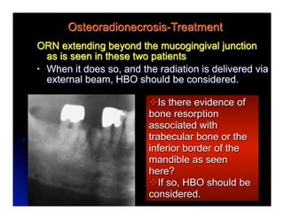
SlideShare a Scribd company logo
Osteoradionecrosis
                      John Beumer III, DDS, MS,
                           Eric Sung, DDS
                 Division of Advanced Prosthodontics
                       UCLA School of Dentistry


All rights reserved. This program of instruction is covered by copyright ©. No
part of this program of instruction may be reproduced, recorded, or transmitted,
by any means, electronic, digital, photographic, mechanical, etc., or by any
information storage or retrieval system, without prior permission of the authors.
Osteoradionecrosis
Definition – Exposure of bone within the radiation
treatment volume of 2-6 months duration or longer
Pathogenesis – 3 Hypotheses
Meyer (1970)
  v   Irradiated bone subjected to a traumatic event could lead to a
       superimposed infection similar to osteomyelitis


Marx (1983)
  v   3H theory of ORN. Following radiation, formulation of hypoxic-
       hypocellular-hypovascular tissue (3H) then tissue breakdown, and finally
       a nonhealing wound as ORN


Delanian and Lefaix (2004)
  v   ORN is due to the activation and dysregulation of fibroblastic activity. A
       cascade of cytokines and free radicals leads to the destruction of
       endothelial cells and vascular thrombosis, which leads to necrosis of
       micro-vessels, local ischemia and tissue loss. The combination of death
       of osteoblasts, failure of osteoblasts to repopulate, continued
       osteoclastic activity and excessive proliferation of myofibroblasts, results
       in bone matrix being replaced by fibrous tissues.
Staging System
Schwartz and Kagan (2002)
  v Stage I – Superficial
    involvement of the mandible
    with minimal soft tissue
    ulceration: only the exposed
    cortical bone is necrotic

  v Stage   II – Both the exposed
    cortical bone and the underlying
    medullary bone are necrotic

  v Stage   III – Full diffuse
    involvement of the mandible,
    including a full thickness
    segment of bone.
Osteoradionecrosis - Sequellae
    Why is this such an important sequellae
    of radiation therapy?


Because advanced cases may lead to fistula
formation as is seen in this patient . . . .
Osteoradionecrosis - Sequellae
prolonged exposures of bone intra-orally,


pathologic fractures of the mandible   and . . .
Osteoradionecrosis - Sequellae


. . . mandibular discontinuity defects with
 accompanying mandibular deviation,
 disruption of speech, mastication and
 saliva control, and facial deformity.
Osteoradionecrosis – Key Facts
Principle cause – Loss of vasculature
  v   Severe narrowing of the lumens of the facial and inferior alveolar
       arteries
  v   Loss of the fine vasculature of the bone marrow and the Haversian
       systems of lamellar bone
  v   Bone matrix replaced by fibrous tissues.


Affects the mandible to a much greater degree
  than the maxilla
  v   More than 90% of all osteoradionecrosis arise in the mandible
  v   Most osteoradionecrosis (ORN) arising from the maxilla heals with
       conservative treatment and does not require hyperbaric oxygen.
       Exceptions – ORN’s secondary to chemoRT or Neutron beam therapy.
Osteoradionecrosis - Key Facts
The incidence varies depending on total dosage, clinical tumor
volume, fractionation, modality and dental disease factor

v   As low as 3% (Beumer et al, 1972) and as high as 37% (Grant and Fletcher,
     1966) when high energy photons are used

v   Higher when brachytherapy is used

v   Extremely high when neutron radiation therapy is used (NRT) (Marunick
     2000)

v   Fewer than 5% of all osteoradionecrosis occurs in edentulous patients
     (Beumer et al, 1984).


v   Increased risk associated with concomitant chemotherapy (Kuhnt et al,
     2006; Hehr et al, 2006)
Osteoradionecrosis - Contributing Factors
                    High Energy Photons

 v   CRT and Radiation fields – If external beam is used
      alone, the more the body of the mandible in the field
      the greater the risk of osteoradionecrosis (ORN).

 v   IMRT may decrease the risk

 v   Dose to Bone – Below 6500 cGy or equivalent the risk
      is very low and almost all that develop in this dosage
      range heal with conservative measures and do not
      require hyperbaric oxygen therapy.

 v   ChemoRT - Increases the risk of ORN and treatment
      is less predictable.
Changing methods of radiation delivery
Conventional radiation therapy (CRT)
  l   200 cGy per fraction
  l   Total doses
        l   7000 cGy definitive dose
        l   5000-6000 cGy post op


 Intensity modulated radiation therapy (IMRT)

                                        This technique uses multiple
                                        radiation beams of non-uniform
                                        intensities. The beams are
                                        modulated to the required intensity
                                        maps for delivering highly conformal
                                        doses of radiation to the treatment
                                        targets, while limiting dose to
                                        structures adjacent
Osteoradionecrosis - Contributing Factors
                   High Energy Photons
IMRT - Volume of the mandible receiving doses in excess of
  6500 cGy is reduced
Osteoradionecrosis - Contributing
            Factors
     High Energy Photons with CRT

Dose to bone
v 6500-7000   cGy – rate of bone necrosis is
   5-15%
v 7000-7500 cGy – rate of bone necrosis is
   15-50%
v 7500 and above - rate of bone necrosis is >
   50%

     Will these numbers hold with IMRT?
Osteoradionecrosis - Contributing

 Chemotherapy increases the risk!!!
   vA full course of concomitant
   chemotherapy increases the Biologic
   equivalent dose (BED) by 700-1000 cGy


*If the patient received concomitant chemotherapy,
a serious bone necrosis leading to resection of the
mandible can occur at doses as low as 5000 cGy.
Osteoradionecrosis - Contributing Factors
    Modality – Brachytherapy
    External beam vs combined external beam and
    brachytherapy. When the dose is boosted
    locally with brachytherapy with multiple sources
    positioned close to or on the mandible, the dose
    to bone locally can be quite high and the patient
    is at great risk of developing an ORN.


  *When the dose to the local bone exceeds 7500
  cGy, the risk of osteoradionecrosis is 50% or more.
Incidence of Osteoradionecrosis
   According to Radiation Dose to Bone
                       Incidence of bone necrosis
Dose to bone (cGy)       Dentulous Pts    Edentulous
<6500                        0/36 (0%)      0/3 (0%)
6500-7500                    8/29 (28%)     1/15 (7%)
7500                       11/13 (85%)     2/4 (50%)
Total                      19/78 (24%)     3/22 (14%)


 Source: Morrish et al, Cancer 47, l980
Osteoradionecrosis
Predisposing and precipitating factors
   1960’s - 2000
    1.   Postradiation extraction of teeth within the
         radiation field
*   2.   Spontaneous associated with dental infection
    3.   Preradiation extractions
    4.   Spontaneous: Dose delivered beyond tolerance
         of normal tissues
    5.   Denture irritation
    6.   Placement of osseointegrated implants into
         irradiated bone

    *Most common factors until chemoRT
Osteoradionecrosis
Predisposing and precipitating factors since 2000

    1.   Spontaneous associated with dental infection
*   2.   Spontaneous: Dose delivered beyond tolerance of
         normal tissues
    3.   Preradiation extractions
    4.   Postradiation extraction of teeth within the radiation
         field
    5.   Denture irritation
    6.   Placement of osseointegrated implants into
         irradiated bone


    *Most common factors since chemoRT
Osteoradionecrosis – predisposing factors (cont’d)
Postradiation extraction of teeth within the
 radiation treatment volume-Maxilla


     In most patients, maxillary teeth in the radiation field
     can be extracted post radiation, with little risk of
     osteoradionecrosis. Even if an ORN develops as
     shown here, the nonvital areas of bone sequestrate
     naturally, leaving only small local bony deformities.
Osteoradionecrosis – predisposing factors (cont’d)
Postradiation extraction of teeth within the
 radiation treatment volume-Maxilla


 Both these osteos developed following extraction of teeth after
 radiation therapy. Both patients received in excess of 6600 cGy
 to the extraction sites. Conservative treatment was employed
 (local irrigation, removal of loose sequestra of bone etc.) and
 both healed several months after onset.
Osteoradionecrosis – predisposing factors (cont’d)
  Postradiation extraction of teeth within the
   radiation treatment volume-Maxilla


Exceptions:
v Patients treated with concomitant chemotherapy
v Patients treated with neutron radiation therapy (NRT)

       ORN’s associated with these treatment modalities
       are frequently intractable and do not respond to
       any form of treatment including HBO.
Osteoradionecrosis-predisposing factors
Postradiation extraction of teeth within the
 radiation treatment volume-Mandible


Post radiation extraction of
mandibular teeth in the field of
radiation when the patient receives
in excess of 6500 cGy is risky and
often leads to osteoradionecrosis.
Osteoradionecrosis-predisposing factors
Postradiation extraction of teeth within the
 radiation treatment volume-Mandible


All four of these patients presented with squamous
carcinomas of either the oral tongue or floor of the
mouth and were treated with external beam photons
and opposed mandibular fields. All received in excess
of 6600 cGy and developed ORN after postradiation
extractions.

All patients developed ORN before the introduction of hyperbaric oxygen and
all eventually had major portions of their mandibles resected resulting in
discontinuity defects in order to control the infection.
Postradiation extractions*
Ill advised in the mandible if the dose to bone is
   above 5500 cGy unless accompanied by HBO
   or the pentoxyfilline-tocopherol protocol
 Incidence of osteoradionecrosis secondary to postradiation extractions
                                 Patients               Osteos

  Grant and Fletcher, 1966     16                   7     44%
  Beumer et al, 1984           40                  11     28%
  Morrish et al, 1980          18                  11     61%
  Marx, et al, 1984            37                  11     30%

  Totals                      111                  40      36%

 *In these clinical reports the dose to bone was in excess of
 6500 cGy and the patients did not receive HBO.
Osteoradionecrosis – Predisposing factors
Preradiation extraction of teeth within the
radiation treatment volume
A                                        In contrast, most
                                         ORN secondary to
                                         preradiation
                                         extraction, such as
                                         these, heal with
                                         conservative
                      B                  measures.


Most are confined within the attached gingiva
(Stage I) and the gingival fibers help maintain the
attachment of the periosteum to underlying bone,
providing blood supply to the underlying bone and
preventing expansion of the bone exposure.
Osteoradionecrosis – Predisposing factors
Preradiation extraction of teeth within the
radiation treatment volume
                                          Both patients shown
A                                         developed ORN at
                                          preradiation extraction
                                          sites. Both had been
                                          treated with external beam
                                          photons and the extraction
                                          sites had received in
                                          excess of 7100 cGy.
                        B
                                          B


Most ORN that develops at preradiation
extraction sites stays localized within
the zone of attached tissue and can be
treated successfully with conservative
measures (patient B).                     Resolution of this ORN
                                          took 8 months.
Preradiation extractions


     Number of patients                            120
     Osteoradionecrosis directly associated with
       extraction sites                             12


Only one of the 12 patients developing ORN associated
with preradiation extractions required mandibular
resection and developed a discontinuity defect of the
mandible.

                                          From Beumer et al, 1984
Osteoradionecrosis – predisposing factors
  Spontaneous associated with acute
  periodontal infections


                      All five of these patients presented with
                      similar histories- bone exposure sites
                      treated in excess of 6600 cGy, previous
history of pain and swelling, followed by a foul smelling
discharge, with eventual pain relief accompanied by
exposure of bone.
Spontaneous associated with acute
           periodontal infections
v Osteoradionecrosis of the mandible that develops as a
result of periodontal infections after radiation therapy is most
serious (particularly if the Radiotherapy was delivered with
external beam) because many result in discontinuity defects.
v Upon initial presentation most ORN developing in this way
extends deep within the body of the mandible (Stage II or III),
and often the lingual or buccal periosteum is dissected away
as the bone exposure enlarges.


The periosteum is the prime source of blood supply and its loss
results in divitalization of the underlying bone, leading in most
patients, to surgical resection of a portion of the mandibular body.
Osteoradionecrosis – predisposing factors
         Secondary to denture irritation

                               A                    B


     Most ORN secondary to denture irritation is
     not serious as long as it is confined to the
     residual attached gingiva (Stage I)(A), and
     heal with conservative measures.
Osteoradionecrosis – predisposing factors
         Secondary to denture irritation

                                A

                                                         B


 Both these ORN’s developed in association with denture wear.
 In patient “A” the bone exposure developed 7 months after
 insertion at the site of a preradiation dental extraction and was
 probably caused by a particle of food becoming lodged beneath
 the denture. In patient “B” the probable cause was an
 overextended denture flange. Both patients received over 6900
 cGy via external beam but both healed with conservative
 measures and did not require hyperbaric oxygen.
Osteoradionecrosis – predisposing factors
      Spontaneous – Dose delivered beyond
      normal tissue tolerance


Both these patients were treated with a combination of
external beam photons and brachytherapy and developed
ORN adjacent to the sites of placement of iridium implants.
Both received over 8500 cGy to the lingual surface of the
mandible.
Osteoradionecrosis – predisposing factors
     Spontaneous – Dose delivered beyond
     normal tissue tolerance


Both these patients were treated with concomitant
chemoradiation and the BED (biologic equivalent dose) to the
posterior body of the mandible approached 8000 cGy
Osteoradionecrosis – predisposing factors
      Spontaneous – Dose delivered beyond
      normal tissue tolerance


This patient was treated with concomitant chemoradiation and
the BED (biologic equivalent dose) to the maxilla approached
8000 cGy. Eventually the entire maxilla was lost.
Osteoradionecrosis – predisposing factors
   Osseointegrated implants


Only a limited number of anectodal case reports
are available for review. The inicidence will
probably be similar to that seen in patients
undergoing post radiation extraction.
Osteoradionecrosis – predisposing factors
Osseointegrated implants


 v This patient received 6600 cGy via opposed mandibular fields for
         a squamous carcinoma of the oral tongue. Implants were
         placed 6 years after completion of therapy.

 v Three years after placement the patient developed an infection on the
        lingual side of the left posterior implant (arrow). Eventually an oral
        cutaneous fistula developed followed by a pathologic       fracture of
        the mandible.

  The mandible was resected and reconstructed with a fibula free flap.
Osteoradionecrosis
Goal of treatment – Resolution with
 maintenance of mandibular continuity
Osteoradionecrosis –Treatment Options
v   Conservative – Local irrigation, regular followup, removal of
     loose sequestra and debridement as appropriate
v   Hyperbaric oxygen protocol - HBO combined with surgical
     debridement or resection
v   Pentoxifylline – tocopherol protocol


     Stage I
     If the ORN is confined within the
     zone of attached gingiva and not
     directly associated with the
     dentition, conservative measures
     are recommended.
Osteoradionecrosis –Treatment Options
v   Conservative – Local irrigation, regular followup, removal of
     loose sequestra and debridement as appropriate
v   Hyperbaric oxygen (HBO) combined with surgical debridement,
     resection and closure
v   Pentoxyfilline – tocopherol protocol


Stage II and III
Does the exposure extend
beyond the mucogingival
junction? Is there evidence of
bone resorption along the inferior
border of the mandible? If so,
either the HBO protocol or
pentoxifylline tocophoral protocol
should be considered .
Osteoradionecrosis –Treatment Options
v   Conservative – Local irrigation, regular followup, removal of
     loose sequestra and debridement as appropriate
v   Hyperbaric oxygen (HBO) combined with surgical debridement,
     resection and closure
v   Pentoxyfilline – tocopherol protocol

What has been its clinical course? Is the exposure getting worse?
If so, either the HBO protocol or the pentoxifylline tocophoral
protocol should be considered .


     Initial presentation           3 moths later
Osteoradionecrosis –Treatment Options
v   Conservative – Local irrigation, regular followup, removal of
     loose sequestra and debridement as appropriate
v   Hyperbaric oxygen (HBO) combined with surgical debridement,
     resection and closure
v   Pentoxyfilline – tocopherol protocol

     What was the mode of therapy? If the dose
     to the primary was boosted with
     brachytherapy, what was the dosimetry of
     the implant.

     vIf unfavorable, either the HBO protocol or
     pentoxifylline protocol should be considered.

     vIf favorable and the external beam dose was
     below 5500 cGy, conservative therapy can be used.
Osteoradionecrosis –Treatment Options
v   Conservative – Local irrigation, regular followup, removal of
     loose sequestra and debridement as appropriate
v   Hyperbaric oxygen (HBO) combined with surgical debridement,
     resection and closure
v   Pentoxyfilline – tocopherol protocol


vRadical neck on the side of the ORN, continued alcohol and
tobacco abuse etc? These factors would encourage the use of
the HBO protocol or the pentoxifylline-tocopherol protocol.

vIf adjunctive or concomitant chemotherapy is used in
combination with radiation, the HBO protocol or the
pentoxifylline-tocopherol protocol should be considered.
Osteoradionecrosis
Predictors of treatment outcomes
  v Initial   presentation
     v Stage  I - Does the ORN extend beyond or is it within
        the zone of keratinized attached mucosa?
     v This is an important prognostic factor because, in
        general, ORN that extends beyond this zone has a
        poor treatment outcome.
Osteoradionecrosis
Predictors of treatment outcomes
  v Initial
         Presentation and precipitating factors-
    Osteoradionecrosis that is precipitated by:
      v Postradiation extractions – Generally extend beyond the
         zone of attached keratinized mucosa and extend into the
         underlying medulary bone. These by definition would be
         Stage II
      v Periodontal infections – Generally extend beyond this
         zone. Most would be Stage II


 These initiating factors generally imply a poor outcome
Osteoradionecrosis
Predictors of treatment outcomes
  vInitial Presentation and precipitating factors-
    Osteoradionecrosis that is precipitated by:
     vPreradiation extractions – Generally stay within this
       zone. Most are Stage I
     vDenture irritation – Generally stay within this zone.
       Most are Stage I.


These initiating factors generally imply a good outcome
Osteoradionecrosis
Predictors of treatment outcomes
  vMode of radiation treatment
    vCombined external beam and brachytherapy:
      Generally such ORN is locally confined and heals with
      conservative therapy if the dosimetry of brachytherapy
      is favorable.
    v External beam alone: If the ORN occurs in the
      mandible in a high dose area the outcomes are less
      favorable.
    vConcomitant chemoRT: ORN’s in these patients
      generally do not respond to conservative measures,
      HBO or the pentoxyfilline – tocopherol protocols and
      require resection and reconstruction with a free flap
Osteoradionecrosis
Goal of treatment – Resolution with
 maintenance of mandibular continuity
Predictors of treatment
 outcomes
  vDose and Treatment Volume
    vThe higher the dose and the larger the treatment
      volume the less predictable the outcome.
Osteoradionecrosis-Treatment
ORN extending beyond the mucogingival junction
  as is seen in these two patients
• When it does so, and the radiation is delivered via
  external beam, HBO should be considered.

                         vIs there evidence of
                         bone resorption
                         associated with
                         trabecular bone or the
                         inferior border of the
                         mandible as seen
                         here?
                         vIf so, HBO should be
                         considered.
Osteoradionecrosis-Treatment
ORN extending beyond the mucogingival junction
  as is seen in these two patients
• When it does so, and the radiation is delivered via
  external beam, HBO should be considered.
Osteoradionecrosis-Treatment Decisions
        and Clinical Examples
Is there evidence of bone resorption associated
with trabecular bone or the inferior border of the
mandible as seen here?
If so, HBO should be considered.


Eventually patient suffered a pathologic fracture of the mandible.
Osteoradionecrosis-Treatment Decisions
Clinical course - Has the bone exposure
  enlarged to extend beyond the mucogingival
  junction?
• If so, HBO should be considered.


 Initial presentation   3 months later
Osteoradionecrosis-Treatment Decisions
Brachytherapy, tumor dose and the dose to the
  local bone - What was the dosimetry of the
  implant?

                   If the dosimetry associated with
                   the implant is favorable, i.e., the
                   total dose to the inferior border
                   and the buccal plate is below
                   5500 cGy, conservative
                   measures are recommended
                   even if the ORN extends
                   beyond the mucogingival
                   junction.
Post-radiation dental disease
Post radiation
   extractions
and HBO
l Dose to Mandible is
   greater than 5500
   cGy
l Expensive and
   time-consuming
Treatment of Osteoradionecrosis - Summary
Role of Conservative Therapy
  v ORN     confined within the zone of attached
     keratinized mucosa that is stable or improving.
  v ORN in patients treated with brachytherapy with
     favorable dose distribution.
  v Initial treatment for an ORN when the dose to
     bone is below 6500 cGy and delivered with
     external beam and conventional fractionation.
Treatment of Osteoradionecrosis - Summary
Role of Hyperbaric oxygen
  v ORN that extends beyond the mucogingival junction in
     patients treated solely with external beam therapy where
     the dose to bone is in excess of 6500 cGy using
     conventional fractionation.
  v ORN precipitated by periodontal or periapical infections
     when the patient has been treated with high dose external
     beam therapy.
  v ORN associated with brachytherapy that extends beyond
     the mucogingival junction that does not respond to
     conservative therapy and where the dosimetry associated
     with the implant is unfavorable.
  v ORN resulting in significant resorption of bone extending
     to the inferior border of the mandible.
Osteoradionecrosis-Treatment Decisions

Other factors to consider*
  v Radical neck dissection on the side of
     the ORN. The facial artery, which
     supplies feeding vessels to the lingual
     periosteum, is removed during this
     procedure.
  v Continued alcohol and tobacco abuse
  v Poor oral compliance


*Thesefactors would favor the use of the
HBO or pentoxyfilline-tocopherol protocols.
Osteoradionecrosis
Role of surgical debridement
  v Incombination with HBO or the pentoxifylline
    - tocopherol protocol
Role of surgical resection and
 reconstruction
  v When  conservative therapies have failed
  v When patient presents with history of high
     dose radiation in combination with
     concomitant chemotherapy
Osteoradionecrosis – Role of Conservative Therapy
                 Clinical Examples
                This patient was S/P 5500 cGy delivered with
                external beam plus 2500-3500 cGy via an
                iridium implant for a squamous carcinoma of
                the right lateral tongue and floor of the mouth.
                Two years later he developed an ORN on the
                lingual surface of the mandible.
               Evaluation of the dosimetry of the implant
               revealed most of the treatment volume
               confined to the lingual surface of the mandible

                               Exam revealed the ORN to be
                               undermined by the oral epithelium and
                               loose. Removal was accomplished
                               with cotton pliers revealing normal
                               epithelium beneath. Eventually all the
                               teeth exfoliated and the remaining
                               bone exposures sequestrated
                               spontaneously.
Osteoradionecrosis-Role of Conservative Therapy
       Clinical Examples
This patient received 5500 cGy with external beam with the
linear accelerator plus 2500-3500 cGy via an iridium implant.
Two years later he developed an ORN on the left lingual surface
of the mandible.
                           Nonvital bone was removed with a
                           high speed air rotor with copius
                           irrigation. After several episodes
                           mucosal coverage was attained.


Close examination
reveals that the
bone exposure was
being undermined
by oral epithelium.
Exception
                 High Dose Rate Implants
l   Technique sensitive
     l   It is very easy to deliver excessive doses
          when using this technique
Osteoradionecrosis-Role of Conservative Therapy
                   Clinical Examples
•   This patient is S/P 7100 cGy with external beam photons for
    treatment of a squamous carcinoma of the soft palate.
    Opposed lateral facial fields were employed.
•   Six months after therapy he developed an ORN at the site of
    a preradiation extraction.
•   The ORN was confined to the attached gingiva.
                        After 8 months of
                        conservative
                        therapy the ORN
                        had resolved.
Osteoradionecrosis – Treatment with HBO
              Clinical Examples
   • ORN associated with external beam
     •   If the ORN extends beyond the mucogingival
         junction and the dose to bone is above 6500cGy
         (with conventional fractionation) or its biological
         equivalent, hyperbaric oxygen combined with
         surgical sequestrectomy is recommended.
Osteoradionecrosis – Treatment with HBO
                   Clinical Examples
This patient presented S/P 7100 cGy delivered with external
beam for a squamous carcinoma,with an ORN of the right lingual
surface of the mandible. Note that the exposure extends beyond
the mucogingival junction.
                      Following a course of hyperbaric oxygen
                      (30 treatments), surgical excision of the
                      nonvital bone and extraction of teeth in
                      the local area was accomplished.


            Another ten HBO treatments were administered. The wound
            re-epithelialized and mandibular continuity was maintained.
Osteoradionecrosis-Treatment with HBO
                           Clinical Examples
             l This patient presented with a bone exposure after
                therapy at a preradiation extraction site. He had
                received over 9500 cGy to the right body of the
                mandible. He continued to smoke and drink
                heavily.
                                  The necrosis continued to
 The exposure began to            enlarge and eventually the
 enlarge and so the patient       entire body of the mandible
 was referred for a course of     was resected and later
 HBO.                             reconstructed.


HBO is effective when the tissues are still viable but delay in
the face of high dosage may compromise the chance of
success and result in greater bone loss.
Osteoradionecrosis-Treatment with HBO
                        Clinical Examples
                   This patient presented with a small ORN on
                   the lingual of #29 and #30 secondary to a
                   periodontal abscess. Four years earlier she
                   had received 5500 cGy with external beam
                   plus another 4500 cGy via an iridium implant.
                   Note the bone loss associated
                   with these teeth


Hygiene was excellent and
the patient had ceased
using tobacco or alcohol.
Note that the labial gingiva
appears quite healthy.
Osteoradionecrosis – Treatment with HBO
                  Clinical Examples
                                           Conservative
                                           therapy was
                                           initiated but the
                                           ORN increased in
                                           size.

•HBO combined with surgical debridement was recommended
but the patient refused. The exposure progressed until the
lingual plate from molar to molar was exposed and the patient
developed an orocutaneous fistula.

•The patient finally accepted HBO and a series of
treatments were initiated combined with biweekly irrigations
and debridement.
Osteoradionecrosis-Treatment with HBO
        Eventually a large segment of nonvital
        bone was undermined by oral epithelium,
        became loose and was removed with
        cotton pliers. The oral cutaneous fistula
        closed.
             Appearing beneath, was a bed
             of granulation tissue and oral
             mucous membrane. However,
             small localized areas of exposed
             bone still remained.
Osteoradionecrosis - Treatment with HBO
                   l   One month later most of the
                        granulation tissue had been
                        covered with oral epithelium


                             During the next 18 months
                           the small residual areas of
                           exposed bone became
                           undermined by oral
                           epithelium and sequestrated
                           sometimes helped along with
                           a high speed air rotor.

  Three years later most of the teeth had exfoliated and the
patient was fitted with a removable partial denture.
Eventually all the mandibular teeth exfoliated. Note the
telangiectasia associated with oral mucosa and the atrophy of
the tongue. A complete denture was fabricated for the
purposes of lip support and esthetics and well tolerated by the
patient.
Why was the outcome successful?
•The dosimetry of the implant was favorable.
Although the implant delivered a high dose to a
large volume of tissue, the dose to the inferior
border and buccal cortical bone of the mandible
was low (only slightly above 5500 cGy)
•The external beam dose was low – 5500 cGy. At this level the
periosteum and marrow retain significant vascular elements.

•The patient was compliant. The patient
stopped smoking and her oral hygiene was
superb. She never missed an irrigation
appointment.
  Hyperbaric oxygen. These
treatments revascularized the
marginally necrotic tissues and
facilitated the patient’s response to
the local infections.
Osteoradionecrosis –Treatment with Surgical
                    This patient developed an ORN following
                    postradiation extractions. He had
                    received 6600 cGy for a squamous
                    carcinoma of the right lateral tongue.
                However, a localtreatment was initiatated
                   Conservative surgical sequestrectomy
                was attempted which resulted in the stable
                   and the bone exposure remained
                exposure extending beyond the zone of
                   and began to improve.
                attached gingiva. The exposure rapidly
                expanded, eventually extending lingually to
                the inferior border of the mandible.

                           Pathologic fracture and an
                           orocutaneous fistula soon
                           followed and the mandibular
                           body was eventually resected in
Clinical Examples          order to resolve the infections.
Why was the outcome so unsuccessful?
•   The patient continued to smoke and use alcohol.
•   All the radiation was delivered with external beam
    photons. The tumor dose was 6600 cGy and the
    dose was distributed homogenously from the
    buccal side to the lingual side of the mandibular
    body.
•   At these dosage levels the vascularity and
    cellularity of the periosteum was severely
    compromised and no longer provided sufficient
    vascular support to the underlying bone.
    However, as long as the periosteum remained
    attached to underlying bone, the bone remained
    viable.
•   When the mucoperiosteum was dissected away
Why was the outcome so unsuccessful?
•    As long as the exposure was confined within the
     attached mucosa, it did not spread because the
     gingival fibers helped to maintain contact
     between the periosteum and cortical bone.

•    Once the exposure had spread beyond the
     attached mucosa, the attachment of periosteum
     to the underlying bone became very tenuous.
     The slightest infectious insult will dissect the
     atrophied periosteum from the underlying bone.
     When dissection occurs, the bone looses its
     primary remaining blood supply and becomes
     essentially nonvital. HBO will not salvage such a
     mandible and its resection is inevitable.
Osteoradionecrosis – Case report
l   This patient presented with an ORN 18 months post radiation.
     It is not painful, there are no draining fistulas and a panorex
     reveals little change in the density of the trabecular bone or
     the cortex of the inferior border of the mandible. The exposure
     has been present for about 3 months and extends beyond the
     mucogingival junction lingually. What radiation data do you
     need to acquire before developing a plan of treatment?

      Mode of therapy
      Dose to bone
      Dosimetry

     What patient data do you need to obtain?

       Oral Compliance
       Smoking habits
       Medical history
Osteoradionecrosis – Case report
l   The patient received 5000 cGy to the tumor volume and the
     neck with external beam high energy photons. The dose to
     the primary was boosted with an iridium implant with an
     additional 2500-3500 cGy. The CT generated dosimetry is
     shown here. What treatment do you recommend?
                      Conservative therapy was employed. Bone
                      undermined by oral mucosa, was periodically
                      removed with a high speed air rotor. Within 9
                      months the nonvital bone had been removed and
                      mucosal integrity was restored.
Osteoradionecrosis - Case report
 l   This patient was treated with external beam high energy
      photons and brachytherapy for a squamous carcinoma of the
      right lateral tongue. Two years after completion she
      developed an acute periodontal infection which eventually led
      to dehiscence of the lingual gingiva and an
      osteoradionecrosis. What information do you need to
      develop a plan of treatment for this patient?
Radiation data
        • Fields of external beam
        • Dosimetry of the implant
        • Dose to bone of the buccal
          half of the mandible

Other data
            Oral compliance
           Smoking habits
           Updated MH
Osteoradionecrosis – Case Report
•   Dosimetry was very favorable. Since the implants had
    been placed into the lateral border of the tongue, the
    dose delivered to the middle of the mandibular body
    by the implant was minimal.
• The dosage delivered to lingual plate was about 2000 cGy.
• Oral hygiene was poor, however, and the patient
     continued to use tobacco.


What treatment would you recommend for this patient?
Osteoradionecrosis-Case Report
    Conservative therapy was adopted because the
    external beam dose to the mandible was low
    and the dosimetry of the implant was favorable.


The premolar and the cuspid exfoliated and the
nonvital bone sequestrated within 9 months.
Eventually all of the remaining teeth exfoliated.
Osteoradionecrosis – Surgical Management
     Free vascularized flaps and myocutaneous
     flaps can be used to cover exposed areas of
     bone following surgical sequestrectomy.


The nonvital bone is removed surgically and a flap
is used to cover the remaining bone. In this patient
a pectoralis myocutaneous flap was used.
Osteoradionecrosis – Surgical Management
    Reconstruction with fibula free flaps


Potential problems with free flap reconstruction
vSemi-occluded vessels in the neck with fibrotic and
     fragile walls.
vComplication rate -50%
                             Suh et al, 2010
vRelapse rate about 25 %
Osteoradionecrosis:
     Treatment Philosophies
Marx
 •   Dr. Marx and his colleagues believe
     almost all osteoradionecrosis of the
     mandible would benefit from hyperbaric
     oxygen treatment.

 •   What follows is the protocol developed
     by Dr. Marx used to treat patients with
     osteoradionecrosis.
Hyperbaric Oxygen (per Marx)
  • Stage I – These patients present with ORN
    but without pathologic fracture of the
    mandible, orocutaneous fistula or
    radiographic evidence of bone resorption of
    the inferior border of the mandible.
     •   Thirty HBO treatments (2.4 atmospheres, 100% oxygen
         for 90 minutes)
     •   At the end of 30 treatments, if improvement is noted,
         another 10 treatments are administered.


If no improvement is noted, the patient is considered
a nonresponder and is advanced to Stage II.
Hyperbaric Oxygen (per Marx)
  • Stage II
     •   Nonresponders are taken to surgery and
         surgical debridement of the local area is
         performed. Nonvital bone is removed via a
         transalveolar sequestrectomy and the labial
         and lingual mucoperiosteal flaps are closed in
         three layers over a base of bleeding
         bone.
     •   An additional 10 HBO treatments are added.

If the wound dehisces, the patient is considered
a nonresponder and advanced to Stage III.
Hyperbaric Oxygen (per Marx)
• Stage III
  •   Nonresponders from Stage II therapy, and patients
      presenting with orocutaneous fistula, pathologic fracture of
      the mandible or bone resorption of the inferior border of
      the mandible, are considered Stage III patients.

  •   Following the initial 30 treatments, bony segments of the
      nonvital mandibular bone are resected. Soft tissue deficits
      if present are restored with local or distant flaps and
      orocutaneous fistulas are closed

  •   Another 10 HBO treatments are administered and the
      patient is advanced to Stage IIIR
Hyperbaric Oxygen (per Marx)

• Stage IIIR
  •   Ten weeks after resection the
      mandible is reconstructed with a
      free bone graft, using a
      transcutaneous exposure.

  •   Mandibular fixation is achieved and
      the patient given another 10 HBO
      treatments.
Hyperbaric Oxygen

l   Permanence
     l Patients  restudied 4
        years after HBO
        demonstrated the same
        angiogenesis as those
        studied immediately
        after completion.
     l New hyperbaric induced
        vessels do not appear to
        involute after cessation
        of HBO faster than the
        normal rate of aging.
Osteoradionecrosis:
         Reconstruction after Resection
             v        Free grafts vs free
                       vascularized grafts
Both methods achieve successful results in the hands of
experienced surgeons although the success rates associated with
the use of free grafts is enhanced by the use of hyperbaric
oxygen.


Potential problems with free flaps:
v Semi-occluded vessels in the neck with fibrotic and
      fragile walls.
vRelapse
Osteoradionecrosis:
         Reconstruction after Resection
Patient is S/P resection of the mandible for an ORN. He had
received in excess of 6800 cGy to the body of the mandible with
external beam.
                                      The defect was restored with a
                                      block bone graft from the iliac
                                      crest


Hyperbaric
oxygen was
unsuccessful in
saving the
continuity of the
mandible.                                        One year S/P bone graft
Osteoradionecrosis:
          Reconstruction after Resection
Patient is S/P resection of the mandible for an osteoradionecrosis.
He had received in excess of 6800 cGy to the body of the
mandible.
                                     The defect was restored with a
                                     block bone graft from the iliac
                                     crest


Six months later
osseointegrated
implants were
successfully
placed into the
grafted bone.                                   One year S/P bone graft
Osteoradionecrosis – Prevention
Since Osteoradionecrosis (ORN) is a process that
primarily affects the mandible . . . . . and
since mandibular teeth are the prime precipitators
of ORN we recommend the following strategies.
Prevention strategies
  Remove mandibular teeth with significant periodontal or
  advanced caries in the field prior to therapy in high
  dose areas.
  Remove mandibular teeth adjacent to a prospective
  iridium implant.
Osteoradionecrosis – Prevention
Since Osteoradionecrosis (ORN) is a process that
primarily affects the mandible and . . . . .
since mandibular teeth are the prime precipitators
of ORN we recommend the following strategies.

Prevention strategies
  Remove all teeth within the gross tumor volume and the
  patient is potentially noncompliant particularly in
  patients who receive concomitant chemotherapy.
Soft Tissue Necrosis
Definition – A non-neoplastic mucosal ulceration
occurring in the postradiation field and which does not
expose bone
 Most occur within a year of therapy and develop at the site of
 radioactive implants or peroral cone application sites.


The key issue is to determine whether these ulcerations
 represent recurrent tumor. Clinical symptoms useful in making
 the diagnosis are:
a) Soft tissue necroses lack an inflammatory halo
b) Soft tissue necrosis is much more painful than tumor recurrence
c) Soft tissue necroses are not indurated, tumor recurrences are
   indurated
Soft Tissue Necrosis
Treatment
 •   Establish the diagnosis
 •   Local excision and primary closure
 •   Local supportive measures and
     followup
 •   Hyperbaric oxygen
Soft Tissue Necrosis
     Patient received 5500 cGy via external beam and another
     2500-3000 cGy with a radium needle implant for a squamous
     cell carcinoma of the lateral border of the tongue.


v   Nine months after therapy he developed this ulceration at the
     site of the tumor.
v   Cytology and biopsy were negative and a diagnosis of radiation
     soft tissue necrosis was assumed.
v   The lesion epithelialized 4 months later
Soft Tissue Necrosis
Soft tissue necrosis secondary to chemoradiation


 The remaining soft palate musculature was scarred and
 atrophic and elevated poorly. There was little lateral wall
 movement. The defect at the junction of the hard and soft
 palate was easily obturated, but not the posterior
 velopharyngeal defect
Chemoradiation
v Late effects of radiation are more
   profound, i.e. risk of osteoradionecrosis is
   higher at the lower doses.
v Treatment outcomes are less predictable
  v The  usual methods of treatment
     recommended in this program of instruction
     based on dosage and initial clinical
     presentation may not apply and may not be as
     effective when chemoradiation has been used.
  v High relapse rate when using free flaps for
     reconstruction
Neutron Radiation Therapy
                 (NRT)
Treatment outcomes are less predictable and
the incidence of ORN is higher
•   The usual methods of treatment recommended in this
    program of instruction may not apply and may not be as
    effective.
Neutron Radiation Therapy (NRT)
•   Treatment outcomes are less
    predictable and the incidence of
    ORN is higher
    •   In a report by Marunick et al (2000), 4 of 9 patients
        treated for malignant salivary gland tumors arising in
        the maxilla or the paranasal sinuses, developed
        osteoradionecrosis.
    •   None responded to the methods of treatment
        described in this program and eventually lead to
        loss of the entire palate in most of the patients.
Osteoradionecrosis Secondary to
            Chemoradiation
vSpontaneous  – in most instances no known
      causative factors
vNot responsive to HBO
ORN secondary to ChemoRT


l   Dramatically higher rates in the maxilla
     with poorer outcomes.
v Visit ffofr.org for hundreds of additional lectures
   on Complete Dentures, Implant Dentistry,
   Removable Partial Dentures, Esthetic Dentistry
   and Maxillofacial Prosthetics.
v The lectures are free.
v Our objective is to create the best and most
   comprehensive online programs of instruction in
   Prosthodontics

More Related Content

What's hot

ORAL SUBMUCOUS FIBROSIS- PPT
ORAL SUBMUCOUS FIBROSIS- PPTORAL SUBMUCOUS FIBROSIS- PPT
ORAL SUBMUCOUS FIBROSIS- PPT
K BHATTACHARJEE
 
Masticatory space infection
Masticatory space infectionMasticatory space infection
Masticatory space infection
Dr. swati sahu
 
Oroantral Fistula
Oroantral FistulaOroantral Fistula
Oroantral Fistula
jiveshmunankarmi
 
Lefort 1 fracture
Lefort 1 fracture Lefort 1 fracture
Lefort 1 fracture
Dr Pratiksha Malhotra
 
Vestibuloplasty
VestibuloplastyVestibuloplasty
Vestibuloplasty
vrushupatel
 
Condylar fractures
Condylar fracturesCondylar fractures
Condylar fractures
Zeeshan Arif
 
Basic principles of oral and maxillofacial surgery
Basic principles of oral and maxillofacial surgeryBasic principles of oral and maxillofacial surgery
Basic principles of oral and maxillofacial surgery
Dr Bhavik Miyani
 
Mixed radiopaque & radiolucent lesions of jaw
Mixed radiopaque & radiolucent lesions of jaw Mixed radiopaque & radiolucent lesions of jaw
Mixed radiopaque & radiolucent lesions of jaw
Dr. Samarth Johari
 
Bsso
BssoBsso
mandibular molar Impactions
mandibular molar Impactionsmandibular molar Impactions
mandibular molar ImpactionsNishant Tewari
 
Oral Submucous Fibrosis
Oral Submucous FibrosisOral Submucous Fibrosis
Oral Submucous FibrosisVibhuti Kaul
 
POTENTIALLY MALIGNANT DISORDER
POTENTIALLY MALIGNANT DISORDERPOTENTIALLY MALIGNANT DISORDER
POTENTIALLY MALIGNANT DISORDER
AnweshaBiswas13
 
Giant cell lesion’s of jaw
Giant cell lesion’s of jawGiant cell lesion’s of jaw
Giant cell lesion’s of jawRipan Das
 
Mandibular third moalr impaction
Mandibular third moalr impactionMandibular third moalr impaction
Mandibular third moalr impaction
Ashish Soni
 
Aetiology and management of trismus
Aetiology and management of trismusAetiology and management of trismus
Aetiology and management of trismus
Hope Inegbenosun
 
Mandibular Angle Fractures
Mandibular Angle FracturesMandibular Angle Fractures
Mandibular Angle Fractures
Ahmed Adawy
 
Orofacial pain
Orofacial painOrofacial pain
Orofacial pain
Saleh Bakry
 
Oral Submucous Fibrosis and its Management.
Oral Submucous Fibrosis and its Management.Oral Submucous Fibrosis and its Management.
Oral Submucous Fibrosis and its Management.
Maxfac Center
 

What's hot (20)

ORAL SUBMUCOUS FIBROSIS- PPT
ORAL SUBMUCOUS FIBROSIS- PPTORAL SUBMUCOUS FIBROSIS- PPT
ORAL SUBMUCOUS FIBROSIS- PPT
 
Masticatory space infection
Masticatory space infectionMasticatory space infection
Masticatory space infection
 
Oroantral Fistula
Oroantral FistulaOroantral Fistula
Oroantral Fistula
 
Lefort 1 fracture
Lefort 1 fracture Lefort 1 fracture
Lefort 1 fracture
 
Vestibuloplasty
VestibuloplastyVestibuloplasty
Vestibuloplasty
 
Condylar fractures
Condylar fracturesCondylar fractures
Condylar fractures
 
Basic principles of oral and maxillofacial surgery
Basic principles of oral and maxillofacial surgeryBasic principles of oral and maxillofacial surgery
Basic principles of oral and maxillofacial surgery
 
Mixed radiopaque & radiolucent lesions of jaw
Mixed radiopaque & radiolucent lesions of jaw Mixed radiopaque & radiolucent lesions of jaw
Mixed radiopaque & radiolucent lesions of jaw
 
Bsso
BssoBsso
Bsso
 
mandibular molar Impactions
mandibular molar Impactionsmandibular molar Impactions
mandibular molar Impactions
 
Zygomatic fractures
Zygomatic fracturesZygomatic fractures
Zygomatic fractures
 
Oral Submucous Fibrosis
Oral Submucous FibrosisOral Submucous Fibrosis
Oral Submucous Fibrosis
 
POTENTIALLY MALIGNANT DISORDER
POTENTIALLY MALIGNANT DISORDERPOTENTIALLY MALIGNANT DISORDER
POTENTIALLY MALIGNANT DISORDER
 
5. Vestibuloplasty.pptx
5. Vestibuloplasty.pptx5. Vestibuloplasty.pptx
5. Vestibuloplasty.pptx
 
Giant cell lesion’s of jaw
Giant cell lesion’s of jawGiant cell lesion’s of jaw
Giant cell lesion’s of jaw
 
Mandibular third moalr impaction
Mandibular third moalr impactionMandibular third moalr impaction
Mandibular third moalr impaction
 
Aetiology and management of trismus
Aetiology and management of trismusAetiology and management of trismus
Aetiology and management of trismus
 
Mandibular Angle Fractures
Mandibular Angle FracturesMandibular Angle Fractures
Mandibular Angle Fractures
 
Orofacial pain
Orofacial painOrofacial pain
Orofacial pain
 
Oral Submucous Fibrosis and its Management.
Oral Submucous Fibrosis and its Management.Oral Submucous Fibrosis and its Management.
Oral Submucous Fibrosis and its Management.
 

Viewers also liked

Osteoradionecrosis
OsteoradionecrosisOsteoradionecrosis
Osteoradionecrosis
Muhammad Khan
 
Osteorradionecrosis.
Osteorradionecrosis.Osteorradionecrosis.
Osteorradionecrosis.
Dr. Ramón de J. Villar Prieto
 
osteomyelitis & osteoradionecrosis
 osteomyelitis & osteoradionecrosis osteomyelitis & osteoradionecrosis
osteomyelitis & osteoradionecrosisvasanramkumar
 
Osteonecrosis of the jaws
Osteonecrosis of the jawsOsteonecrosis of the jaws
Osteonecrosis of the jaws
Ninian Peckitt
 
Osteomyelitis of the jaws
Osteomyelitis of the jawsOsteomyelitis of the jaws
Osteomyelitis of the jaws
islam kassem
 
Osteomyelitis of jaws
Osteomyelitis of jawsOsteomyelitis of jaws
Osteomyelitis of jaws
stutisaxena
 
Acute and Chronic Osteomyelitis - Infection of Bone
Acute and Chronic Osteomyelitis - Infection of BoneAcute and Chronic Osteomyelitis - Infection of Bone
Acute and Chronic Osteomyelitis - Infection of Bone
Rahul Singh
 
Osteomyelitis
OsteomyelitisOsteomyelitis
Osteomyelitis
shahnawaz65
 
Dental extractions in irradiated patients
Dental extractions in irradiated patientsDental extractions in irradiated patients
Dental extractions in irradiated patientsUjwal Gautam
 
Osteoradionecrosis - Dr. Amit T. Suryawanshi (MDS).Kolhapur, Sangli & Pune.
Osteoradionecrosis - Dr. Amit T. Suryawanshi (MDS).Kolhapur, Sangli & Pune.Osteoradionecrosis - Dr. Amit T. Suryawanshi (MDS).Kolhapur, Sangli & Pune.
Osteoradionecrosis - Dr. Amit T. Suryawanshi (MDS).Kolhapur, Sangli & Pune.
All Good Things
 
Osteomyelitis of jaws
Osteomyelitis of jawsOsteomyelitis of jaws
Osteomyelitis of jaws
Anushan Madushanka
 

Viewers also liked (20)

Osteoradionecrosis
OsteoradionecrosisOsteoradionecrosis
Osteoradionecrosis
 
Osteorradionecrosis.
Osteorradionecrosis.Osteorradionecrosis.
Osteorradionecrosis.
 
osteomyelitis & osteoradionecrosis
 osteomyelitis & osteoradionecrosis osteomyelitis & osteoradionecrosis
osteomyelitis & osteoradionecrosis
 
Osteonecrosis of the jaws
Osteonecrosis of the jawsOsteonecrosis of the jaws
Osteonecrosis of the jaws
 
Complete dentures for irradiated patients
Complete dentures for irradiated patientsComplete dentures for irradiated patients
Complete dentures for irradiated patients
 
(Replace) 11. palatal resections alterations at surgery to enhance the prosth...
(Replace) 11. palatal resections alterations at surgery to enhance the prosth...(Replace) 11. palatal resections alterations at surgery to enhance the prosth...
(Replace) 11. palatal resections alterations at surgery to enhance the prosth...
 
Osteomyelitis of the jaws
Osteomyelitis of the jawsOsteomyelitis of the jaws
Osteomyelitis of the jaws
 
17. (new)implant retained maxillary obturators
17. (new)implant retained maxillary obturators17. (new)implant retained maxillary obturators
17. (new)implant retained maxillary obturators
 
Osteomyelitis of jaws
Osteomyelitis of jawsOsteomyelitis of jaws
Osteomyelitis of jaws
 
Acute and Chronic Osteomyelitis - Infection of Bone
Acute and Chronic Osteomyelitis - Infection of BoneAcute and Chronic Osteomyelitis - Infection of Bone
Acute and Chronic Osteomyelitis - Infection of Bone
 
Osteomyelitis
OsteomyelitisOsteomyelitis
Osteomyelitis
 
Osteomyelitis
OsteomyelitisOsteomyelitis
Osteomyelitis
 
Osteomyelitis
OsteomyelitisOsteomyelitis
Osteomyelitis
 
Maintenance, restorative care and postradiation dental disease
Maintenance, restorative care and postradiation dental diseaseMaintenance, restorative care and postradiation dental disease
Maintenance, restorative care and postradiation dental disease
 
Dental extractions in irradiated patients
Dental extractions in irradiated patientsDental extractions in irradiated patients
Dental extractions in irradiated patients
 
Osteoradionecrosis - Dr. Amit T. Suryawanshi (MDS).Kolhapur, Sangli & Pune.
Osteoradionecrosis - Dr. Amit T. Suryawanshi (MDS).Kolhapur, Sangli & Pune.Osteoradionecrosis - Dr. Amit T. Suryawanshi (MDS).Kolhapur, Sangli & Pune.
Osteoradionecrosis - Dr. Amit T. Suryawanshi (MDS).Kolhapur, Sangli & Pune.
 
1.radiation of h&n tumors
1.radiation of h&n tumors1.radiation of h&n tumors
1.radiation of h&n tumors
 
Osteomyelitis of jaws
Osteomyelitis of jawsOsteomyelitis of jaws
Osteomyelitis of jaws
 
4.radiation effects – salivary glands, bone and teeth
4.radiation effects – salivary glands, bone and teeth4.radiation effects – salivary glands, bone and teeth
4.radiation effects – salivary glands, bone and teeth
 
5.preradiation dental assessment
5.preradiation dental assessment5.preradiation dental assessment
5.preradiation dental assessment
 

Similar to 9.(new)osteoradionecrosis

Osteoradionecrosis
OsteoradionecrosisOsteoradionecrosis
Osteoradionecrosis
AravindNair71
 
per flap with vest deep.pptx
per flap with vest deep.pptxper flap with vest deep.pptx
per flap with vest deep.pptx
mayankgupta672202
 
Implants in irradiated jaw
Implants in irradiated jawImplants in irradiated jaw
Implants in irradiated jaw
Zubair_ahmd
 
JOURNAL CLUB PRESENTATION IN PROSTHODONTICS on HYPERBARIC OXYGEN THERAPY FOR ...
JOURNAL CLUB PRESENTATION IN PROSTHODONTICS on HYPERBARIC OXYGEN THERAPY FOR ...JOURNAL CLUB PRESENTATION IN PROSTHODONTICS on HYPERBARIC OXYGEN THERAPY FOR ...
JOURNAL CLUB PRESENTATION IN PROSTHODONTICS on HYPERBARIC OXYGEN THERAPY FOR ...
NAMITHA ANAND
 
Peri implant Diseases and its management
Peri implant Diseases and its managementPeri implant Diseases and its management
Peri implant Diseases and its management
Jignesh Patel
 
osteoradionecrosis-160118051248 new.pptx
osteoradionecrosis-160118051248 new.pptxosteoradionecrosis-160118051248 new.pptx
osteoradionecrosis-160118051248 new.pptx
ShaliniN51
 
OSTEORADIONECROSIS.pptx
OSTEORADIONECROSIS.pptxOSTEORADIONECROSIS.pptx
OSTEORADIONECROSIS.pptx
Dr. Sumit KUMAR
 
15. Osteoradionecrosis and HBO therapy
15. Osteoradionecrosis and HBO therapy15. Osteoradionecrosis and HBO therapy
15. Osteoradionecrosis and HBO therapy
Meghali Diwaker
 
Dr. Rahul VC Tiwari - Fellowship In Orthognathic Surgery - Jubilee Mission Me...
Dr. Rahul VC Tiwari - Fellowship In Orthognathic Surgery - Jubilee Mission Me...Dr. Rahul VC Tiwari - Fellowship In Orthognathic Surgery - Jubilee Mission Me...
Dr. Rahul VC Tiwari - Fellowship In Orthognathic Surgery - Jubilee Mission Me...
CLOVE Dental OMNI Hospitals Andhra Hospital
 
Periimplantitis
PeriimplantitisPeriimplantitis
Periimplantitis
JahnaviYadav5
 
105th publication sjodr- 1st name
105th publication  sjodr- 1st name105th publication  sjodr- 1st name
105th publication sjodr- 1st name
CLOVE Dental OMNI Hospitals Andhra Hospital
 
Infection 3
Infection 3Infection 3
OSTEORADIONECROSIS.pptx
OSTEORADIONECROSIS.pptxOSTEORADIONECROSIS.pptx
OSTEORADIONECROSIS.pptx
Adyasha3
 
Frontal osteoma - HARSH AMIN
Frontal osteoma - HARSH AMINFrontal osteoma - HARSH AMIN
Frontal osteoma - HARSH AMIN
Dr. HARSH AMIN
 
journal club on Use of Er:YAG Laser to Decontaminate Infected Dental Implant ...
journal club on Use of Er:YAG Laser to Decontaminate InfectedDental Implant ...journal club on Use of Er:YAG Laser to Decontaminate InfectedDental Implant ...
journal club on Use of Er:YAG Laser to Decontaminate Infected Dental Implant ...
Shilpa Shiv
 
Treatment of Aneurysmal Bone Cysts by Minimally Invasive Curettage and Alloge...
Treatment of Aneurysmal Bone Cysts by Minimally Invasive Curettage and Alloge...Treatment of Aneurysmal Bone Cysts by Minimally Invasive Curettage and Alloge...
Treatment of Aneurysmal Bone Cysts by Minimally Invasive Curettage and Alloge...
Prof Freih Abu Hassan البروفيسور فريح ابوحسان
 
Treatment of Aneurysmal Bone Cysts by Minimally Invasive Curettage and Alloge...
Treatment of Aneurysmal Bone Cysts by Minimally Invasive Curettage and Alloge...Treatment of Aneurysmal Bone Cysts by Minimally Invasive Curettage and Alloge...
Treatment of Aneurysmal Bone Cysts by Minimally Invasive Curettage and Alloge...
Prof Freih Abu Hassan البروفيسور فريح ابوحسان
 

Similar to 9.(new)osteoradionecrosis (20)

Osteoradionecrosis
OsteoradionecrosisOsteoradionecrosis
Osteoradionecrosis
 
8.implant in irradiated patients
8.implant in irradiated patients8.implant in irradiated patients
8.implant in irradiated patients
 
per flap with vest deep.pptx
per flap with vest deep.pptxper flap with vest deep.pptx
per flap with vest deep.pptx
 
Implants in irradiated jaw
Implants in irradiated jawImplants in irradiated jaw
Implants in irradiated jaw
 
JOURNAL CLUB PRESENTATION IN PROSTHODONTICS on HYPERBARIC OXYGEN THERAPY FOR ...
JOURNAL CLUB PRESENTATION IN PROSTHODONTICS on HYPERBARIC OXYGEN THERAPY FOR ...JOURNAL CLUB PRESENTATION IN PROSTHODONTICS on HYPERBARIC OXYGEN THERAPY FOR ...
JOURNAL CLUB PRESENTATION IN PROSTHODONTICS on HYPERBARIC OXYGEN THERAPY FOR ...
 
Peri implant Diseases and its management
Peri implant Diseases and its managementPeri implant Diseases and its management
Peri implant Diseases and its management
 
osteoradionecrosis-160118051248 new.pptx
osteoradionecrosis-160118051248 new.pptxosteoradionecrosis-160118051248 new.pptx
osteoradionecrosis-160118051248 new.pptx
 
OSTEORADIONECROSIS.pptx
OSTEORADIONECROSIS.pptxOSTEORADIONECROSIS.pptx
OSTEORADIONECROSIS.pptx
 
15. Osteoradionecrosis and HBO therapy
15. Osteoradionecrosis and HBO therapy15. Osteoradionecrosis and HBO therapy
15. Osteoradionecrosis and HBO therapy
 
Dr. Rahul VC Tiwari - Fellowship In Orthognathic Surgery - Jubilee Mission Me...
Dr. Rahul VC Tiwari - Fellowship In Orthognathic Surgery - Jubilee Mission Me...Dr. Rahul VC Tiwari - Fellowship In Orthognathic Surgery - Jubilee Mission Me...
Dr. Rahul VC Tiwari - Fellowship In Orthognathic Surgery - Jubilee Mission Me...
 
Periimplantitis
PeriimplantitisPeriimplantitis
Periimplantitis
 
Complete Dentures for Irradiated Patients
Complete Dentures for Irradiated PatientsComplete Dentures for Irradiated Patients
Complete Dentures for Irradiated Patients
 
105th publication sjodr- 1st name
105th publication  sjodr- 1st name105th publication  sjodr- 1st name
105th publication sjodr- 1st name
 
Infection 3
Infection 3Infection 3
Infection 3
 
OSTEORADIONECROSIS.pptx
OSTEORADIONECROSIS.pptxOSTEORADIONECROSIS.pptx
OSTEORADIONECROSIS.pptx
 
Frontal osteoma - HARSH AMIN
Frontal osteoma - HARSH AMINFrontal osteoma - HARSH AMIN
Frontal osteoma - HARSH AMIN
 
journal club on Use of Er:YAG Laser to Decontaminate Infected Dental Implant ...
journal club on Use of Er:YAG Laser to Decontaminate InfectedDental Implant ...journal club on Use of Er:YAG Laser to Decontaminate InfectedDental Implant ...
journal club on Use of Er:YAG Laser to Decontaminate Infected Dental Implant ...
 
Treatment of Aneurysmal Bone Cysts by Minimally Invasive Curettage and Alloge...
Treatment of Aneurysmal Bone Cysts by Minimally Invasive Curettage and Alloge...Treatment of Aneurysmal Bone Cysts by Minimally Invasive Curettage and Alloge...
Treatment of Aneurysmal Bone Cysts by Minimally Invasive Curettage and Alloge...
 
Treatment of Aneurysmal Bone Cysts by Minimally Invasive Curettage and Alloge...
Treatment of Aneurysmal Bone Cysts by Minimally Invasive Curettage and Alloge...Treatment of Aneurysmal Bone Cysts by Minimally Invasive Curettage and Alloge...
Treatment of Aneurysmal Bone Cysts by Minimally Invasive Curettage and Alloge...
 
3.radiation effects mucosa, taste, jaw opening
3.radiation effects mucosa, taste, jaw opening3.radiation effects mucosa, taste, jaw opening
3.radiation effects mucosa, taste, jaw opening
 

More from www.ffofr.org - Foundation for Oral Facial Rehabilitiation

Digital Design of Mandibular Removable Partial Dentures
Digital Design of Mandibular Removable Partial DenturesDigital Design of Mandibular Removable Partial Dentures
Digital Design of Mandibular Removable Partial Dentures
www.ffofr.org - Foundation for Oral Facial Rehabilitiation
 
Digital design of maxillary of rpd's
Digital design of maxillary of rpd'sDigital design of maxillary of rpd's
Prosthodontics Procedures and Complications - Posterior Quadrants
 Prosthodontics Procedures and Complications - Posterior Quadrants Prosthodontics Procedures and Complications - Posterior Quadrants
Prosthodontics Procedures and Complications - Posterior Quadrants
www.ffofr.org - Foundation for Oral Facial Rehabilitiation
 
Restoration of posterior quadrants
Restoration of posterior quadrantsRestoration of posterior quadrants
Restoration of endodontically treated teeth
Restoration of endodontically treated teethRestoration of endodontically treated teeth
Restoration of endodontically treated teeth
www.ffofr.org - Foundation for Oral Facial Rehabilitiation
 
Provisional restorations
Provisional restorationsProvisional restorations
Secondard impression materials
Secondard impression materialsSecondard impression materials
Fluid control and tissue managemtent
Fluid control and tissue managemtentFluid control and tissue managemtent
Ceramics in fixed prosthodontics considerations for use in dental practice
Ceramics in fixed prosthodontics   considerations for use in dental practiceCeramics in fixed prosthodontics   considerations for use in dental practice
Ceramics in fixed prosthodontics considerations for use in dental practice
www.ffofr.org - Foundation for Oral Facial Rehabilitiation
 
Dental cements and cementation procedures
Dental cements and cementation proceduresDental cements and cementation procedures
Dental cements and cementation procedures
www.ffofr.org - Foundation for Oral Facial Rehabilitiation
 
Single tooth defects in the posterior quadrants
Single tooth defects in the posterior quadrantsSingle tooth defects in the posterior quadrants
Single tooth defects in the posterior quadrants
www.ffofr.org - Foundation for Oral Facial Rehabilitiation
 
Dental implants cement retention vs screw retention
Dental implants   cement retention vs screw retentionDental implants   cement retention vs screw retention
Dental implants cement retention vs screw retention
www.ffofr.org - Foundation for Oral Facial Rehabilitiation
 

More from www.ffofr.org - Foundation for Oral Facial Rehabilitiation (20)

Digital Design of Mandibular Removable Partial Dentures
Digital Design of Mandibular Removable Partial DenturesDigital Design of Mandibular Removable Partial Dentures
Digital Design of Mandibular Removable Partial Dentures
 
Digital design of maxillary of rpd's
Digital design of maxillary of rpd'sDigital design of maxillary of rpd's
Digital design of maxillary of rpd's
 
Prosthodontics Procedures and Complications - Posterior Quadrants
 Prosthodontics Procedures and Complications - Posterior Quadrants Prosthodontics Procedures and Complications - Posterior Quadrants
Prosthodontics Procedures and Complications - Posterior Quadrants
 
Single tooth
Single toothSingle tooth
Single tooth
 
Restoration of posterior quadrants
Restoration of posterior quadrantsRestoration of posterior quadrants
Restoration of posterior quadrants
 
Implants and rp ds
Implants and rp dsImplants and rp ds
Implants and rp ds
 
Computer guided
Computer guidedComputer guided
Computer guided
 
Angled implants
Angled implantsAngled implants
Angled implants
 
Restoration of endodontically treated teeth
Restoration of endodontically treated teethRestoration of endodontically treated teeth
Restoration of endodontically treated teeth
 
Provisional restorations
Provisional restorationsProvisional restorations
Provisional restorations
 
Secondard impression materials
Secondard impression materialsSecondard impression materials
Secondard impression materials
 
Fluid control and tissue managemtent
Fluid control and tissue managemtentFluid control and tissue managemtent
Fluid control and tissue managemtent
 
Ceramics in fixed prosthodontics considerations for use in dental practice
Ceramics in fixed prosthodontics   considerations for use in dental practiceCeramics in fixed prosthodontics   considerations for use in dental practice
Ceramics in fixed prosthodontics considerations for use in dental practice
 
Dental cements and cementation procedures
Dental cements and cementation proceduresDental cements and cementation procedures
Dental cements and cementation procedures
 
Single tooth defects in the posterior quadrants
Single tooth defects in the posterior quadrantsSingle tooth defects in the posterior quadrants
Single tooth defects in the posterior quadrants
 
Dental implants cement retention vs screw retention
Dental implants   cement retention vs screw retentionDental implants   cement retention vs screw retention
Dental implants cement retention vs screw retention
 
12.resin bonded prostheses
12.resin bonded prostheses12.resin bonded prostheses
12.resin bonded prostheses
 
11.tp & fpd designs
11.tp & fpd designs11.tp & fpd designs
11.tp & fpd designs
 
10.rest rct
10.rest rct10.rest rct
10.rest rct
 
9.dental cements
9.dental cements9.dental cements
9.dental cements
 

9.(new)osteoradionecrosis

  • 1. Osteoradionecrosis John Beumer III, DDS, MS, Eric Sung, DDS Division of Advanced Prosthodontics UCLA School of Dentistry All rights reserved. This program of instruction is covered by copyright ©. No part of this program of instruction may be reproduced, recorded, or transmitted, by any means, electronic, digital, photographic, mechanical, etc., or by any information storage or retrieval system, without prior permission of the authors.
  • 2. Osteoradionecrosis Definition – Exposure of bone within the radiation treatment volume of 2-6 months duration or longer
  • 3. Pathogenesis – 3 Hypotheses Meyer (1970) v Irradiated bone subjected to a traumatic event could lead to a superimposed infection similar to osteomyelitis Marx (1983) v 3H theory of ORN. Following radiation, formulation of hypoxic- hypocellular-hypovascular tissue (3H) then tissue breakdown, and finally a nonhealing wound as ORN Delanian and Lefaix (2004) v ORN is due to the activation and dysregulation of fibroblastic activity. A cascade of cytokines and free radicals leads to the destruction of endothelial cells and vascular thrombosis, which leads to necrosis of micro-vessels, local ischemia and tissue loss. The combination of death of osteoblasts, failure of osteoblasts to repopulate, continued osteoclastic activity and excessive proliferation of myofibroblasts, results in bone matrix being replaced by fibrous tissues.
  • 4. Staging System Schwartz and Kagan (2002) v Stage I – Superficial involvement of the mandible with minimal soft tissue ulceration: only the exposed cortical bone is necrotic v Stage II – Both the exposed cortical bone and the underlying medullary bone are necrotic v Stage III – Full diffuse involvement of the mandible, including a full thickness segment of bone.
  • 5. Osteoradionecrosis - Sequellae Why is this such an important sequellae of radiation therapy? Because advanced cases may lead to fistula formation as is seen in this patient . . . .
  • 6. Osteoradionecrosis - Sequellae prolonged exposures of bone intra-orally, pathologic fractures of the mandible and . . .
  • 7. Osteoradionecrosis - Sequellae . . . mandibular discontinuity defects with accompanying mandibular deviation, disruption of speech, mastication and saliva control, and facial deformity.
  • 8. Osteoradionecrosis – Key Facts Principle cause – Loss of vasculature v Severe narrowing of the lumens of the facial and inferior alveolar arteries v Loss of the fine vasculature of the bone marrow and the Haversian systems of lamellar bone v Bone matrix replaced by fibrous tissues. Affects the mandible to a much greater degree than the maxilla v More than 90% of all osteoradionecrosis arise in the mandible v Most osteoradionecrosis (ORN) arising from the maxilla heals with conservative treatment and does not require hyperbaric oxygen. Exceptions – ORN’s secondary to chemoRT or Neutron beam therapy.
  • 9. Osteoradionecrosis - Key Facts The incidence varies depending on total dosage, clinical tumor volume, fractionation, modality and dental disease factor v As low as 3% (Beumer et al, 1972) and as high as 37% (Grant and Fletcher, 1966) when high energy photons are used v Higher when brachytherapy is used v Extremely high when neutron radiation therapy is used (NRT) (Marunick 2000) v Fewer than 5% of all osteoradionecrosis occurs in edentulous patients (Beumer et al, 1984). v Increased risk associated with concomitant chemotherapy (Kuhnt et al, 2006; Hehr et al, 2006)
  • 10. Osteoradionecrosis - Contributing Factors High Energy Photons v CRT and Radiation fields – If external beam is used alone, the more the body of the mandible in the field the greater the risk of osteoradionecrosis (ORN). v IMRT may decrease the risk v Dose to Bone – Below 6500 cGy or equivalent the risk is very low and almost all that develop in this dosage range heal with conservative measures and do not require hyperbaric oxygen therapy. v ChemoRT - Increases the risk of ORN and treatment is less predictable.
  • 11. Changing methods of radiation delivery Conventional radiation therapy (CRT) l 200 cGy per fraction l Total doses l 7000 cGy definitive dose l 5000-6000 cGy post op Intensity modulated radiation therapy (IMRT) This technique uses multiple radiation beams of non-uniform intensities. The beams are modulated to the required intensity maps for delivering highly conformal doses of radiation to the treatment targets, while limiting dose to structures adjacent
  • 12. Osteoradionecrosis - Contributing Factors High Energy Photons IMRT - Volume of the mandible receiving doses in excess of 6500 cGy is reduced
  • 13. Osteoradionecrosis - Contributing Factors High Energy Photons with CRT Dose to bone v 6500-7000 cGy – rate of bone necrosis is 5-15% v 7000-7500 cGy – rate of bone necrosis is 15-50% v 7500 and above - rate of bone necrosis is > 50% Will these numbers hold with IMRT?
  • 14. Osteoradionecrosis - Contributing Chemotherapy increases the risk!!! vA full course of concomitant chemotherapy increases the Biologic equivalent dose (BED) by 700-1000 cGy *If the patient received concomitant chemotherapy, a serious bone necrosis leading to resection of the mandible can occur at doses as low as 5000 cGy.
  • 15. Osteoradionecrosis - Contributing Factors Modality – Brachytherapy External beam vs combined external beam and brachytherapy. When the dose is boosted locally with brachytherapy with multiple sources positioned close to or on the mandible, the dose to bone locally can be quite high and the patient is at great risk of developing an ORN. *When the dose to the local bone exceeds 7500 cGy, the risk of osteoradionecrosis is 50% or more.
  • 16. Incidence of Osteoradionecrosis According to Radiation Dose to Bone Incidence of bone necrosis Dose to bone (cGy) Dentulous Pts Edentulous <6500 0/36 (0%) 0/3 (0%) 6500-7500 8/29 (28%) 1/15 (7%) 7500 11/13 (85%) 2/4 (50%) Total 19/78 (24%) 3/22 (14%) Source: Morrish et al, Cancer 47, l980
  • 17. Osteoradionecrosis Predisposing and precipitating factors 1960’s - 2000 1. Postradiation extraction of teeth within the radiation field * 2. Spontaneous associated with dental infection 3. Preradiation extractions 4. Spontaneous: Dose delivered beyond tolerance of normal tissues 5. Denture irritation 6. Placement of osseointegrated implants into irradiated bone *Most common factors until chemoRT
  • 18. Osteoradionecrosis Predisposing and precipitating factors since 2000 1. Spontaneous associated with dental infection * 2. Spontaneous: Dose delivered beyond tolerance of normal tissues 3. Preradiation extractions 4. Postradiation extraction of teeth within the radiation field 5. Denture irritation 6. Placement of osseointegrated implants into irradiated bone *Most common factors since chemoRT
  • 19. Osteoradionecrosis – predisposing factors (cont’d) Postradiation extraction of teeth within the radiation treatment volume-Maxilla In most patients, maxillary teeth in the radiation field can be extracted post radiation, with little risk of osteoradionecrosis. Even if an ORN develops as shown here, the nonvital areas of bone sequestrate naturally, leaving only small local bony deformities.
  • 20. Osteoradionecrosis – predisposing factors (cont’d) Postradiation extraction of teeth within the radiation treatment volume-Maxilla Both these osteos developed following extraction of teeth after radiation therapy. Both patients received in excess of 6600 cGy to the extraction sites. Conservative treatment was employed (local irrigation, removal of loose sequestra of bone etc.) and both healed several months after onset.
  • 21. Osteoradionecrosis – predisposing factors (cont’d) Postradiation extraction of teeth within the radiation treatment volume-Maxilla Exceptions: v Patients treated with concomitant chemotherapy v Patients treated with neutron radiation therapy (NRT) ORN’s associated with these treatment modalities are frequently intractable and do not respond to any form of treatment including HBO.
  • 22. Osteoradionecrosis-predisposing factors Postradiation extraction of teeth within the radiation treatment volume-Mandible Post radiation extraction of mandibular teeth in the field of radiation when the patient receives in excess of 6500 cGy is risky and often leads to osteoradionecrosis.
  • 23. Osteoradionecrosis-predisposing factors Postradiation extraction of teeth within the radiation treatment volume-Mandible All four of these patients presented with squamous carcinomas of either the oral tongue or floor of the mouth and were treated with external beam photons and opposed mandibular fields. All received in excess of 6600 cGy and developed ORN after postradiation extractions. All patients developed ORN before the introduction of hyperbaric oxygen and all eventually had major portions of their mandibles resected resulting in discontinuity defects in order to control the infection.
  • 24. Postradiation extractions* Ill advised in the mandible if the dose to bone is above 5500 cGy unless accompanied by HBO or the pentoxyfilline-tocopherol protocol Incidence of osteoradionecrosis secondary to postradiation extractions Patients Osteos Grant and Fletcher, 1966 16 7 44% Beumer et al, 1984 40 11 28% Morrish et al, 1980 18 11 61% Marx, et al, 1984 37 11 30% Totals 111 40 36% *In these clinical reports the dose to bone was in excess of 6500 cGy and the patients did not receive HBO.
  • 25. Osteoradionecrosis – Predisposing factors Preradiation extraction of teeth within the radiation treatment volume A In contrast, most ORN secondary to preradiation extraction, such as these, heal with conservative B measures. Most are confined within the attached gingiva (Stage I) and the gingival fibers help maintain the attachment of the periosteum to underlying bone, providing blood supply to the underlying bone and preventing expansion of the bone exposure.
  • 26. Osteoradionecrosis – Predisposing factors Preradiation extraction of teeth within the radiation treatment volume Both patients shown A developed ORN at preradiation extraction sites. Both had been treated with external beam photons and the extraction sites had received in excess of 7100 cGy. B B Most ORN that develops at preradiation extraction sites stays localized within the zone of attached tissue and can be treated successfully with conservative measures (patient B). Resolution of this ORN took 8 months.
  • 27. Preradiation extractions Number of patients 120 Osteoradionecrosis directly associated with extraction sites 12 Only one of the 12 patients developing ORN associated with preradiation extractions required mandibular resection and developed a discontinuity defect of the mandible. From Beumer et al, 1984
  • 28. Osteoradionecrosis – predisposing factors Spontaneous associated with acute periodontal infections All five of these patients presented with similar histories- bone exposure sites treated in excess of 6600 cGy, previous history of pain and swelling, followed by a foul smelling discharge, with eventual pain relief accompanied by exposure of bone.
  • 29. Spontaneous associated with acute periodontal infections v Osteoradionecrosis of the mandible that develops as a result of periodontal infections after radiation therapy is most serious (particularly if the Radiotherapy was delivered with external beam) because many result in discontinuity defects. v Upon initial presentation most ORN developing in this way extends deep within the body of the mandible (Stage II or III), and often the lingual or buccal periosteum is dissected away as the bone exposure enlarges. The periosteum is the prime source of blood supply and its loss results in divitalization of the underlying bone, leading in most patients, to surgical resection of a portion of the mandibular body.
  • 30. Osteoradionecrosis – predisposing factors Secondary to denture irritation A B Most ORN secondary to denture irritation is not serious as long as it is confined to the residual attached gingiva (Stage I)(A), and heal with conservative measures.
  • 31. Osteoradionecrosis – predisposing factors Secondary to denture irritation A B Both these ORN’s developed in association with denture wear. In patient “A” the bone exposure developed 7 months after insertion at the site of a preradiation dental extraction and was probably caused by a particle of food becoming lodged beneath the denture. In patient “B” the probable cause was an overextended denture flange. Both patients received over 6900 cGy via external beam but both healed with conservative measures and did not require hyperbaric oxygen.
  • 32. Osteoradionecrosis – predisposing factors Spontaneous – Dose delivered beyond normal tissue tolerance Both these patients were treated with a combination of external beam photons and brachytherapy and developed ORN adjacent to the sites of placement of iridium implants. Both received over 8500 cGy to the lingual surface of the mandible.
  • 33. Osteoradionecrosis – predisposing factors Spontaneous – Dose delivered beyond normal tissue tolerance Both these patients were treated with concomitant chemoradiation and the BED (biologic equivalent dose) to the posterior body of the mandible approached 8000 cGy
  • 34. Osteoradionecrosis – predisposing factors Spontaneous – Dose delivered beyond normal tissue tolerance This patient was treated with concomitant chemoradiation and the BED (biologic equivalent dose) to the maxilla approached 8000 cGy. Eventually the entire maxilla was lost.
  • 35. Osteoradionecrosis – predisposing factors Osseointegrated implants Only a limited number of anectodal case reports are available for review. The inicidence will probably be similar to that seen in patients undergoing post radiation extraction.
  • 36. Osteoradionecrosis – predisposing factors Osseointegrated implants v This patient received 6600 cGy via opposed mandibular fields for a squamous carcinoma of the oral tongue. Implants were placed 6 years after completion of therapy. v Three years after placement the patient developed an infection on the lingual side of the left posterior implant (arrow). Eventually an oral cutaneous fistula developed followed by a pathologic fracture of the mandible. The mandible was resected and reconstructed with a fibula free flap.
  • 37. Osteoradionecrosis Goal of treatment – Resolution with maintenance of mandibular continuity
  • 38. Osteoradionecrosis –Treatment Options v Conservative – Local irrigation, regular followup, removal of loose sequestra and debridement as appropriate v Hyperbaric oxygen protocol - HBO combined with surgical debridement or resection v Pentoxifylline – tocopherol protocol Stage I If the ORN is confined within the zone of attached gingiva and not directly associated with the dentition, conservative measures are recommended.
  • 39. Osteoradionecrosis –Treatment Options v Conservative – Local irrigation, regular followup, removal of loose sequestra and debridement as appropriate v Hyperbaric oxygen (HBO) combined with surgical debridement, resection and closure v Pentoxyfilline – tocopherol protocol Stage II and III Does the exposure extend beyond the mucogingival junction? Is there evidence of bone resorption along the inferior border of the mandible? If so, either the HBO protocol or pentoxifylline tocophoral protocol should be considered .
  • 40. Osteoradionecrosis –Treatment Options v Conservative – Local irrigation, regular followup, removal of loose sequestra and debridement as appropriate v Hyperbaric oxygen (HBO) combined with surgical debridement, resection and closure v Pentoxyfilline – tocopherol protocol What has been its clinical course? Is the exposure getting worse? If so, either the HBO protocol or the pentoxifylline tocophoral protocol should be considered . Initial presentation 3 moths later
  • 41. Osteoradionecrosis –Treatment Options v Conservative – Local irrigation, regular followup, removal of loose sequestra and debridement as appropriate v Hyperbaric oxygen (HBO) combined with surgical debridement, resection and closure v Pentoxyfilline – tocopherol protocol What was the mode of therapy? If the dose to the primary was boosted with brachytherapy, what was the dosimetry of the implant. vIf unfavorable, either the HBO protocol or pentoxifylline protocol should be considered. vIf favorable and the external beam dose was below 5500 cGy, conservative therapy can be used.
  • 42. Osteoradionecrosis –Treatment Options v Conservative – Local irrigation, regular followup, removal of loose sequestra and debridement as appropriate v Hyperbaric oxygen (HBO) combined with surgical debridement, resection and closure v Pentoxyfilline – tocopherol protocol vRadical neck on the side of the ORN, continued alcohol and tobacco abuse etc? These factors would encourage the use of the HBO protocol or the pentoxifylline-tocopherol protocol. vIf adjunctive or concomitant chemotherapy is used in combination with radiation, the HBO protocol or the pentoxifylline-tocopherol protocol should be considered.
  • 43. Osteoradionecrosis Predictors of treatment outcomes v Initial presentation v Stage I - Does the ORN extend beyond or is it within the zone of keratinized attached mucosa? v This is an important prognostic factor because, in general, ORN that extends beyond this zone has a poor treatment outcome.
  • 44. Osteoradionecrosis Predictors of treatment outcomes v Initial Presentation and precipitating factors- Osteoradionecrosis that is precipitated by: v Postradiation extractions – Generally extend beyond the zone of attached keratinized mucosa and extend into the underlying medulary bone. These by definition would be Stage II v Periodontal infections – Generally extend beyond this zone. Most would be Stage II These initiating factors generally imply a poor outcome
  • 45. Osteoradionecrosis Predictors of treatment outcomes vInitial Presentation and precipitating factors- Osteoradionecrosis that is precipitated by: vPreradiation extractions – Generally stay within this zone. Most are Stage I vDenture irritation – Generally stay within this zone. Most are Stage I. These initiating factors generally imply a good outcome
  • 46. Osteoradionecrosis Predictors of treatment outcomes vMode of radiation treatment vCombined external beam and brachytherapy: Generally such ORN is locally confined and heals with conservative therapy if the dosimetry of brachytherapy is favorable. v External beam alone: If the ORN occurs in the mandible in a high dose area the outcomes are less favorable. vConcomitant chemoRT: ORN’s in these patients generally do not respond to conservative measures, HBO or the pentoxyfilline – tocopherol protocols and require resection and reconstruction with a free flap
  • 47. Osteoradionecrosis Goal of treatment – Resolution with maintenance of mandibular continuity Predictors of treatment outcomes vDose and Treatment Volume vThe higher the dose and the larger the treatment volume the less predictable the outcome.
  • 48. Osteoradionecrosis-Treatment ORN extending beyond the mucogingival junction as is seen in these two patients • When it does so, and the radiation is delivered via external beam, HBO should be considered. vIs there evidence of bone resorption associated with trabecular bone or the inferior border of the mandible as seen here? vIf so, HBO should be considered.
  • 49. Osteoradionecrosis-Treatment ORN extending beyond the mucogingival junction as is seen in these two patients • When it does so, and the radiation is delivered via external beam, HBO should be considered.
  • 50. Osteoradionecrosis-Treatment Decisions and Clinical Examples Is there evidence of bone resorption associated with trabecular bone or the inferior border of the mandible as seen here? If so, HBO should be considered. Eventually patient suffered a pathologic fracture of the mandible.
  • 51. Osteoradionecrosis-Treatment Decisions Clinical course - Has the bone exposure enlarged to extend beyond the mucogingival junction? • If so, HBO should be considered. Initial presentation 3 months later
  • 52. Osteoradionecrosis-Treatment Decisions Brachytherapy, tumor dose and the dose to the local bone - What was the dosimetry of the implant? If the dosimetry associated with the implant is favorable, i.e., the total dose to the inferior border and the buccal plate is below 5500 cGy, conservative measures are recommended even if the ORN extends beyond the mucogingival junction.
  • 53. Post-radiation dental disease Post radiation extractions and HBO l Dose to Mandible is greater than 5500 cGy l Expensive and time-consuming
  • 54. Treatment of Osteoradionecrosis - Summary Role of Conservative Therapy v ORN confined within the zone of attached keratinized mucosa that is stable or improving. v ORN in patients treated with brachytherapy with favorable dose distribution. v Initial treatment for an ORN when the dose to bone is below 6500 cGy and delivered with external beam and conventional fractionation.
  • 55. Treatment of Osteoradionecrosis - Summary Role of Hyperbaric oxygen v ORN that extends beyond the mucogingival junction in patients treated solely with external beam therapy where the dose to bone is in excess of 6500 cGy using conventional fractionation. v ORN precipitated by periodontal or periapical infections when the patient has been treated with high dose external beam therapy. v ORN associated with brachytherapy that extends beyond the mucogingival junction that does not respond to conservative therapy and where the dosimetry associated with the implant is unfavorable. v ORN resulting in significant resorption of bone extending to the inferior border of the mandible.
  • 56. Osteoradionecrosis-Treatment Decisions Other factors to consider* v Radical neck dissection on the side of the ORN. The facial artery, which supplies feeding vessels to the lingual periosteum, is removed during this procedure. v Continued alcohol and tobacco abuse v Poor oral compliance *Thesefactors would favor the use of the HBO or pentoxyfilline-tocopherol protocols.
  • 57. Osteoradionecrosis Role of surgical debridement v Incombination with HBO or the pentoxifylline - tocopherol protocol Role of surgical resection and reconstruction v When conservative therapies have failed v When patient presents with history of high dose radiation in combination with concomitant chemotherapy
  • 58. Osteoradionecrosis – Role of Conservative Therapy Clinical Examples This patient was S/P 5500 cGy delivered with external beam plus 2500-3500 cGy via an iridium implant for a squamous carcinoma of the right lateral tongue and floor of the mouth. Two years later he developed an ORN on the lingual surface of the mandible. Evaluation of the dosimetry of the implant revealed most of the treatment volume confined to the lingual surface of the mandible Exam revealed the ORN to be undermined by the oral epithelium and loose. Removal was accomplished with cotton pliers revealing normal epithelium beneath. Eventually all the teeth exfoliated and the remaining bone exposures sequestrated spontaneously.
  • 59. Osteoradionecrosis-Role of Conservative Therapy Clinical Examples This patient received 5500 cGy with external beam with the linear accelerator plus 2500-3500 cGy via an iridium implant. Two years later he developed an ORN on the left lingual surface of the mandible. Nonvital bone was removed with a high speed air rotor with copius irrigation. After several episodes mucosal coverage was attained. Close examination reveals that the bone exposure was being undermined by oral epithelium.
  • 60. Exception High Dose Rate Implants l Technique sensitive l It is very easy to deliver excessive doses when using this technique
  • 61. Osteoradionecrosis-Role of Conservative Therapy Clinical Examples • This patient is S/P 7100 cGy with external beam photons for treatment of a squamous carcinoma of the soft palate. Opposed lateral facial fields were employed. • Six months after therapy he developed an ORN at the site of a preradiation extraction. • The ORN was confined to the attached gingiva. After 8 months of conservative therapy the ORN had resolved.
  • 62. Osteoradionecrosis – Treatment with HBO Clinical Examples • ORN associated with external beam • If the ORN extends beyond the mucogingival junction and the dose to bone is above 6500cGy (with conventional fractionation) or its biological equivalent, hyperbaric oxygen combined with surgical sequestrectomy is recommended.
  • 63. Osteoradionecrosis – Treatment with HBO Clinical Examples This patient presented S/P 7100 cGy delivered with external beam for a squamous carcinoma,with an ORN of the right lingual surface of the mandible. Note that the exposure extends beyond the mucogingival junction. Following a course of hyperbaric oxygen (30 treatments), surgical excision of the nonvital bone and extraction of teeth in the local area was accomplished. Another ten HBO treatments were administered. The wound re-epithelialized and mandibular continuity was maintained.
  • 64. Osteoradionecrosis-Treatment with HBO Clinical Examples l This patient presented with a bone exposure after therapy at a preradiation extraction site. He had received over 9500 cGy to the right body of the mandible. He continued to smoke and drink heavily. The necrosis continued to The exposure began to enlarge and eventually the enlarge and so the patient entire body of the mandible was referred for a course of was resected and later HBO. reconstructed. HBO is effective when the tissues are still viable but delay in the face of high dosage may compromise the chance of success and result in greater bone loss.
  • 65. Osteoradionecrosis-Treatment with HBO Clinical Examples This patient presented with a small ORN on the lingual of #29 and #30 secondary to a periodontal abscess. Four years earlier she had received 5500 cGy with external beam plus another 4500 cGy via an iridium implant. Note the bone loss associated with these teeth Hygiene was excellent and the patient had ceased using tobacco or alcohol. Note that the labial gingiva appears quite healthy.
  • 66. Osteoradionecrosis – Treatment with HBO Clinical Examples Conservative therapy was initiated but the ORN increased in size. •HBO combined with surgical debridement was recommended but the patient refused. The exposure progressed until the lingual plate from molar to molar was exposed and the patient developed an orocutaneous fistula. •The patient finally accepted HBO and a series of treatments were initiated combined with biweekly irrigations and debridement.
  • 67. Osteoradionecrosis-Treatment with HBO Eventually a large segment of nonvital bone was undermined by oral epithelium, became loose and was removed with cotton pliers. The oral cutaneous fistula closed. Appearing beneath, was a bed of granulation tissue and oral mucous membrane. However, small localized areas of exposed bone still remained.
  • 68. Osteoradionecrosis - Treatment with HBO l One month later most of the granulation tissue had been covered with oral epithelium During the next 18 months the small residual areas of exposed bone became undermined by oral epithelium and sequestrated sometimes helped along with a high speed air rotor. Three years later most of the teeth had exfoliated and the patient was fitted with a removable partial denture.
  • 69. Eventually all the mandibular teeth exfoliated. Note the telangiectasia associated with oral mucosa and the atrophy of the tongue. A complete denture was fabricated for the purposes of lip support and esthetics and well tolerated by the patient.
  • 70. Why was the outcome successful? •The dosimetry of the implant was favorable. Although the implant delivered a high dose to a large volume of tissue, the dose to the inferior border and buccal cortical bone of the mandible was low (only slightly above 5500 cGy) •The external beam dose was low – 5500 cGy. At this level the periosteum and marrow retain significant vascular elements. •The patient was compliant. The patient stopped smoking and her oral hygiene was superb. She never missed an irrigation appointment. Hyperbaric oxygen. These treatments revascularized the marginally necrotic tissues and facilitated the patient’s response to the local infections.
  • 71. Osteoradionecrosis –Treatment with Surgical This patient developed an ORN following postradiation extractions. He had received 6600 cGy for a squamous carcinoma of the right lateral tongue. However, a localtreatment was initiatated Conservative surgical sequestrectomy was attempted which resulted in the stable and the bone exposure remained exposure extending beyond the zone of and began to improve. attached gingiva. The exposure rapidly expanded, eventually extending lingually to the inferior border of the mandible. Pathologic fracture and an orocutaneous fistula soon followed and the mandibular body was eventually resected in Clinical Examples order to resolve the infections.
  • 72. Why was the outcome so unsuccessful? • The patient continued to smoke and use alcohol. • All the radiation was delivered with external beam photons. The tumor dose was 6600 cGy and the dose was distributed homogenously from the buccal side to the lingual side of the mandibular body. • At these dosage levels the vascularity and cellularity of the periosteum was severely compromised and no longer provided sufficient vascular support to the underlying bone. However, as long as the periosteum remained attached to underlying bone, the bone remained viable. • When the mucoperiosteum was dissected away
  • 73. Why was the outcome so unsuccessful? • As long as the exposure was confined within the attached mucosa, it did not spread because the gingival fibers helped to maintain contact between the periosteum and cortical bone. • Once the exposure had spread beyond the attached mucosa, the attachment of periosteum to the underlying bone became very tenuous. The slightest infectious insult will dissect the atrophied periosteum from the underlying bone. When dissection occurs, the bone looses its primary remaining blood supply and becomes essentially nonvital. HBO will not salvage such a mandible and its resection is inevitable.
  • 74. Osteoradionecrosis – Case report l This patient presented with an ORN 18 months post radiation. It is not painful, there are no draining fistulas and a panorex reveals little change in the density of the trabecular bone or the cortex of the inferior border of the mandible. The exposure has been present for about 3 months and extends beyond the mucogingival junction lingually. What radiation data do you need to acquire before developing a plan of treatment? Mode of therapy Dose to bone Dosimetry What patient data do you need to obtain? Oral Compliance Smoking habits Medical history
  • 75. Osteoradionecrosis – Case report l The patient received 5000 cGy to the tumor volume and the neck with external beam high energy photons. The dose to the primary was boosted with an iridium implant with an additional 2500-3500 cGy. The CT generated dosimetry is shown here. What treatment do you recommend? Conservative therapy was employed. Bone undermined by oral mucosa, was periodically removed with a high speed air rotor. Within 9 months the nonvital bone had been removed and mucosal integrity was restored.
  • 76. Osteoradionecrosis - Case report l This patient was treated with external beam high energy photons and brachytherapy for a squamous carcinoma of the right lateral tongue. Two years after completion she developed an acute periodontal infection which eventually led to dehiscence of the lingual gingiva and an osteoradionecrosis. What information do you need to develop a plan of treatment for this patient? Radiation data • Fields of external beam • Dosimetry of the implant • Dose to bone of the buccal half of the mandible Other data Oral compliance Smoking habits Updated MH
  • 77. Osteoradionecrosis – Case Report • Dosimetry was very favorable. Since the implants had been placed into the lateral border of the tongue, the dose delivered to the middle of the mandibular body by the implant was minimal. • The dosage delivered to lingual plate was about 2000 cGy. • Oral hygiene was poor, however, and the patient continued to use tobacco. What treatment would you recommend for this patient?
  • 78. Osteoradionecrosis-Case Report Conservative therapy was adopted because the external beam dose to the mandible was low and the dosimetry of the implant was favorable. The premolar and the cuspid exfoliated and the nonvital bone sequestrated within 9 months. Eventually all of the remaining teeth exfoliated.
  • 79. Osteoradionecrosis – Surgical Management Free vascularized flaps and myocutaneous flaps can be used to cover exposed areas of bone following surgical sequestrectomy. The nonvital bone is removed surgically and a flap is used to cover the remaining bone. In this patient a pectoralis myocutaneous flap was used.
  • 80. Osteoradionecrosis – Surgical Management Reconstruction with fibula free flaps Potential problems with free flap reconstruction vSemi-occluded vessels in the neck with fibrotic and fragile walls. vComplication rate -50% Suh et al, 2010 vRelapse rate about 25 %
  • 81. Osteoradionecrosis: Treatment Philosophies Marx • Dr. Marx and his colleagues believe almost all osteoradionecrosis of the mandible would benefit from hyperbaric oxygen treatment. • What follows is the protocol developed by Dr. Marx used to treat patients with osteoradionecrosis.
  • 82. Hyperbaric Oxygen (per Marx) • Stage I – These patients present with ORN but without pathologic fracture of the mandible, orocutaneous fistula or radiographic evidence of bone resorption of the inferior border of the mandible. • Thirty HBO treatments (2.4 atmospheres, 100% oxygen for 90 minutes) • At the end of 30 treatments, if improvement is noted, another 10 treatments are administered. If no improvement is noted, the patient is considered a nonresponder and is advanced to Stage II.
  • 83. Hyperbaric Oxygen (per Marx) • Stage II • Nonresponders are taken to surgery and surgical debridement of the local area is performed. Nonvital bone is removed via a transalveolar sequestrectomy and the labial and lingual mucoperiosteal flaps are closed in three layers over a base of bleeding bone. • An additional 10 HBO treatments are added. If the wound dehisces, the patient is considered a nonresponder and advanced to Stage III.
  • 84. Hyperbaric Oxygen (per Marx) • Stage III • Nonresponders from Stage II therapy, and patients presenting with orocutaneous fistula, pathologic fracture of the mandible or bone resorption of the inferior border of the mandible, are considered Stage III patients. • Following the initial 30 treatments, bony segments of the nonvital mandibular bone are resected. Soft tissue deficits if present are restored with local or distant flaps and orocutaneous fistulas are closed • Another 10 HBO treatments are administered and the patient is advanced to Stage IIIR
  • 85. Hyperbaric Oxygen (per Marx) • Stage IIIR • Ten weeks after resection the mandible is reconstructed with a free bone graft, using a transcutaneous exposure. • Mandibular fixation is achieved and the patient given another 10 HBO treatments.
  • 86. Hyperbaric Oxygen l Permanence l Patients restudied 4 years after HBO demonstrated the same angiogenesis as those studied immediately after completion. l New hyperbaric induced vessels do not appear to involute after cessation of HBO faster than the normal rate of aging.
  • 87. Osteoradionecrosis: Reconstruction after Resection v Free grafts vs free vascularized grafts Both methods achieve successful results in the hands of experienced surgeons although the success rates associated with the use of free grafts is enhanced by the use of hyperbaric oxygen. Potential problems with free flaps: v Semi-occluded vessels in the neck with fibrotic and fragile walls. vRelapse
  • 88. Osteoradionecrosis: Reconstruction after Resection Patient is S/P resection of the mandible for an ORN. He had received in excess of 6800 cGy to the body of the mandible with external beam. The defect was restored with a block bone graft from the iliac crest Hyperbaric oxygen was unsuccessful in saving the continuity of the mandible. One year S/P bone graft
  • 89. Osteoradionecrosis: Reconstruction after Resection Patient is S/P resection of the mandible for an osteoradionecrosis. He had received in excess of 6800 cGy to the body of the mandible. The defect was restored with a block bone graft from the iliac crest Six months later osseointegrated implants were successfully placed into the grafted bone. One year S/P bone graft
  • 90. Osteoradionecrosis – Prevention Since Osteoradionecrosis (ORN) is a process that primarily affects the mandible . . . . . and since mandibular teeth are the prime precipitators of ORN we recommend the following strategies. Prevention strategies Remove mandibular teeth with significant periodontal or advanced caries in the field prior to therapy in high dose areas. Remove mandibular teeth adjacent to a prospective iridium implant.
  • 91. Osteoradionecrosis – Prevention Since Osteoradionecrosis (ORN) is a process that primarily affects the mandible and . . . . . since mandibular teeth are the prime precipitators of ORN we recommend the following strategies. Prevention strategies Remove all teeth within the gross tumor volume and the patient is potentially noncompliant particularly in patients who receive concomitant chemotherapy.
  • 92. Soft Tissue Necrosis Definition – A non-neoplastic mucosal ulceration occurring in the postradiation field and which does not expose bone Most occur within a year of therapy and develop at the site of radioactive implants or peroral cone application sites. The key issue is to determine whether these ulcerations represent recurrent tumor. Clinical symptoms useful in making the diagnosis are: a) Soft tissue necroses lack an inflammatory halo b) Soft tissue necrosis is much more painful than tumor recurrence c) Soft tissue necroses are not indurated, tumor recurrences are indurated
  • 93. Soft Tissue Necrosis Treatment • Establish the diagnosis • Local excision and primary closure • Local supportive measures and followup • Hyperbaric oxygen
  • 94. Soft Tissue Necrosis Patient received 5500 cGy via external beam and another 2500-3000 cGy with a radium needle implant for a squamous cell carcinoma of the lateral border of the tongue. v Nine months after therapy he developed this ulceration at the site of the tumor. v Cytology and biopsy were negative and a diagnosis of radiation soft tissue necrosis was assumed. v The lesion epithelialized 4 months later
  • 95. Soft Tissue Necrosis Soft tissue necrosis secondary to chemoradiation The remaining soft palate musculature was scarred and atrophic and elevated poorly. There was little lateral wall movement. The defect at the junction of the hard and soft palate was easily obturated, but not the posterior velopharyngeal defect
  • 96. Chemoradiation v Late effects of radiation are more profound, i.e. risk of osteoradionecrosis is higher at the lower doses. v Treatment outcomes are less predictable v The usual methods of treatment recommended in this program of instruction based on dosage and initial clinical presentation may not apply and may not be as effective when chemoradiation has been used. v High relapse rate when using free flaps for reconstruction
  • 97. Neutron Radiation Therapy (NRT) Treatment outcomes are less predictable and the incidence of ORN is higher • The usual methods of treatment recommended in this program of instruction may not apply and may not be as effective.
  • 98. Neutron Radiation Therapy (NRT) • Treatment outcomes are less predictable and the incidence of ORN is higher • In a report by Marunick et al (2000), 4 of 9 patients treated for malignant salivary gland tumors arising in the maxilla or the paranasal sinuses, developed osteoradionecrosis. • None responded to the methods of treatment described in this program and eventually lead to loss of the entire palate in most of the patients.
  • 99. Osteoradionecrosis Secondary to Chemoradiation vSpontaneous – in most instances no known causative factors vNot responsive to HBO
  • 100. ORN secondary to ChemoRT l Dramatically higher rates in the maxilla with poorer outcomes.
  • 101. v Visit ffofr.org for hundreds of additional lectures on Complete Dentures, Implant Dentistry, Removable Partial Dentures, Esthetic Dentistry and Maxillofacial Prosthetics. v The lectures are free. v Our objective is to create the best and most comprehensive online programs of instruction in Prosthodontics