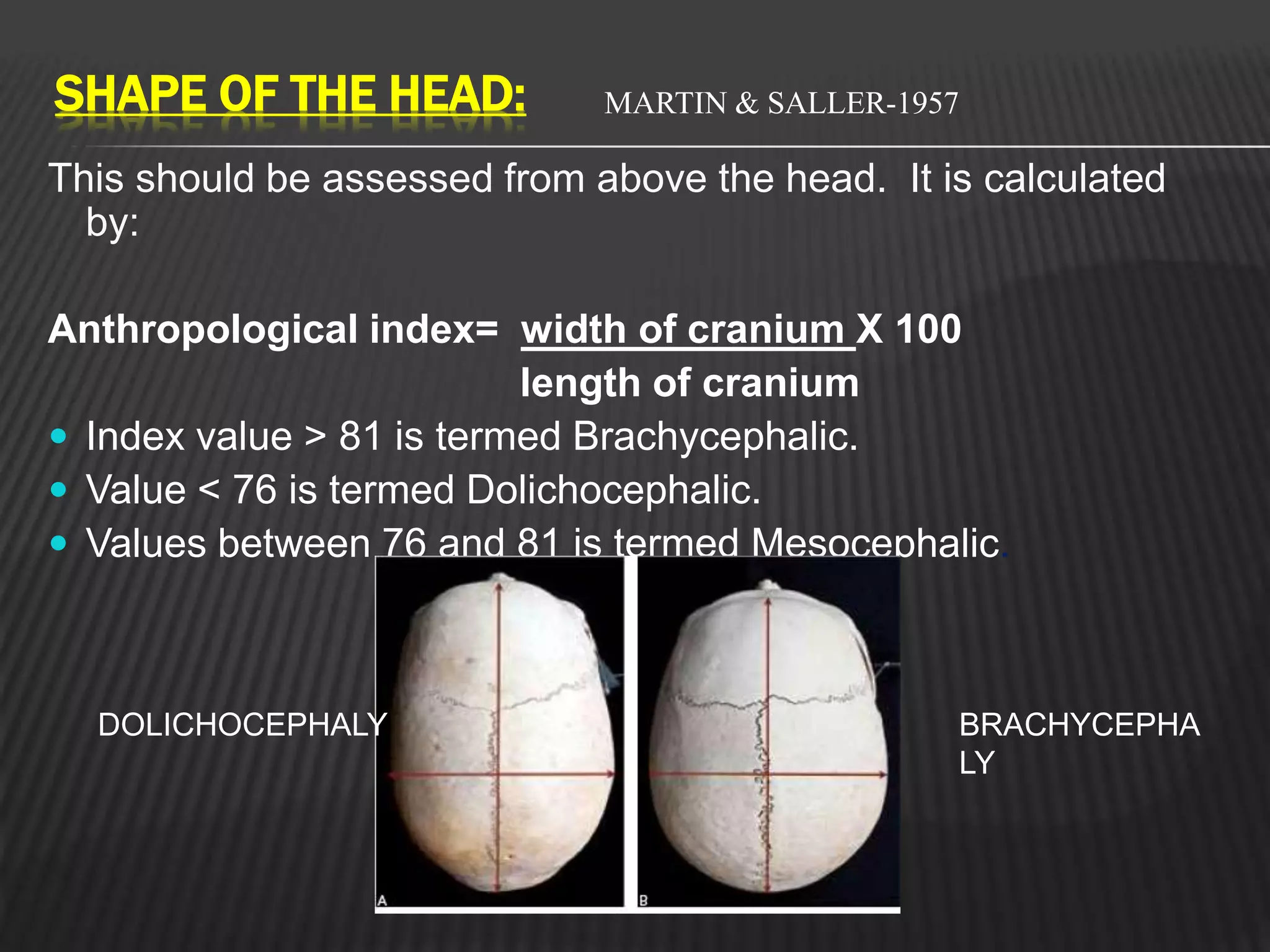
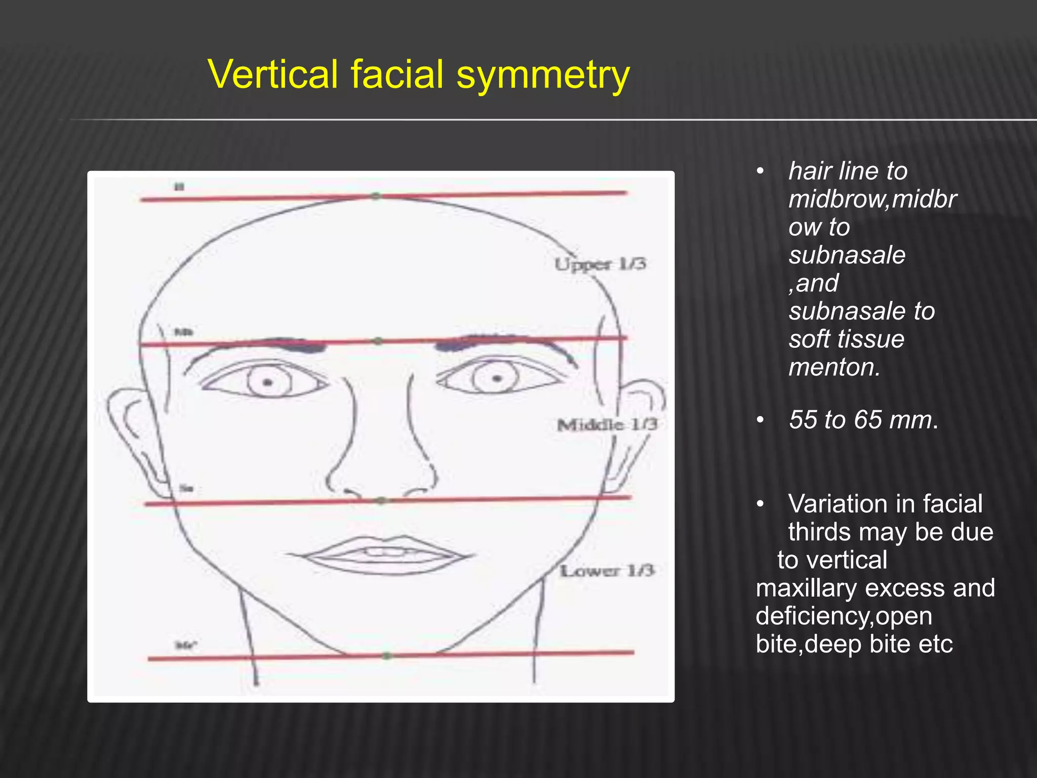
K.SANTOSHI
II MDS
ORTHODONTIC DIAGNOSIS
CONTENTS
Introduction
Steps in diagnosis and treatment planning
Diagnostic aids
Essential Aids
Case history
Clinical examination
General examination
Extra-oral examination
Intra-oral examination
Functional analysis
Study casts
Radiographs-Periapical, Bitewing,Panoramic.
Facial photographs
Supplemental Aids
The supplemental diagnostic aids include;
1-Specialized radiographs ex;
a-cephlometric radiographs
b-occlusal intra-oral films
c-selected lateral jaw views
d-cone shift technique
2.Electromyographic examination of muscle activity
3. Hand wrist radiographs to assess bone age or
maturation age
Conclusion
References
INTRODUCTION
Orthodontic diagnosis deals with recognition of the various
characteristics of the malocclusion.
It involves collection of data in a systematic manner to
help in identifying the nature and cause of the problem.
Orthodontic diagnosis should be based on sound scientific
knowledge combined at times with clinical experience and
common sense.
A proper diagnosis is essential for better treatment plan .
Orthodontic diagnosis – rakosi , graber
3
Contemporary orthodontic 5th edition proffit
Orthodontic diagnosis – rakosi , graber 7
DIAGNOSTIC AIDS
11
NAME OF THE PATIENT: For identification, For better communication and Medicolegal
legal records.
AGE : Chronologica age – Growth modification procedures
SEX : Timing of growth spurt and esthetics
OCCUPATION: Economic status , Occupational hazards
ADDRESS: for correspondance , to know the endemic and pandemic outburts
ETHENIC ORIGIN: ethnic facial charecteristics
CONTACT NUMBER:
CHIEF COMPLAINT: recorded in patients own words and in order
of preference & priority.
Most common logical reasons for orthodontic treatment will be
1)Impaired dento-facial esthetics leading to psychologicalproblems
2)Impaired function (chewing, speech and oral hygiene
maintenance) 3)Concern about alignment & occlusion of teeth.
4)Desire to enhance esthetics to improve quality of life.
Contemporary orthodontic 4th edition proffit
9
Medical history
H / O of previoushospitalization.
H / O chronic diseases like diabetes, cardiacproblems.
H / O allergy specially LATEX &NICKEL.
H / O bloodtransfusion & drugs
Tonsillectomy/Adenoidectomy
Epilepsy
PRENATAL HISTORY
Medications during pregnancy
Delivery- Full term/ Premature
Type - Normal/ Forceps/Caesarian
TMJ ankylosis due to prenatal trauma by
forceps delivery
POST NATAL HISTORY
FEEDING METHODS-(Breast or Bottlle
and
INJURIES- To Dento-Alveolar and Oro-Facial
structures,
HABITS ANY ABNORMAL ORAL HABITS.
ORAL HABITS
THUMB SUCKING
EXTRA ORAL EXAMINATION
Digits in acute thumb suckers Digits in chronic thumb
suckers
Reddens Fibrous
,rounghened
Clean , chapped Hypotonic upper lip
EFFECTS OF DIGIT- SUCKING
1. MAXILLA-
Proclination of maxillary incisors
Increased arch length
Increased anterior placement of apical base
of maxilla
Constricted maxilla
Increased clinical crown length of incisors
Counter-clockwise rotation of occlusal plane
Atypical root resorption of primary incisors
Trauma to the incisors
MANDIBLE -
Retroclination of lower incisors
Increased inter-molar width
Distal placement of mandible
INTER-ARCH RELATIONSHIP –
Decreased inter-incisal angle
Increased overjet
Posterior cross-bite
Anterior open-bite
Narrow nasal floor
TONGUE THRUSTING
Placement of the tongue tip forward between incisors during swallowing
Proffit
SIMPLE TONGUE THRUST
Contraction of lips, mentalis and mandibular elevators
Teeth are in occlusion as tongue protrudes into open bite
Open bite
Hypertrophy of tonsils which are not enlarged enough
Diminishes with the age
Treatment is simple
Good prognosis
COMPLEX TONGUE THRUST
Contractions of lips , facial and mentalis muscles
Lack of contractions of mandibular elevators
Teeth apart during swallow
History of chronic nasorespiratory disease and allergies
More diffuse open bite
Inflamed tonsils
Does not diminish with age
Poor prognosis
RETAINED INFANTILE SWALLOW
Strong contractions of lips and facial musculature
especially
buccinator.
Anterior and lateral thrusting
Inexpressive face
Difficulty in mastication
Poor prognosis
MALOCCLUSION
Proclination of upper anteriors
Anterior or posterior open bite
Protrusion of anterior segment of both arches
Constricted maxillary arch
Posterior cross bite
Spacing
MOUTH BREATHING
CLINICAL FEATURES
• Long and narrow face ( Adenoid
face)
• Narrow nose and nasal passage
• Short and flaccid upper lip
• Contracted upper arch with possibility
of posterior cross bite
• Excessive eruption of posteriors
• Constricted maxillary arch
• Excessive overjet
• Anterior openbite
• Marginal gingivitis
LIP HABITS
BRUXISM
FAMILY HISTORY
H / O cleft lip &palate
Hereditary dysgnathias include-
 Class II Div 2
 Skeletal open bite
 Bimaxillary protrusion
 Skeletal classIII
PHYSICAL GROWTH EVALUATION
 The best clinical effects are achieved in good growers and
poorest results are achieved in poor growers.
 By good growers, clinicians mean a patient with an amount,
rate, direction and pattern of growth that facilitates treatment.
 The most favourable time to attack many orthodontic problems
with skeletal manifestation is during growth acceleration in
puberty.
CONTEMPORARY ORTHODONTICS WILLIAM. R. PROFFIT 5TH
EDITION
 Thus, predicting nature and timing of onset of pubertal growth is
important in planning orthodontic therapy.
GENERAL EXAMINATION
1.BUILT :
Asthetic: thin built and usually possess narrow dental arches
Pletoric: obese built & generally have broad dental arches
Atheletic: neither thin nor obese normally built and normal dental
arches.
GENERAL EXAMINATION
2. BODY TYPE:
Sheldon in 1940 described body build as :
 Endomorphs- short and obese.
 Ectomorphs- long and thin.
 Mesomorphs- between endo and ectomorphs.
GENERAL EXAMINATION
 3.GAIT :
This is examined as the patient walks in the clinic. Any
neuromuscular defects should be made out in this
evaluation.
 4.POSTURE:
While evaluation of posture; look for kyphosis, lordosis or
scoliosis. There has been association of vertebral
abnormalities with facial disharmonies.
 5.HEIGHT AND WEIGHT:
They give a clue to physical maturation and growth of the
patient which may have dentofacial correlation.
28
SHAPE OF THE HEAD:
This should be assessed from above the head. It is calculated
by:
Anthropological index= width of cranium X 100
length of cranium
 Index value > 81 is termed Brachycephalic.
 Value < 76 is termed Dolichocephalic.
 Values between 76 and 81 is termed Mesocephalic.
DOLICHOCEPHALY BRACHYCEPHA
LY
MARTIN & SALLER-1957
Martin, R., and K. Saller (1957). Lehrbuch der
anthropologie. Gustav Fischer Verlag, Stuttgart
FACIAL INDEX
Defined as ratio between
morphological facial height &
bizygomatic distance.
Given by Martin & Saller in1957.
Morphologic facial index
=Morphologic facial height
Bizygomatic width
Contemporary orthodontic 4th edition proffit
31
FACIAL SYMMETRY
 The patient’s facial symmetry is examined to determine
disproportions of the face in transverse and vertical planes.
Gross facial asymmetry can occur as a result of:
 A. congenital defects
 B.hemi-facial atrophy/hypertrophy
 C.unilateral condylar ankylosis and hyperplasia
Vertical facial symmetry
• hair line to
midbrow,midbr
ow to
subnasale
,and
subnasale to
soft tissue
menton.
• 55 to 65 mm.
• Variation in facial
thirds may be due
to vertical
maxillary excess and
deficiency,open
bite,deep bite etc
Middle fifth of the face - a line
from inner canthus should be
coincided with ala of nose.
Medial two fifths of the face- a line
from the outer canthus of the eye
should be coincided with the gonial
angle of the mandible
Outer two fifths of the face-
measured from the base of the ear
to the helix of the ears
TRANSVERSE FACIAL PROPORTIONS
35
PROFILE ANALYSIS
Drop two lines
 A line joining the forehead and the
soft tissue point A (deepest point in
the curvature of upper lip)
 A line joining point A and the soft
tissue pogonion (most anterior point
of the chin)
N’
Sn
Pg’
PROFILE ANALYSIS
 Depending on the angle formed
Straight-Class I Convex-Class II Concave-Class III
FACIAL DIVERGENCE
 This term was coined by Milo Hellman in 1921.
 It is the anterior or posterior inclination of lower face relative to
forehead
 The facial angle formed by Nasion-Pogonion soft tissue line
and the frankfort horizontal line is used to define as facial
divergance.
FACIAL DIVERGENCE
STRAIGHT/ ORTHOGNATHIC-(90)
ANTERIOR DIVERGANCE-(more than 90)
POSTERIOR DIVERGANCE-(less than 90)
ASSESSMENT OF ANTERO- POSTERIOR JAW RELATIONSHIP
 Estimation is done by placement of the index and the middle
fingers at point A and point B respectively.
40
SKELETAL PATTERN
ASSESSMENT OF VERTICAL SKELETAL
RELATION
1. Average FMA angle- Two planes
meet at the occipital region.
2. Low angle- Two planes meet beyond
the occipital region.
3. High angle- Two planes meet at the
mastoid region in front of the ear.
41
 By the angle formed between Lower
border of the mandible and Frankfort
horizontal plane.
MANDIBULAR PLANE ANGLE
Both planes meet at occipitalregion.
I f they meet beyond it- Low angle or horizontal growth pattern.
I f they meet anterior- High angle or Vertical growth pattern.
Vertical grower Horizontal grower
SOFT TISSUE EXAMINATION
ACCORDING TO BURSTONE(AJO APRIL 1967)
LIP LENGTH
Upper lip length:
From: subnasale to
upper lip inferior
(19 to 22mm)
Lower lip length:
From: lower lip superior to
Menton
(38 to 44mm)
LIP MORPHOLOGY
ACCORDING TO BURSTONE(AJO APRIL 1967)
 Lips might be full,
thick(12 to 20mm) or
might be thin(6 to 10
mm).
 Full and everted are
usually associated with
proclined upper and
lower labial segments.
 Lips that are thin are
usually associated with
retroclined upper and
lower labial teeth.
KORKHA’S LIP STEPS-HORIZONTAL LIP ANALYSIS
POSITIVE LIP STEP NORMAL LIP STEP MARKED NEGATIVE
LIP STEP
LIP COMPETENCE
• In child patient, it is common for lips to be apart at rest.
• As the child progresses through to teens, increase in soft tissue maturation
means lips become competent.
• If older child or certainly adults have incompetent lips they may demonstrate
increased contraction of circumoral musculature to habitually keep lips
together.
• This can be seen as puckering in the skin over the chin caused by excessive
contraction of mentalis muscle
EXAMINATION OF LIPS :
47
Size :NormalShortThinThickEverted
Posture : CompetentIncompetentPotentially Incompetent
Competent
lips
Potentially incompetent
lips
Incompetent
lips
Contemporary orthodontic 4th edition proffit
NASOLABIAL ANGLE
Normal (90-100)/ Acute/Obtuse
ACUTE ANGLE NASO-LABIAL ANGLE OBTUSE ANGLE
Proclined upper anteriors
Prognathic maxilla
Increased angle-
Retrusion of upper lip
Retroclined upper anteriors
Retrognathic maxilla
Contemporary orthodontic 4th edition proffit
Normal 110
Protrusive upper lip
Decreased angle-
MENTOLABIAL SULCUS
49
 It is a fold of soft tissue between lower lip &
chin.
 Affected by- Facial Height
Overjet
Chin Projection.
Deep sulcus – Class II Div 1
Shallow sulcus – Bimaxillary protrusion
NORMAL SHALLOW DEEP
Hyperactive
mentalis activity
produces
puckering effect in
chin region called
as GOLF BALL
APPERANCE
Contemporary orthodontic 4th edition proffit
51
NOSE EXAMINATION
51
Size- One third of total face height.
Microrhinic
Macrorhinic
Nostrils- Oval & bilaterally symmetrical
Types of nose…1.leptorhine 2.mesorhine3.platyrhine
 STRAIGHT BRIDGE CONVEX BRIDGE CROOKED NOSE
Projection- Depends on
Bone over inferior border ofmandible
Soft tissue overchin
Overdevelopment of chin height alters position o
f
l
o
wer lip &
Interferes with lip closure.
Influence on profile-
-Protruding chin with deep mentolabial sulcus- Retrusive lip
profile.
-Negative chin formation with absence of sulcus- Protrusive lip
profile.
Orthodontics – current principles and technique 5th edition
CHIN
Adequate
chin
Excessive
chin
Recessive
chin
54
Orthodontics – current principles and technique 5th edition
VISUALIZED TREATMENT OBJECTIVE
This examination help s us deciding whether any functional
appliance that postures the mandible forward will improve the facial
profile and appearance.
Patient is instructed to swallow ,lick the lips and then relax. His profile
with teeth in habitual occlusion is observed . He is then asked to
bring the mandible forward into a correct sagittal relationship
reducing the overjet
Profile doesnot improve when
Excess anterior facial height
Deficient symphyseal development
Steep mandibular plane
Improved profile seen in
Anteriorly rotated growth patterns
Functional retrusion
Deep overbites
Excessive inter occlusal clearence with
normally positioned maxilla
It helps in predicting treatment changes that would occur in the future for
the patient.
The accuracy of prediction is a combination of the effect of treatment
procedures and accuracy of predicting future growth
They are not very accurate but may act as rough estimate of acurate
outcome
INTRA-ORAL EXAMINATION
 In general any investigation of the teeth and jaws aims to
determine 3 p’s
 Presence
 Position
 Pathology
Other features of teeth needed to be ascertained are
 Shape
 Size
 Developmental stage (if this is related to patient’s age)
PRESENCE
 In mixed dentition mobility of deciduous teeth must be
tested .
 At beginning of intra oral examination it is essential to
count the teeth as it is easy to overlook developmental
absence.
 Symmetry in components of occlusion must be determined
i.e., if a primary tooth is mobile on one side then other side
must be checked for.
POSITION
 Position of erupted and unerupted teeth
 Unerupted teeth -location.
 Darkened toothand if infarction line is on a tooth found on
transillumination, then special investigation like electric pulp
testing should be done.
 Also presence of large restorations, crowns, bridges,
implants, root canal fillings, ankylosed teeth and other dental
anomalies should be noted.
Attrition on incisal edge and a displacement is a functional
indication of orthodontic treatment.
Other health considerations also to look for are:
 Dental caries
 Periodontal disease
 Traumatic injury to teeth
 Tonsils
 The patient is asked to say A-a-h and the tongue is
depressed with a mouth mirror, it is possible to examine size
colour and form of pharyngeal tonsils.
EXAMINATION OF TONGUE
Abnormalities Of Tongue Can Upset The
Muscle Balance And Equilibrium Leading To
Malocclusion.
A Patient Whose Tongue Can Reach The Tip
Of The Nose Is Said To Have A Long Nose.
The Lingual Frenum Should Be Examined
For Tongue Tie
EXAMINATION OF THE PALATE
1. Dolicofacial patients have deep palate.
2. Presence of swellings in the palate
3. Mucosal ulcerations and indentations are a
feature of traumatic deep bite.
4. Presence of cleft in the palate.
5. The third rugae is usually in line with canines.
This is useful in the assessment of maxillary
anterior proclination.
EXAMINATION OF GINGIVA
Gingiva Should Be Examined For
1. Inflammation
2. Recession
3. Mucogingival Lesions
Anterior Gingivitis Common In Mouth Breathers
Due To Dryness Of Mouth Caused By Open Lip
Posture.
EXAMINATION OF FRENAL ATTACHMENTS
The maxillary labial frenum sometimes be thick
fibrous and attached relatively low.
This may lead to midline diastema.
Abnormal frenal attachment are diagnosed by
blench test.
EXAMINATION OF TONSILS AND ADENOIDS
Abnormaly Inflamed Tonsils Cause Alterations In
Tongue And Jaw Posture There By Upsetting The
Oro-facial Balance Leading To Malocclusion
Hard tissue
 Teeth present
 Un-erupted teeth
 Supernumerary teeth
 Missing teeth
 Retained teeth
 Crowding
66
 Rotated teeth
 Spacing
 Texture
 Caries
 Endodontically treated teeth
 Occlusal warefacets
67
PALATAL CONTOUR
68
 PALATAL HEIGHT INDEX (Korkhaus)
 Palatal height X 100
Posterior arch width
 Normal value is 42
39.3 51.3
RELATION OF MANDIBULAR TO MAXILLARY
ARCH
 Maximum incisal opening
 Freeway space
 Curve of spee
 Midline
 Upper
 Lower
 At rest
 In occlusion
69
ANTERIO-POSTERIOR RELATIONSHIP
 Molar relation
 Canine relation
 Incisor relation
 Over Jet & Bite
70
Class I Class II Class III
CANINE RELATION
71
INCISOR RELATIONSHIP
72
BSI1983 BSI198
3
BSI198
3
BSI198
3
Mageet AO. Classification of Skeletal and Dental Malocclusion:
Revisited. StomaEduJ. 2016;3(2)
SAGITTAL PLANE MALOCCLUSION
Pre-normal occlusion
-mandibular dental arch is
placed anteriorly in centric
occlusion
Post-normal occlusion
-mandibular dental arch is
placed more posteriorly in
centric occlusion
VERTICAL PLANE MALOCCLUSION
Deep bite
Vertical overlap between the
maxillary & mandibular teeth
is in excess than normal
Open bite
Exist in anterior or posterior
TRANSVERSE PLANE MALOCCLUSION
- includes various types of CROSS BITES
- mainly due to constriction of dental arches
INFRA-OCCLUSION
Supra occlusion Rotations
FUNCTIONAL EXAMINATION
 Improper functioning of the stomatognathic system can result in
various malocclusions.
 The functional examination should include :
 Assessment of postural rest position and inter occlusal space
 Path of closure
 Assessment of respiration
 Examination of TMJ
 Examination of swallowing
 Examination of speech
77
Assessment of postural rest position and inter – occlusal
clearance
 The postural rest position is the position of the mandible at which
the muscles that close the jaws and those that open them are in a
state of minimal contraction to maintain the posture of the
mandible.
 At the postural rest position, a space exist between the upper and
lower jaws. This space is called the inter occlusal clearance or
freeway space.
 Normally the freeway space is 3mm in canine regions.
78
Methods :
 Phonetics : ‘m’ or ‘c’ or ‘Mississippi’
 Command method : e.g. swallowing
 Non command method : e.g. visualize
 Measurement of inter occlusal clearance
 Direct intra oral procedure : vernier caliper
 Direct extra oral procedure
 Indirect extra oral procedure : e.g. radiographs,
Kinesiography
79
LATEROGNATHY
80
 Center of mandible is not aligned with facial midline in rest &
in occlusion
 True neuromuscular or anatomical asymmetry
 Lateral cross bite with laterognathy is True Cross bite.
LATERO OCCLUSION
81
 Skeletal midline shift of mandible can be observed only in
occlusion
 In postural position midlines are well aligned
 Deviation is due to tooth guidance.
 Known as Functional non true malocclusion.
EVALUATION OF PATH OF CLOSURE
Path of closure:
Movement of the mandible from Rest position  Habitual occlusion.
a. Forward path of closure : occurs in patients with mild skeletal
prenormalcy or edge to edge incisor contact.
b. Backward path of closure : Class II div 2 cases exhibit premature
incisor contact due to retroclined maxillary incisors.
c. Lateral path of closure : Associated with occlusal prematurity and
a narrow maxillary arch.
82
ASSESSMENT OF RESPIRATION
 Humans may exhibit 3 types of breathing :
-Nasal
-Oral
-Oro-nasal
 Tests to diagnose the type of respiration :
a) Mirror test
b) Cotton test
c) Water test
d) Observation
83
Mirror Test Cotton Test Water Test
84
EXAMINATION OF TMJ
 Patient is examined for symptoms of Temporomandibular joint
problems such as
 Clicking & Crepitus sounds
 Pain in the masticatory muscles
 Limitation of jaw movement
 Hyper mobility and morphological abnormalities.
 The maximum mouth opening (Normal: 40 – 45 mm)
85
SPEECH
-Certain malocclusions may cause defects in speech due to
interference with movement of the tongue and lips.
Speech sounds Problem Related malocclusion
/s/,/z/-sibilants Lisp Anterior open bite,
large gap between
incisors
/t/, /d/ linguoalveolar
stops
Difficulty in production Irregular incisors,
especially lingual
position of maxillary
incisors
/f/, /v/ labiodental
fricatives
Distortion Skeletal class III
th, sh, ch linguodental
fricatives
Distortion Anterior open bite
86
EVALUATION OF SWALLOWING
 The persistence of the infantile swallowing can be a cause for
Malocclusion
 Retained infantile swallow is indicated by the presence of
the following features :
a. Protrusion of the tip of the tongue.
b. Teeth occlude on only one molar in each quadrant
c. Violent contraction of perioral muscles during swallowing.
87
Infantile swallow (Visceral swallow)
 Jaws are apart, with the tongue between the gum pads
 Mandible is stabilized by contraction of the facial muscles and the
interposed tongue
 Swallow is guided & controlled by sensory interchange between the
lips and the tongue.
88
Mature swallow (Somatic swallow)
 Teeth are together
 Mandible is stabilized by contraction of the mandibular elevators.
 Tip of the tongue is held against the palate, above and behind the
incisors
89
METHODS OF EXAMINATION
 Electronic recording
 Electromyographic examination
 Recording of pressure exerted by the tongue
intraorally
 Roentgenocephalometric analysis
 Cineradiography
 Palatography
 Neurophysiologic examination
90
PALATOGRAPHIC EXAMINATION
91
STUDY CASTS
Orthodontic study models are accurate plaster reproductions of teeth and their
surrounding soft tissues .that are essential diagnostic aid that make it possible
to study the arrangement of teeth and the occlusion from all directions .
Uses of study model include:
a) They enable study of occlusion from all aspects
b) Enable accurate measurements to be made in dental arch.they help in
the measurement of arch length, arch width,and tooth size
c) Help in assessment of treatment progress by dentist as well as by patient
d) Help in assessing the nature and severity of malocclusion
e) Helpful in motivation of patient and to explain the treatment plan as well
as progress to patient and parents
g) Useful to transfer records in case patient is treated by another clinician
RADIOGRHIC ANALYSIS
94
LATERAL CEPHALOMETRY
95
CVMI
Degree of proclination of maxillary and mandibular
Incisors
Soft tissue analysis
Supplementary diagnostic aid
Types
Lateral cephalogram:
This is taken with the head in a standardized reproducible
position at a specified distance from x ray source
Frontal cephalogram
Provides the Anterior – Posterior view of skull
It helps in diagnosing Anterio posterior jaw relation
Growth pattern
Details of maxilla and mandible
CVMI
96
Vertrebral growth takes place from the cartilagenous layer on
the superior and inferior surfaces of the vertrebra..
Hassel and Farman (1995) found that the shapes of cervical
vertebrae werefound to differ with different level of skeletal
development
Cervical vertrebra maturation indices were determined
based on the presence of curvature in the inferior border,
shapes of bodies of the dens, C3 and C4 and intervertrebral
spacing.
PANORAMIC RADIOGRAPGH
Helps in diagnosing
presence of any impacted teeth
TMJ problems
any pathology
Enlarged Panoramic-
Accurate imaging in
anterior region.
Distortion in posterior
region.
Contemporary orthodontic 4th edition proffit
98
OCCLUSAL VIEW
Supplementary projection to locate malposed
unerupted teeth.
Palatal cleft
Expansion
Contemporary orthodontic 4th edition proffit 99
MP3
168
CONCLUSION
The patient assessment forms the essential basis of orthodontic
treatment. It is taken in steps starting from a
history/questionnaire and proceeding to a clinical examination
that includes extra-oral and intra-oral examination. The extra-
oral examination is carried out first as this can fundamentally
influence the treatment options. The skeletal pattern, soft tissue
form and the presence or absence of habits must all be
considered. The intra-oral assessment examines the oral health,
individual tooth positions and inter-occlusal relationships. When
this has been completed in conjunction with the extra-oral
examination, a treatment plan can then be formulated
 Contemporary orthodontic 4thedition proffit
 Orthodontic diagnosis – rakosi , graber
 Orthodontics – current principles and technique 5th edition
 Dentistry of child mcdonald
 Šidlauskienė M. Relationships between Malocclusion,
Body Posture, and Nasopharyngeal Pathology in Pre-
Orthodontic Children. Med Sci Monit. 2015;21:1765-
1773.
 Martin,R., and K.saller (1957). Leherbuch der
anthropologie. Gustav Fisher Verlag, Stuttgart.
REFERENCES

Orthodontic diagnosis

  • 1.
  • 2.
    CONTENTS Introduction Steps in diagnosisand treatment planning Diagnostic aids Essential Aids Case history Clinical examination General examination Extra-oral examination Intra-oral examination Functional analysis Study casts Radiographs-Periapical, Bitewing,Panoramic. Facial photographs
  • 3.
    Supplemental Aids The supplementaldiagnostic aids include; 1-Specialized radiographs ex; a-cephlometric radiographs b-occlusal intra-oral films c-selected lateral jaw views d-cone shift technique 2.Electromyographic examination of muscle activity 3. Hand wrist radiographs to assess bone age or maturation age Conclusion References
  • 4.
    INTRODUCTION Orthodontic diagnosis dealswith recognition of the various characteristics of the malocclusion. It involves collection of data in a systematic manner to help in identifying the nature and cause of the problem. Orthodontic diagnosis should be based on sound scientific knowledge combined at times with clinical experience and common sense. A proper diagnosis is essential for better treatment plan . Orthodontic diagnosis – rakosi , graber
  • 5.
  • 6.
    Orthodontic diagnosis –rakosi , graber 7 DIAGNOSTIC AIDS
  • 7.
  • 8.
    NAME OF THEPATIENT: For identification, For better communication and Medicolegal legal records. AGE : Chronologica age – Growth modification procedures SEX : Timing of growth spurt and esthetics OCCUPATION: Economic status , Occupational hazards ADDRESS: for correspondance , to know the endemic and pandemic outburts ETHENIC ORIGIN: ethnic facial charecteristics CONTACT NUMBER:
  • 9.
    CHIEF COMPLAINT: recordedin patients own words and in order of preference & priority. Most common logical reasons for orthodontic treatment will be 1)Impaired dento-facial esthetics leading to psychologicalproblems 2)Impaired function (chewing, speech and oral hygiene maintenance) 3)Concern about alignment & occlusion of teeth. 4)Desire to enhance esthetics to improve quality of life. Contemporary orthodontic 4th edition proffit 9
  • 10.
    Medical history H /O of previoushospitalization. H / O chronic diseases like diabetes, cardiacproblems. H / O allergy specially LATEX &NICKEL. H / O bloodtransfusion & drugs Tonsillectomy/Adenoidectomy Epilepsy PRENATAL HISTORY Medications during pregnancy Delivery- Full term/ Premature Type - Normal/ Forceps/Caesarian TMJ ankylosis due to prenatal trauma by forceps delivery POST NATAL HISTORY FEEDING METHODS-(Breast or Bottlle and INJURIES- To Dento-Alveolar and Oro-Facial structures, HABITS ANY ABNORMAL ORAL HABITS.
  • 11.
  • 12.
    THUMB SUCKING EXTRA ORALEXAMINATION Digits in acute thumb suckers Digits in chronic thumb suckers Reddens Fibrous ,rounghened Clean , chapped Hypotonic upper lip
  • 13.
    EFFECTS OF DIGIT-SUCKING 1. MAXILLA- Proclination of maxillary incisors Increased arch length Increased anterior placement of apical base of maxilla Constricted maxilla Increased clinical crown length of incisors Counter-clockwise rotation of occlusal plane Atypical root resorption of primary incisors Trauma to the incisors
  • 14.
    MANDIBLE - Retroclination oflower incisors Increased inter-molar width Distal placement of mandible INTER-ARCH RELATIONSHIP – Decreased inter-incisal angle Increased overjet Posterior cross-bite Anterior open-bite Narrow nasal floor
  • 15.
    TONGUE THRUSTING Placement ofthe tongue tip forward between incisors during swallowing Proffit SIMPLE TONGUE THRUST Contraction of lips, mentalis and mandibular elevators Teeth are in occlusion as tongue protrudes into open bite Open bite Hypertrophy of tonsils which are not enlarged enough Diminishes with the age Treatment is simple Good prognosis
  • 16.
    COMPLEX TONGUE THRUST Contractionsof lips , facial and mentalis muscles Lack of contractions of mandibular elevators Teeth apart during swallow History of chronic nasorespiratory disease and allergies More diffuse open bite Inflamed tonsils Does not diminish with age Poor prognosis
  • 17.
    RETAINED INFANTILE SWALLOW Strongcontractions of lips and facial musculature especially buccinator. Anterior and lateral thrusting Inexpressive face Difficulty in mastication Poor prognosis
  • 18.
    MALOCCLUSION Proclination of upperanteriors Anterior or posterior open bite Protrusion of anterior segment of both arches Constricted maxillary arch Posterior cross bite Spacing
  • 19.
    MOUTH BREATHING CLINICAL FEATURES •Long and narrow face ( Adenoid face) • Narrow nose and nasal passage • Short and flaccid upper lip • Contracted upper arch with possibility of posterior cross bite • Excessive eruption of posteriors • Constricted maxillary arch • Excessive overjet • Anterior openbite • Marginal gingivitis
  • 20.
  • 21.
    FAMILY HISTORY H /O cleft lip &palate Hereditary dysgnathias include-  Class II Div 2  Skeletal open bite  Bimaxillary protrusion  Skeletal classIII
  • 22.
    PHYSICAL GROWTH EVALUATION The best clinical effects are achieved in good growers and poorest results are achieved in poor growers.  By good growers, clinicians mean a patient with an amount, rate, direction and pattern of growth that facilitates treatment.  The most favourable time to attack many orthodontic problems with skeletal manifestation is during growth acceleration in puberty. CONTEMPORARY ORTHODONTICS WILLIAM. R. PROFFIT 5TH EDITION
  • 23.
     Thus, predictingnature and timing of onset of pubertal growth is important in planning orthodontic therapy.
  • 25.
    GENERAL EXAMINATION 1.BUILT : Asthetic:thin built and usually possess narrow dental arches Pletoric: obese built & generally have broad dental arches Atheletic: neither thin nor obese normally built and normal dental arches.
  • 26.
    GENERAL EXAMINATION 2. BODYTYPE: Sheldon in 1940 described body build as :  Endomorphs- short and obese.  Ectomorphs- long and thin.  Mesomorphs- between endo and ectomorphs.
  • 27.
    GENERAL EXAMINATION  3.GAIT: This is examined as the patient walks in the clinic. Any neuromuscular defects should be made out in this evaluation.  4.POSTURE: While evaluation of posture; look for kyphosis, lordosis or scoliosis. There has been association of vertebral abnormalities with facial disharmonies.  5.HEIGHT AND WEIGHT: They give a clue to physical maturation and growth of the patient which may have dentofacial correlation.
  • 28.
  • 29.
    SHAPE OF THEHEAD: This should be assessed from above the head. It is calculated by: Anthropological index= width of cranium X 100 length of cranium  Index value > 81 is termed Brachycephalic.  Value < 76 is termed Dolichocephalic.  Values between 76 and 81 is termed Mesocephalic. DOLICHOCEPHALY BRACHYCEPHA LY MARTIN & SALLER-1957
  • 30.
    Martin, R., andK. Saller (1957). Lehrbuch der anthropologie. Gustav Fischer Verlag, Stuttgart
  • 31.
    FACIAL INDEX Defined asratio between morphological facial height & bizygomatic distance. Given by Martin & Saller in1957. Morphologic facial index =Morphologic facial height Bizygomatic width Contemporary orthodontic 4th edition proffit 31
  • 33.
    FACIAL SYMMETRY  Thepatient’s facial symmetry is examined to determine disproportions of the face in transverse and vertical planes. Gross facial asymmetry can occur as a result of:  A. congenital defects  B.hemi-facial atrophy/hypertrophy  C.unilateral condylar ankylosis and hyperplasia
  • 34.
    Vertical facial symmetry •hair line to midbrow,midbr ow to subnasale ,and subnasale to soft tissue menton. • 55 to 65 mm. • Variation in facial thirds may be due to vertical maxillary excess and deficiency,open bite,deep bite etc
  • 35.
    Middle fifth ofthe face - a line from inner canthus should be coincided with ala of nose. Medial two fifths of the face- a line from the outer canthus of the eye should be coincided with the gonial angle of the mandible Outer two fifths of the face- measured from the base of the ear to the helix of the ears TRANSVERSE FACIAL PROPORTIONS 35
  • 36.
    PROFILE ANALYSIS Drop twolines  A line joining the forehead and the soft tissue point A (deepest point in the curvature of upper lip)  A line joining point A and the soft tissue pogonion (most anterior point of the chin) N’ Sn Pg’
  • 37.
    PROFILE ANALYSIS  Dependingon the angle formed Straight-Class I Convex-Class II Concave-Class III
  • 38.
    FACIAL DIVERGENCE  Thisterm was coined by Milo Hellman in 1921.  It is the anterior or posterior inclination of lower face relative to forehead  The facial angle formed by Nasion-Pogonion soft tissue line and the frankfort horizontal line is used to define as facial divergance.
  • 39.
    FACIAL DIVERGENCE STRAIGHT/ ORTHOGNATHIC-(90) ANTERIORDIVERGANCE-(more than 90) POSTERIOR DIVERGANCE-(less than 90)
  • 40.
    ASSESSMENT OF ANTERO-POSTERIOR JAW RELATIONSHIP  Estimation is done by placement of the index and the middle fingers at point A and point B respectively. 40 SKELETAL PATTERN
  • 41.
    ASSESSMENT OF VERTICALSKELETAL RELATION 1. Average FMA angle- Two planes meet at the occipital region. 2. Low angle- Two planes meet beyond the occipital region. 3. High angle- Two planes meet at the mastoid region in front of the ear. 41  By the angle formed between Lower border of the mandible and Frankfort horizontal plane.
  • 42.
    MANDIBULAR PLANE ANGLE Bothplanes meet at occipitalregion. I f they meet beyond it- Low angle or horizontal growth pattern. I f they meet anterior- High angle or Vertical growth pattern. Vertical grower Horizontal grower
  • 43.
    SOFT TISSUE EXAMINATION ACCORDINGTO BURSTONE(AJO APRIL 1967) LIP LENGTH Upper lip length: From: subnasale to upper lip inferior (19 to 22mm) Lower lip length: From: lower lip superior to Menton (38 to 44mm)
  • 44.
    LIP MORPHOLOGY ACCORDING TOBURSTONE(AJO APRIL 1967)  Lips might be full, thick(12 to 20mm) or might be thin(6 to 10 mm).  Full and everted are usually associated with proclined upper and lower labial segments.  Lips that are thin are usually associated with retroclined upper and lower labial teeth.
  • 45.
    KORKHA’S LIP STEPS-HORIZONTALLIP ANALYSIS POSITIVE LIP STEP NORMAL LIP STEP MARKED NEGATIVE LIP STEP
  • 46.
    LIP COMPETENCE • Inchild patient, it is common for lips to be apart at rest. • As the child progresses through to teens, increase in soft tissue maturation means lips become competent. • If older child or certainly adults have incompetent lips they may demonstrate increased contraction of circumoral musculature to habitually keep lips together. • This can be seen as puckering in the skin over the chin caused by excessive contraction of mentalis muscle
  • 47.
    EXAMINATION OF LIPS: 47 Size :NormalShortThinThickEverted Posture : CompetentIncompetentPotentially Incompetent Competent lips Potentially incompetent lips Incompetent lips Contemporary orthodontic 4th edition proffit
  • 48.
    NASOLABIAL ANGLE Normal (90-100)/Acute/Obtuse ACUTE ANGLE NASO-LABIAL ANGLE OBTUSE ANGLE Proclined upper anteriors Prognathic maxilla Increased angle- Retrusion of upper lip Retroclined upper anteriors Retrognathic maxilla Contemporary orthodontic 4th edition proffit Normal 110 Protrusive upper lip Decreased angle-
  • 49.
    MENTOLABIAL SULCUS 49  Itis a fold of soft tissue between lower lip & chin.  Affected by- Facial Height Overjet Chin Projection. Deep sulcus – Class II Div 1 Shallow sulcus – Bimaxillary protrusion NORMAL SHALLOW DEEP
  • 50.
    Hyperactive mentalis activity produces puckering effectin chin region called as GOLF BALL APPERANCE Contemporary orthodontic 4th edition proffit 51
  • 51.
    NOSE EXAMINATION 51 Size- Onethird of total face height. Microrhinic Macrorhinic Nostrils- Oval & bilaterally symmetrical Types of nose…1.leptorhine 2.mesorhine3.platyrhine  STRAIGHT BRIDGE CONVEX BRIDGE CROOKED NOSE
  • 52.
    Projection- Depends on Boneover inferior border ofmandible Soft tissue overchin Overdevelopment of chin height alters position o f l o wer lip & Interferes with lip closure. Influence on profile- -Protruding chin with deep mentolabial sulcus- Retrusive lip profile. -Negative chin formation with absence of sulcus- Protrusive lip profile. Orthodontics – current principles and technique 5th edition CHIN
  • 53.
  • 54.
    VISUALIZED TREATMENT OBJECTIVE Thisexamination help s us deciding whether any functional appliance that postures the mandible forward will improve the facial profile and appearance. Patient is instructed to swallow ,lick the lips and then relax. His profile with teeth in habitual occlusion is observed . He is then asked to bring the mandible forward into a correct sagittal relationship reducing the overjet
  • 55.
    Profile doesnot improvewhen Excess anterior facial height Deficient symphyseal development Steep mandibular plane Improved profile seen in Anteriorly rotated growth patterns Functional retrusion Deep overbites Excessive inter occlusal clearence with normally positioned maxilla
  • 56.
    It helps inpredicting treatment changes that would occur in the future for the patient. The accuracy of prediction is a combination of the effect of treatment procedures and accuracy of predicting future growth They are not very accurate but may act as rough estimate of acurate outcome
  • 57.
    INTRA-ORAL EXAMINATION  Ingeneral any investigation of the teeth and jaws aims to determine 3 p’s  Presence  Position  Pathology Other features of teeth needed to be ascertained are  Shape  Size  Developmental stage (if this is related to patient’s age)
  • 58.
    PRESENCE  In mixeddentition mobility of deciduous teeth must be tested .  At beginning of intra oral examination it is essential to count the teeth as it is easy to overlook developmental absence.  Symmetry in components of occlusion must be determined i.e., if a primary tooth is mobile on one side then other side must be checked for.
  • 59.
    POSITION  Position oferupted and unerupted teeth  Unerupted teeth -location.  Darkened toothand if infarction line is on a tooth found on transillumination, then special investigation like electric pulp testing should be done.  Also presence of large restorations, crowns, bridges, implants, root canal fillings, ankylosed teeth and other dental anomalies should be noted.
  • 60.
    Attrition on incisaledge and a displacement is a functional indication of orthodontic treatment. Other health considerations also to look for are:  Dental caries  Periodontal disease  Traumatic injury to teeth  Tonsils  The patient is asked to say A-a-h and the tongue is depressed with a mouth mirror, it is possible to examine size colour and form of pharyngeal tonsils.
  • 61.
    EXAMINATION OF TONGUE AbnormalitiesOf Tongue Can Upset The Muscle Balance And Equilibrium Leading To Malocclusion. A Patient Whose Tongue Can Reach The Tip Of The Nose Is Said To Have A Long Nose. The Lingual Frenum Should Be Examined For Tongue Tie
  • 62.
    EXAMINATION OF THEPALATE 1. Dolicofacial patients have deep palate. 2. Presence of swellings in the palate 3. Mucosal ulcerations and indentations are a feature of traumatic deep bite. 4. Presence of cleft in the palate. 5. The third rugae is usually in line with canines. This is useful in the assessment of maxillary anterior proclination.
  • 63.
    EXAMINATION OF GINGIVA GingivaShould Be Examined For 1. Inflammation 2. Recession 3. Mucogingival Lesions Anterior Gingivitis Common In Mouth Breathers Due To Dryness Of Mouth Caused By Open Lip Posture.
  • 64.
    EXAMINATION OF FRENALATTACHMENTS The maxillary labial frenum sometimes be thick fibrous and attached relatively low. This may lead to midline diastema. Abnormal frenal attachment are diagnosed by blench test.
  • 65.
    EXAMINATION OF TONSILSAND ADENOIDS Abnormaly Inflamed Tonsils Cause Alterations In Tongue And Jaw Posture There By Upsetting The Oro-facial Balance Leading To Malocclusion
  • 66.
    Hard tissue  Teethpresent  Un-erupted teeth  Supernumerary teeth  Missing teeth  Retained teeth  Crowding 66
  • 67.
     Rotated teeth Spacing  Texture  Caries  Endodontically treated teeth  Occlusal warefacets 67
  • 68.
    PALATAL CONTOUR 68  PALATALHEIGHT INDEX (Korkhaus)  Palatal height X 100 Posterior arch width  Normal value is 42 39.3 51.3
  • 69.
    RELATION OF MANDIBULARTO MAXILLARY ARCH  Maximum incisal opening  Freeway space  Curve of spee  Midline  Upper  Lower  At rest  In occlusion 69
  • 70.
    ANTERIO-POSTERIOR RELATIONSHIP  Molarrelation  Canine relation  Incisor relation  Over Jet & Bite 70 Class I Class II Class III
  • 71.
  • 72.
    INCISOR RELATIONSHIP 72 BSI1983 BSI198 3 BSI198 3 BSI198 3 MageetAO. Classification of Skeletal and Dental Malocclusion: Revisited. StomaEduJ. 2016;3(2)
  • 73.
    SAGITTAL PLANE MALOCCLUSION Pre-normalocclusion -mandibular dental arch is placed anteriorly in centric occlusion Post-normal occlusion -mandibular dental arch is placed more posteriorly in centric occlusion
  • 74.
    VERTICAL PLANE MALOCCLUSION Deepbite Vertical overlap between the maxillary & mandibular teeth is in excess than normal Open bite Exist in anterior or posterior
  • 75.
    TRANSVERSE PLANE MALOCCLUSION -includes various types of CROSS BITES - mainly due to constriction of dental arches
  • 76.
  • 77.
    FUNCTIONAL EXAMINATION  Improperfunctioning of the stomatognathic system can result in various malocclusions.  The functional examination should include :  Assessment of postural rest position and inter occlusal space  Path of closure  Assessment of respiration  Examination of TMJ  Examination of swallowing  Examination of speech 77
  • 78.
    Assessment of posturalrest position and inter – occlusal clearance  The postural rest position is the position of the mandible at which the muscles that close the jaws and those that open them are in a state of minimal contraction to maintain the posture of the mandible.  At the postural rest position, a space exist between the upper and lower jaws. This space is called the inter occlusal clearance or freeway space.  Normally the freeway space is 3mm in canine regions. 78
  • 79.
    Methods :  Phonetics: ‘m’ or ‘c’ or ‘Mississippi’  Command method : e.g. swallowing  Non command method : e.g. visualize  Measurement of inter occlusal clearance  Direct intra oral procedure : vernier caliper  Direct extra oral procedure  Indirect extra oral procedure : e.g. radiographs, Kinesiography 79
  • 80.
    LATEROGNATHY 80  Center ofmandible is not aligned with facial midline in rest & in occlusion  True neuromuscular or anatomical asymmetry  Lateral cross bite with laterognathy is True Cross bite.
  • 81.
    LATERO OCCLUSION 81  Skeletalmidline shift of mandible can be observed only in occlusion  In postural position midlines are well aligned  Deviation is due to tooth guidance.  Known as Functional non true malocclusion.
  • 82.
    EVALUATION OF PATHOF CLOSURE Path of closure: Movement of the mandible from Rest position  Habitual occlusion. a. Forward path of closure : occurs in patients with mild skeletal prenormalcy or edge to edge incisor contact. b. Backward path of closure : Class II div 2 cases exhibit premature incisor contact due to retroclined maxillary incisors. c. Lateral path of closure : Associated with occlusal prematurity and a narrow maxillary arch. 82
  • 83.
    ASSESSMENT OF RESPIRATION Humans may exhibit 3 types of breathing : -Nasal -Oral -Oro-nasal  Tests to diagnose the type of respiration : a) Mirror test b) Cotton test c) Water test d) Observation 83
  • 84.
    Mirror Test CottonTest Water Test 84
  • 85.
    EXAMINATION OF TMJ Patient is examined for symptoms of Temporomandibular joint problems such as  Clicking & Crepitus sounds  Pain in the masticatory muscles  Limitation of jaw movement  Hyper mobility and morphological abnormalities.  The maximum mouth opening (Normal: 40 – 45 mm) 85
  • 86.
    SPEECH -Certain malocclusions maycause defects in speech due to interference with movement of the tongue and lips. Speech sounds Problem Related malocclusion /s/,/z/-sibilants Lisp Anterior open bite, large gap between incisors /t/, /d/ linguoalveolar stops Difficulty in production Irregular incisors, especially lingual position of maxillary incisors /f/, /v/ labiodental fricatives Distortion Skeletal class III th, sh, ch linguodental fricatives Distortion Anterior open bite 86
  • 87.
    EVALUATION OF SWALLOWING The persistence of the infantile swallowing can be a cause for Malocclusion  Retained infantile swallow is indicated by the presence of the following features : a. Protrusion of the tip of the tongue. b. Teeth occlude on only one molar in each quadrant c. Violent contraction of perioral muscles during swallowing. 87
  • 88.
    Infantile swallow (Visceralswallow)  Jaws are apart, with the tongue between the gum pads  Mandible is stabilized by contraction of the facial muscles and the interposed tongue  Swallow is guided & controlled by sensory interchange between the lips and the tongue. 88
  • 89.
    Mature swallow (Somaticswallow)  Teeth are together  Mandible is stabilized by contraction of the mandibular elevators.  Tip of the tongue is held against the palate, above and behind the incisors 89
  • 90.
    METHODS OF EXAMINATION Electronic recording  Electromyographic examination  Recording of pressure exerted by the tongue intraorally  Roentgenocephalometric analysis  Cineradiography  Palatography  Neurophysiologic examination 90
  • 91.
  • 92.
    STUDY CASTS Orthodontic studymodels are accurate plaster reproductions of teeth and their surrounding soft tissues .that are essential diagnostic aid that make it possible to study the arrangement of teeth and the occlusion from all directions .
  • 93.
    Uses of studymodel include: a) They enable study of occlusion from all aspects b) Enable accurate measurements to be made in dental arch.they help in the measurement of arch length, arch width,and tooth size c) Help in assessment of treatment progress by dentist as well as by patient d) Help in assessing the nature and severity of malocclusion e) Helpful in motivation of patient and to explain the treatment plan as well as progress to patient and parents g) Useful to transfer records in case patient is treated by another clinician
  • 94.
  • 95.
    LATERAL CEPHALOMETRY 95 CVMI Degree ofproclination of maxillary and mandibular Incisors Soft tissue analysis Supplementary diagnostic aid Types Lateral cephalogram: This is taken with the head in a standardized reproducible position at a specified distance from x ray source Frontal cephalogram Provides the Anterior – Posterior view of skull It helps in diagnosing Anterio posterior jaw relation Growth pattern Details of maxilla and mandible
  • 96.
    CVMI 96 Vertrebral growth takesplace from the cartilagenous layer on the superior and inferior surfaces of the vertrebra.. Hassel and Farman (1995) found that the shapes of cervical vertebrae werefound to differ with different level of skeletal development Cervical vertrebra maturation indices were determined based on the presence of curvature in the inferior border, shapes of bodies of the dens, C3 and C4 and intervertrebral spacing.
  • 98.
    PANORAMIC RADIOGRAPGH Helps indiagnosing presence of any impacted teeth TMJ problems any pathology Enlarged Panoramic- Accurate imaging in anterior region. Distortion in posterior region. Contemporary orthodontic 4th edition proffit 98
  • 99.
    OCCLUSAL VIEW Supplementary projectionto locate malposed unerupted teeth. Palatal cleft Expansion Contemporary orthodontic 4th edition proffit 99
  • 100.
  • 101.
    CONCLUSION The patient assessmentforms the essential basis of orthodontic treatment. It is taken in steps starting from a history/questionnaire and proceeding to a clinical examination that includes extra-oral and intra-oral examination. The extra- oral examination is carried out first as this can fundamentally influence the treatment options. The skeletal pattern, soft tissue form and the presence or absence of habits must all be considered. The intra-oral assessment examines the oral health, individual tooth positions and inter-occlusal relationships. When this has been completed in conjunction with the extra-oral examination, a treatment plan can then be formulated
  • 102.
     Contemporary orthodontic4thedition proffit  Orthodontic diagnosis – rakosi , graber  Orthodontics – current principles and technique 5th edition  Dentistry of child mcdonald  Šidlauskienė M. Relationships between Malocclusion, Body Posture, and Nasopharyngeal Pathology in Pre- Orthodontic Children. Med Sci Monit. 2015;21:1765- 1773.  Martin,R., and K.saller (1957). Leherbuch der anthropologie. Gustav Fisher Verlag, Stuttgart. REFERENCES

Editor's Notes

  • #36  Describes the ideal transverse relationships of the face. The face is divided sagitally into five equal fifths from helix to helix of outer ears. Each of the segment equals one eye distance in width.
  • #41 Ideally maxillary base is 2-3 mm forward when in occlusion.
  • #42  Low angle – horizonta growing face High angle- vertical growing face
  • #47 Competent: Slight contact of lip when the musculature is relaxed.  Incompetent: Anatomically short lips which do not contact when musculature is relaxed. Lip seal is achieved only by active contraction of the orbicularis oris and mentalis muscle.  Potentially competent: Normal lips which fail to form the lip seal due to proclined upper incisors.  Everted lips: Hypertropied lips with weak muscular tonicity.
  • #53 .
  • #71 Class II ½ unit  End on Relation
  • #72 Class I: mesial incline of the upper canine overlaps the distal slope of the lower canine (The maxillary canine occludes between the mandibular canine and 1st premolar). Class II: Distal slope of the maxillary canine occludes or contact the mesial slope of the lower canine.
  • #73 Class I: When the mandibular incisor edge lie or below the cingulum plateau of the maxillary incisor (BSI, 1983), the overjet is 2-4 mm. Class II: When the mandibular incisors edges lie posterior to the cingulum plateau of the maxillary incisors (BSI, 1983). It could be:- Class II/1: Proclined maxillary incisors with overjet more than 4 mm. Class II/2a: Retroclined maxillary centrals and proclined laterals, or both central and lateral incisors are retroclined with normal or reduced overjet. Class II/2b: Retroclined maxillary centrals and proclined laterals, or both central and lateral incisors are retroclined but with increased overjet. Class III: When the mandibular incisors edges lie anterior to the cingulum plateau of the maxillary incisors (BSI, 1983). Class III type 1: Positive overjet but less than 2 mm. Class III type 2: Edge to edge incisors relationship. Class III type 3a: Negative overjet. Class III type 3b: Negative overjet but patient can make edge to edge (pseudo Class III). The author believes that incisor classification could also be used for esthetic considerations.
  • #92 Palatography involves recording the contact surfaces of the tongue with the palate and teeth while patient performs function( Speech/ Swallowing) Thin uniform layer of contrasting impression material is applied over the patients tongue with spatula.. Once the consonants produced or tongue movements carried out, palatogram can document photographically using surface mirror Accurate S Pronounciation  Mandible pushed forward slightly. Tongue rests on Teeth and Alveolar processes & a groove is formed in the center through which air stream is directed onto central incisors. Lisping  Tongue is protruded & clearly visible b/w anterior teeth.