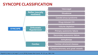
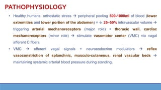
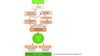
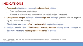
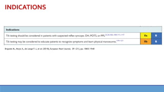
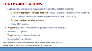
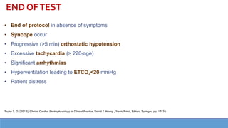
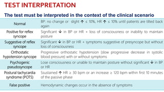
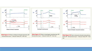
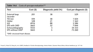
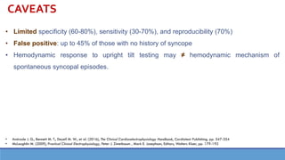
TILT TABLE TEST
PHUNG HUY HOANG, MD MSc
Department of Cardiac Arrhythmias
115 People’s Hospital
March, 2020
SYNCOPE CLASSIFICATION
SYNCOPE
Reflex (neurally
mediated)
Vasovagal
Situational
Carotid sinus syndrome
Orthostatic
Hypotension
Drug-induced OH
Volume depletion
Primary autonomic failure
Secondary autonomic failure
Cardiac
Arrhythmias
Cardiac structural disease
Cardiopulmonary and great vessels
Brignole M., Moya A., de Lange F. J., et al. (2018), European Heart Journal, 39 (21), pp. 1883-1948
PATHOPHYSIOLOGY
• Healthy humans: orthostatic stress → peripheral pooling 500-1000ml of blood (lower
extremities and lower portion of the abdomen) =  25–50% intravascular volume →
triggering arterial mechanoreceptors (major role) + thoracic wall, cardiac
mechanoreceptors (minor role) → stimulate vasomotor center (VMC) via vagal
afferent C fibers.
• VMC → efferent vagal signals + neuroendocrine modulators → reflex
vasoconstriction of splanchnic, musculo-cutaneous, renal vascular beds →
maintaining systemic arterial blood pressure during standing.
Freeman R. (2018), Harrison's Principles of Internal Medicine, J. Larry Jameson, et al.,
Editors, McGraw Hill, pp. 122-129
Prasad S., Wazni O., Chung M., et al. (2007), Handbook of Cardiac Electrophysiology, Andrea Natale , Oussama Wazni, Editors, Informa Healthcare, pp. 147-164.
Rev Port Cardiol (Engl Ed) 2019 Sep;38(9):661-673
INDICATIONS
• Recurrent episodes of syncope of undetermined etiology
• Absence of structural heart disease
• Presence of structural heart disease + cardiac causes of syncope excluded.
• Unexplained single syncopal episode/high-risk settings (potential risk for physical
injury, occupational hazard)
• Discriminate suspected reflex vs orthostatic hypotension syncope
• Identify patients with documented bradyarrhythmia during reflex syncope to
determine whether a vasodepressor response is present
• Andrade J. G., Bennett M. T., Deyell M. W., et al. (2016), The Clinical Cardiacelectrophysiology Handbook, Cardiotext Publishing, pp. 347-354
• Benditt D. (2018), UpToDate.
INDICATIONS
Brignole M., Moya A., de Lange F. J., et al. (2018), European Heart Journal, 39 (21), pp. 1883-1948
CONTRA-INDICATIONS
• Hypotension/Isoproterenol may cause myocardial or cerebral ischemia
o Critical obstructive cardiac disease (critical proximal coronary artery stenosis,
critical valvular stenosis, or severe left ventricular outflow obstruction)
o Critical cerebrovascular stenosis
o Recent MI, strokes
• Pregnant women (hypotension → potentially harmful to fetus)
• Inability to cooperate
• Weight exceeds safe table operation
• Uncontrolled hypertension
• Abedin Z., Conner R. (2007), Essential Cardiac Electrophysiology With Self Assessment, Blackwell Publishing, pp. 218-228
• Benditt D. (2018), UpToDate
• Brignole M., Moya A., de Lange F. J., et al. (2018), European Heart Journal, 39 (21), pp. e43-e80
PATIENT PREPARATION + EQUIPMENT
• Fast at least 2-4h
• IV catheter prior to the procedure (administration of fluids and/or medications ie,
isoproterenol or nitroglycerin)
• Continuous ECG, beat-to-beat blood pressure monitoring
• Room free of distractions and overstimulation (avoid autonomic triggers)
• Motorized tilt-table:
• with a foot board and safety restraints
• capable of smoothly and rapidly moving the patient passively between supine
position & head-up 60-80o and quickly (< 10 seconds) → avoid the consequences
of prolonged hypotension
• Brignole M., Moya A., de Lange F. J., et al. (2018), European Heart Journal, 39 (21), pp. e43-e80
• Taylor S. G. (2015), Clinical Cardiac Electrophysiology in Clinical Practice, David T. Huang , Travis Prinzi, Editors, Springer, pp. 17-36
• Benditt D. (2018), UpToDate
Prasad S., Burkhardt D., Dresing T. (2007), Handbook of Cardiac Electrophysiology,
Andrea Natale , Oussama Wazni, Editors, Informa Healthcare, pp. 247-257
PROCEDURE
Pre-tilt
phase
Passive
phase
Drug
provocation
phase
PROCEDURE
Pre-tilt phase
Passive
phase
Drug
provocation
phase
• Monitored/supine position: obtain baseline HR, BP
• Ensure*:
• supine pre-tilt phase of ≥ 5min when there is no venous cannulation
• ≥ 20min when there is venous cannulation.
*Brignole M., Moya A., de Lange F. J., et al. (2018), European Heart Journal, 39 (21), pp. e43-e80
European Heart Journal (2018) 39, e43–e80
PROCEDURE
Pre-tilt phase
Passive
phase
Drug
provocation
phase
• Upright with NO isoproterenol or nitroglycerin infusion
• Tilt angle 60-70o
• Min = 20min, Max = 45min
• Increasing angle increases sensitivity but also reduces specificity*
*Andrade J. G., Bennett M. T., Deyell M. W., et al. (2016), The Clinical Cardiacelectrophysiology Handbook, Cardiotext Publishing, pp. 347-354
Pre-tilt phase
Passive
phase
Drug
provocation
phase
• If the patient has remained asymptomatic during the passive phase
• An additional 15 to 20 minutes
• Provocative agents:  diagnostic yield by 20–25%*
PROCEDURE
(*) Abedin Z., Conner R. (2007), Essential Cardiac Electrophysiology With Self Assessment, Blackwell Publishing, pp. 218-228.
Pre-tilt phase
Passive
phase
Drug
provocation
phase
• Either drug (isoproterenol or nitroglycerin) can be used ∈ local practice and clinician
expertise
• Isoproterenol: titrated 1-3 /minute →  HR by 20-25% above baseline (with or
without returning the patient to the supine position)
• Nitroglycerin: fixed dose of 300-400g of sublingual (in upright position)
• Diagnostic rates of NTG ≥ Isoproterenol; NTG = simpler to use and better tolerated(1),(2)
• Isoprosterenol: positive response,  specificity(3)
(1) Raviele A., Giada F., Brignole M., et al. (2000), Am J Cardiol, 85 (10), pp. 1194-8
(2) Delepine S., Prunier F., Leftheriotis G., et al. (2002), Am J Cardiol, 90 (5), pp. 488-91
(3) Prasad S., Wazni O., Chung M., et al. (2007), Handbook of Cardiac Electrophysiology, Andrea Natale , Oussama Wazni, Editors, Informa Healthcare, pp. 147-164
PROCEDURE
Arthur W., Kaye G. C. (2000), Postgraduate Medical Journal, 76 (902), pp. 750-753
END OFTEST
• End of protocol in absence of symptoms
• Syncope occur
• Progressive (>5 min) orthostatic hypotension
• Excessive tachycardia (> 220-age)
• Significant arrhythmias
• Hyperventilation leading to ETCO2<20 mmHg
• Patient distress
Taylor S. G. (2015), Clinical Cardiac Electrophysiology in Clinical Practice, David T. Huang , Travis Prinzi, Editors, Springer, pp. 17-36
TEST INTERPRETATION
Normal
BP: no change or slight  ≤ 10%, HR  ≤ 10% until patients are tilted back
again
Positive for reflex
syncope
Significant  in BP or HR + loss of consciousness or inability to maintain
posture
Suggestive of reflex
syncope
Significant  in BP or HR + symptoms suggestive of presyncope but without
loss of consciousness
Orthostatic
hypotension syncope
Progressive orthostatic hypotension (slow progressive decrease in systolic
blood pressure) with or without symptoms
Psychogenic
pseudosyncope
Lose consciousness or unable to maintain posture without significant  in BP
or HR
Postural tachycardia
syndrome (POTS)
Ssustained  HR ≥ 30 bpm or an increase ≥ 120 bpm within first 10 minutes
of the passive phase
False positive Hemodynamic changes occur in the absence of symptoms
The test must be interpreted in the context of the clinical scenario
Different Types of
Response to Tilt-
Testing
Modified VASIS classification
(Vasovagal Syncope International
Study)
Iana Simova , E-journal of Cardiology Practice, Vol. 13,
N° 35, 2015
ESC 2018
Normal tilt table test results
Tilt-induced reflex syncope
Classical orthostatic hypotension
Delayed orthostatic hypotension
Psychogenic pseudosyncope
Postural orthostatic tachycardiasyndrome
The core features of reflex syncope
differentiating it from OH are a
latency after head-up tilt, a ‘convex’
BP decrease, and a decrease in HR
The hallmarks of classical OH are no
BP latency after head-up tilt, a
(upwards) concave shape of the
decrease, and if HR changes, it
increases
Prasad S., Wazni O., Chung M., et al. (2007), Handbook of Cardiac Electrophysiology, Andrea Natale , Oussama Wazni, Editors, Informa Healthcare, pp. 147-164.
CAVEATS
• Limited specificity (60-80%), sensitivity (30-70%), and reproducibility (70%)
• False positive: up to 45% of those with no history of syncope
• Hemodynamic response to upright tilt testing may ≠ hemodynamic mechanism of
spontaneous syncopal episodes.
• Andrade J. G., Bennett M. T., Deyell M. W., et al. (2016), The Clinical Cardiacelectrophysiology Handbook, Cardiotext Publishing, pp. 347-354
• McLaughlin M. (2009), Practical Clinical Electrophysiology, Peter J. Zimetbaum , Mark E. Josephson, Editors, Wolters Kluer, pp. 179-192
COMPLICATIONS
• MINIMAL!
• NO reported deaths during test1
• Ranging from nausea/vomiting, headaches (NTG), palpitations (Isoprosterenol) to the
effects of a syncopal episode (quick return to supine position + raising legs = enough to
restore consciousness in most patients)
• Rarely ventricular arrhythmias (especially with isoproterenol provocation)2
• Self-limited atrial fibrillation2
1. Brignole M., Moya A., de Lange F. J., et al. (2018), European Heart Journal, 39 (21), pp. e43-e80
2. Prasad S., Wazni O., Chung M., et al. (2007), Handbook of Cardiac Electrophysiology, Andrea Natale , Oussama Wazni, Editors, Informa Healthcare, pp. 147-164
1. Abedin Z., Conner R. (2007), "Syncope", Essential Cardiac Electrophysiology With Self Assessment, Blackwell Publishing, pp. 218-228.
2. Arthur W., Kaye G.C. (2000), "The pathophysiology of common causes of syncope", Postgraduate Medical Journal, 76 (902), pp. 750-753.
3. Brignole M., Moya A., de Lange F.J., et al. (2018), "Practical Instructions for the 2018 ESC Guidelines for the diagnosis and management of
syncope", European Heart Journal, 39 (21), pp. e43-e80.
4. Delepine S., Prunier F., Leftheriotis G., et al. (2002), "Comparison between isoproterenol and nitroglycerin sensitized head-upright tilt in patients
with unexplained syncope and negative or positive passive head-up tilt response", Am J Cardiol, 90 (5), pp. 488-91.
5. McLaughlin M. (2009), "Syncope", Practical Clinical Electrophysiology, Peter J. Zimetbaum , Mark E. Josephson, Editors, Wolters Kluer, pp. 179-
192.
6. Prasad S., Burkhardt D., Dresing T. (2007), "Head-up tilt (HUT) table testing", Handbook of Cardiac Electrophysiology, Andrea Natale ,
Oussama Wazni, Editors, Informa Healthcare, pp. 247-257.
7. Prasad S., Wazni O., Chung M., et al. (2007), "Syncope events, definitions, causes, and features", Handbook of Cardiac Electrophysiology,
Andrea Natale , Oussama Wazni, Editors, Informa Healthcare, pp. 147-164.
8. Raviele A., Giada F., Brignole M., et al. (2000), "Comparison of diagnostic accuracy of sublingual nitroglycerin test and low-dose isoproterenol
test in patients with unexplained syncope", Am J Cardiol, 85 (10), pp. 1194-8.
9. Taylor S.G. (2015), "Syncope, Tilt Testing, and Cardioversion", Clinical Cardiac Electrophysiology in Clinical Practice, David T. Huang , Travis
Prinzi, Editors, Springer, pp. 17-36.
10. Andrade J.G., Bennett M.T., Deyell M.W., et al. (2016), "Syncope", The Clinical Cardiacelectrophysiology Handbook, Cardiotext Publishing, pp.
347-354.
11. Benditt D. (2018), "Upright tilt table testing in the evaluation of syncope", UpToDate.
12. Brignole M., Moya A., de Lange F.J., et al. (2018), "2018 ESC Guidelines for the diagnosis and management of syncope", European Heart
Journal, 39 (21), pp. 1883-1948.
13. Freeman R. (2018), "Syncope", Harrison's Principles of Internal Medicine, J. Larry Jameson, et al., Editors, McGraw Hill, pp. 122-129.
REFERENCES
TILT TABLE TEST
PHUNG HUY HOANG, MD MSc
Department of Cardiac Arrhythmias
115 People’s Hospital
March, 2020

Tilt table test - Nghiệm pháp bàn nghiêng

  • 1.
    TILT TABLE TEST PHUNGHUY HOANG, MD MSc Department of Cardiac Arrhythmias 115 People’s Hospital March, 2020
  • 2.
    SYNCOPE CLASSIFICATION SYNCOPE Reflex (neurally mediated) Vasovagal Situational Carotidsinus syndrome Orthostatic Hypotension Drug-induced OH Volume depletion Primary autonomic failure Secondary autonomic failure Cardiac Arrhythmias Cardiac structural disease Cardiopulmonary and great vessels Brignole M., Moya A., de Lange F. J., et al. (2018), European Heart Journal, 39 (21), pp. 1883-1948
  • 3.
    PATHOPHYSIOLOGY • Healthy humans:orthostatic stress → peripheral pooling 500-1000ml of blood (lower extremities and lower portion of the abdomen) =  25–50% intravascular volume → triggering arterial mechanoreceptors (major role) + thoracic wall, cardiac mechanoreceptors (minor role) → stimulate vasomotor center (VMC) via vagal afferent C fibers. • VMC → efferent vagal signals + neuroendocrine modulators → reflex vasoconstriction of splanchnic, musculo-cutaneous, renal vascular beds → maintaining systemic arterial blood pressure during standing.
  • 4.
    Freeman R. (2018),Harrison's Principles of Internal Medicine, J. Larry Jameson, et al., Editors, McGraw Hill, pp. 122-129
  • 5.
    Prasad S., WazniO., Chung M., et al. (2007), Handbook of Cardiac Electrophysiology, Andrea Natale , Oussama Wazni, Editors, Informa Healthcare, pp. 147-164.
  • 6.
    Rev Port Cardiol(Engl Ed) 2019 Sep;38(9):661-673
  • 7.
    INDICATIONS • Recurrent episodesof syncope of undetermined etiology • Absence of structural heart disease • Presence of structural heart disease + cardiac causes of syncope excluded. • Unexplained single syncopal episode/high-risk settings (potential risk for physical injury, occupational hazard) • Discriminate suspected reflex vs orthostatic hypotension syncope • Identify patients with documented bradyarrhythmia during reflex syncope to determine whether a vasodepressor response is present • Andrade J. G., Bennett M. T., Deyell M. W., et al. (2016), The Clinical Cardiacelectrophysiology Handbook, Cardiotext Publishing, pp. 347-354 • Benditt D. (2018), UpToDate.
  • 8.
    INDICATIONS Brignole M., MoyaA., de Lange F. J., et al. (2018), European Heart Journal, 39 (21), pp. 1883-1948
  • 9.
    CONTRA-INDICATIONS • Hypotension/Isoproterenol maycause myocardial or cerebral ischemia o Critical obstructive cardiac disease (critical proximal coronary artery stenosis, critical valvular stenosis, or severe left ventricular outflow obstruction) o Critical cerebrovascular stenosis o Recent MI, strokes • Pregnant women (hypotension → potentially harmful to fetus) • Inability to cooperate • Weight exceeds safe table operation • Uncontrolled hypertension • Abedin Z., Conner R. (2007), Essential Cardiac Electrophysiology With Self Assessment, Blackwell Publishing, pp. 218-228 • Benditt D. (2018), UpToDate • Brignole M., Moya A., de Lange F. J., et al. (2018), European Heart Journal, 39 (21), pp. e43-e80
  • 10.
    PATIENT PREPARATION +EQUIPMENT • Fast at least 2-4h • IV catheter prior to the procedure (administration of fluids and/or medications ie, isoproterenol or nitroglycerin) • Continuous ECG, beat-to-beat blood pressure monitoring • Room free of distractions and overstimulation (avoid autonomic triggers) • Motorized tilt-table: • with a foot board and safety restraints • capable of smoothly and rapidly moving the patient passively between supine position & head-up 60-80o and quickly (< 10 seconds) → avoid the consequences of prolonged hypotension • Brignole M., Moya A., de Lange F. J., et al. (2018), European Heart Journal, 39 (21), pp. e43-e80 • Taylor S. G. (2015), Clinical Cardiac Electrophysiology in Clinical Practice, David T. Huang , Travis Prinzi, Editors, Springer, pp. 17-36 • Benditt D. (2018), UpToDate
  • 11.
    Prasad S., BurkhardtD., Dresing T. (2007), Handbook of Cardiac Electrophysiology, Andrea Natale , Oussama Wazni, Editors, Informa Healthcare, pp. 247-257
  • 12.
  • 13.
    PROCEDURE Pre-tilt phase Passive phase Drug provocation phase • Monitored/supineposition: obtain baseline HR, BP • Ensure*: • supine pre-tilt phase of ≥ 5min when there is no venous cannulation • ≥ 20min when there is venous cannulation. *Brignole M., Moya A., de Lange F. J., et al. (2018), European Heart Journal, 39 (21), pp. e43-e80 European Heart Journal (2018) 39, e43–e80
  • 14.
    PROCEDURE Pre-tilt phase Passive phase Drug provocation phase • Uprightwith NO isoproterenol or nitroglycerin infusion • Tilt angle 60-70o • Min = 20min, Max = 45min • Increasing angle increases sensitivity but also reduces specificity* *Andrade J. G., Bennett M. T., Deyell M. W., et al. (2016), The Clinical Cardiacelectrophysiology Handbook, Cardiotext Publishing, pp. 347-354
  • 15.
    Pre-tilt phase Passive phase Drug provocation phase • Ifthe patient has remained asymptomatic during the passive phase • An additional 15 to 20 minutes • Provocative agents:  diagnostic yield by 20–25%* PROCEDURE (*) Abedin Z., Conner R. (2007), Essential Cardiac Electrophysiology With Self Assessment, Blackwell Publishing, pp. 218-228.
  • 16.
    Pre-tilt phase Passive phase Drug provocation phase • Eitherdrug (isoproterenol or nitroglycerin) can be used ∈ local practice and clinician expertise • Isoproterenol: titrated 1-3 /minute →  HR by 20-25% above baseline (with or without returning the patient to the supine position) • Nitroglycerin: fixed dose of 300-400g of sublingual (in upright position) • Diagnostic rates of NTG ≥ Isoproterenol; NTG = simpler to use and better tolerated(1),(2) • Isoprosterenol: positive response,  specificity(3) (1) Raviele A., Giada F., Brignole M., et al. (2000), Am J Cardiol, 85 (10), pp. 1194-8 (2) Delepine S., Prunier F., Leftheriotis G., et al. (2002), Am J Cardiol, 90 (5), pp. 488-91 (3) Prasad S., Wazni O., Chung M., et al. (2007), Handbook of Cardiac Electrophysiology, Andrea Natale , Oussama Wazni, Editors, Informa Healthcare, pp. 147-164 PROCEDURE
  • 17.
    Arthur W., KayeG. C. (2000), Postgraduate Medical Journal, 76 (902), pp. 750-753
  • 18.
    END OFTEST • Endof protocol in absence of symptoms • Syncope occur • Progressive (>5 min) orthostatic hypotension • Excessive tachycardia (> 220-age) • Significant arrhythmias • Hyperventilation leading to ETCO2<20 mmHg • Patient distress Taylor S. G. (2015), Clinical Cardiac Electrophysiology in Clinical Practice, David T. Huang , Travis Prinzi, Editors, Springer, pp. 17-36
  • 19.
    TEST INTERPRETATION Normal BP: nochange or slight  ≤ 10%, HR  ≤ 10% until patients are tilted back again Positive for reflex syncope Significant  in BP or HR + loss of consciousness or inability to maintain posture Suggestive of reflex syncope Significant  in BP or HR + symptoms suggestive of presyncope but without loss of consciousness Orthostatic hypotension syncope Progressive orthostatic hypotension (slow progressive decrease in systolic blood pressure) with or without symptoms Psychogenic pseudosyncope Lose consciousness or unable to maintain posture without significant  in BP or HR Postural tachycardia syndrome (POTS) Ssustained  HR ≥ 30 bpm or an increase ≥ 120 bpm within first 10 minutes of the passive phase False positive Hemodynamic changes occur in the absence of symptoms The test must be interpreted in the context of the clinical scenario
  • 20.
    Different Types of Responseto Tilt- Testing Modified VASIS classification (Vasovagal Syncope International Study) Iana Simova , E-journal of Cardiology Practice, Vol. 13, N° 35, 2015
  • 21.
    ESC 2018 Normal tilttable test results Tilt-induced reflex syncope Classical orthostatic hypotension Delayed orthostatic hypotension Psychogenic pseudosyncope Postural orthostatic tachycardiasyndrome
  • 22.
    The core featuresof reflex syncope differentiating it from OH are a latency after head-up tilt, a ‘convex’ BP decrease, and a decrease in HR The hallmarks of classical OH are no BP latency after head-up tilt, a (upwards) concave shape of the decrease, and if HR changes, it increases
  • 24.
    Prasad S., WazniO., Chung M., et al. (2007), Handbook of Cardiac Electrophysiology, Andrea Natale , Oussama Wazni, Editors, Informa Healthcare, pp. 147-164.
  • 25.
    CAVEATS • Limited specificity(60-80%), sensitivity (30-70%), and reproducibility (70%) • False positive: up to 45% of those with no history of syncope • Hemodynamic response to upright tilt testing may ≠ hemodynamic mechanism of spontaneous syncopal episodes. • Andrade J. G., Bennett M. T., Deyell M. W., et al. (2016), The Clinical Cardiacelectrophysiology Handbook, Cardiotext Publishing, pp. 347-354 • McLaughlin M. (2009), Practical Clinical Electrophysiology, Peter J. Zimetbaum , Mark E. Josephson, Editors, Wolters Kluer, pp. 179-192
  • 26.
    COMPLICATIONS • MINIMAL! • NOreported deaths during test1 • Ranging from nausea/vomiting, headaches (NTG), palpitations (Isoprosterenol) to the effects of a syncopal episode (quick return to supine position + raising legs = enough to restore consciousness in most patients) • Rarely ventricular arrhythmias (especially with isoproterenol provocation)2 • Self-limited atrial fibrillation2 1. Brignole M., Moya A., de Lange F. J., et al. (2018), European Heart Journal, 39 (21), pp. e43-e80 2. Prasad S., Wazni O., Chung M., et al. (2007), Handbook of Cardiac Electrophysiology, Andrea Natale , Oussama Wazni, Editors, Informa Healthcare, pp. 147-164
  • 27.
    1. Abedin Z.,Conner R. (2007), "Syncope", Essential Cardiac Electrophysiology With Self Assessment, Blackwell Publishing, pp. 218-228. 2. Arthur W., Kaye G.C. (2000), "The pathophysiology of common causes of syncope", Postgraduate Medical Journal, 76 (902), pp. 750-753. 3. Brignole M., Moya A., de Lange F.J., et al. (2018), "Practical Instructions for the 2018 ESC Guidelines for the diagnosis and management of syncope", European Heart Journal, 39 (21), pp. e43-e80. 4. Delepine S., Prunier F., Leftheriotis G., et al. (2002), "Comparison between isoproterenol and nitroglycerin sensitized head-upright tilt in patients with unexplained syncope and negative or positive passive head-up tilt response", Am J Cardiol, 90 (5), pp. 488-91. 5. McLaughlin M. (2009), "Syncope", Practical Clinical Electrophysiology, Peter J. Zimetbaum , Mark E. Josephson, Editors, Wolters Kluer, pp. 179- 192. 6. Prasad S., Burkhardt D., Dresing T. (2007), "Head-up tilt (HUT) table testing", Handbook of Cardiac Electrophysiology, Andrea Natale , Oussama Wazni, Editors, Informa Healthcare, pp. 247-257. 7. Prasad S., Wazni O., Chung M., et al. (2007), "Syncope events, definitions, causes, and features", Handbook of Cardiac Electrophysiology, Andrea Natale , Oussama Wazni, Editors, Informa Healthcare, pp. 147-164. 8. Raviele A., Giada F., Brignole M., et al. (2000), "Comparison of diagnostic accuracy of sublingual nitroglycerin test and low-dose isoproterenol test in patients with unexplained syncope", Am J Cardiol, 85 (10), pp. 1194-8. 9. Taylor S.G. (2015), "Syncope, Tilt Testing, and Cardioversion", Clinical Cardiac Electrophysiology in Clinical Practice, David T. Huang , Travis Prinzi, Editors, Springer, pp. 17-36. 10. Andrade J.G., Bennett M.T., Deyell M.W., et al. (2016), "Syncope", The Clinical Cardiacelectrophysiology Handbook, Cardiotext Publishing, pp. 347-354. 11. Benditt D. (2018), "Upright tilt table testing in the evaluation of syncope", UpToDate. 12. Brignole M., Moya A., de Lange F.J., et al. (2018), "2018 ESC Guidelines for the diagnosis and management of syncope", European Heart Journal, 39 (21), pp. 1883-1948. 13. Freeman R. (2018), "Syncope", Harrison's Principles of Internal Medicine, J. Larry Jameson, et al., Editors, McGraw Hill, pp. 122-129. REFERENCES
  • 28.
    TILT TABLE TEST PHUNGHUY HOANG, MD MSc Department of Cardiac Arrhythmias 115 People’s Hospital March, 2020