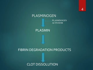
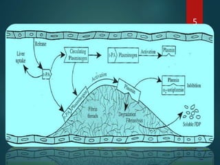
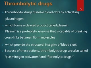
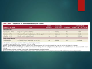
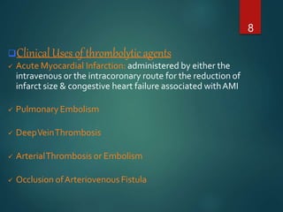
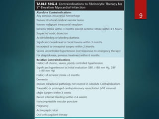
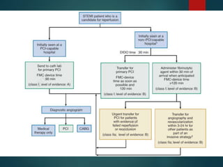
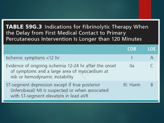
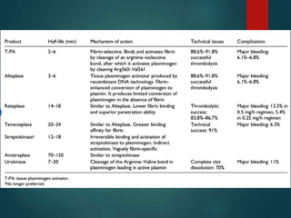
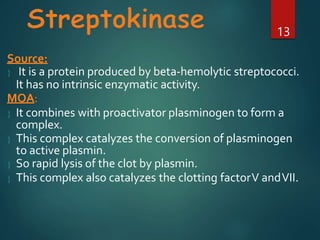
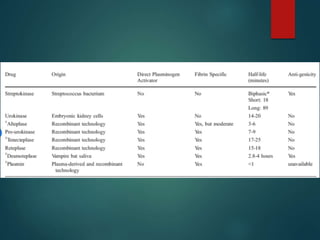
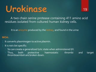
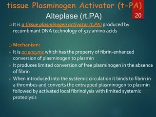
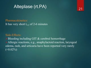
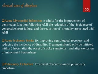
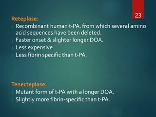
Thrombolytic agents work by activating plasminogen to form plasmin, which breaks down fibrin in blood clots. The document discusses several thrombolytic agents: streptokinase works by forming a complex with plasminogen to catalyze its activation; alteplase is a recombinant tissue plasminogen activator that selectively activates plasminogen bound to fibrin clots; and reteplase and tenecteplase are modified forms of tPA with longer durations of action. Thrombolytic agents are used to treat conditions caused by blood clots such as myocardial infarction, pulmonary embolism, and ischemic stroke.
Study Material
Myocardial infarction (MI), commonly known as a heart attack. MI is a blockage of blood flow to the heart muscle. Myocardial infarction (MI) refers to tissue death (infarction) of the heart muscle (myocardium). It is a type of acute coronary syndrome, which describes a sudden or short-term change in symptoms related to blood flow to the heart. Myocardial infarction is a common presentation of coronary artery disease. The World Health Organization estimated in 2004, that 12.2% of worldwide deaths were from ischemic heart disease.
thrombolytics notes
Thrombolytic therapy is the use of drugs to break up or dissolve blood clots, which are the main cause of both heart attacks and stroke.
A detailed information Thrombolytic or fibrinolytics. Comes under the Drugs affecting the blood and blood forming category. These drugs are used to lyse the clot.
TEST BANK for Operations Management, 14th Edition by William J. Stevenson, Ve...kevinkariuki227
TEST BANK for Operations Management, 14th Edition by William J. Stevenson, Verified Chapters 1 - 19, Complete Newest Version.pdf
Title: Sense of Smell
Presenter: Dr. Faiza, Assistant Professor of Physiology
Qualifications:
MBBS (Best Graduate, AIMC Lahore)
FCPS Physiology
ICMT, CHPE, DHPE (STMU)
MPH (GC University, Faisalabad)
MBA (Virtual University of Pakistan)
Learning Objectives:
Describe the primary categories of smells and the concept of odor blindness.
Explain the structure and location of the olfactory membrane and mucosa, including the types and roles of cells involved in olfaction.
Describe the pathway and mechanisms of olfactory signal transmission from the olfactory receptors to the brain.
Illustrate the biochemical cascade triggered by odorant binding to olfactory receptors, including the role of G-proteins and second messengers in generating an action potential.
Identify different types of olfactory disorders such as anosmia, hyposmia, hyperosmia, and dysosmia, including their potential causes.
Key Topics:
Olfactory Genes:
3% of the human genome accounts for olfactory genes.
400 genes for odorant receptors.
Olfactory Membrane:
Located in the superior part of the nasal cavity.
Medially: Folds downward along the superior septum.
Laterally: Folds over the superior turbinate and upper surface of the middle turbinate.
Total surface area: 5-10 square centimeters.
Olfactory Mucosa:
Olfactory Cells: Bipolar nerve cells derived from the CNS (100 million), with 4-25 olfactory cilia per cell.
Sustentacular Cells: Produce mucus and maintain ionic and molecular environment.
Basal Cells: Replace worn-out olfactory cells with an average lifespan of 1-2 months.
Bowman’s Gland: Secretes mucus.
Stimulation of Olfactory Cells:
Odorant dissolves in mucus and attaches to receptors on olfactory cilia.
Involves a cascade effect through G-proteins and second messengers, leading to depolarization and action potential generation in the olfactory nerve.
Quality of a Good Odorant:
Small (3-20 Carbon atoms), volatile, water-soluble, and lipid-soluble.
Facilitated by odorant-binding proteins in mucus.
Membrane Potential and Action Potential:
Resting membrane potential: -55mV.
Action potential frequency in the olfactory nerve increases with odorant strength.
Adaptation Towards the Sense of Smell:
Rapid adaptation within the first second, with further slow adaptation.
Psychological adaptation greater than receptor adaptation, involving feedback inhibition from the central nervous system.
Primary Sensations of Smell:
Camphoraceous, Musky, Floral, Pepperminty, Ethereal, Pungent, Putrid.
Odor Detection Threshold:
Examples: Hydrogen sulfide (0.0005 ppm), Methyl-mercaptan (0.002 ppm).
Some toxic substances are odorless at lethal concentrations.
Characteristics of Smell:
Odor blindness for single substances due to lack of appropriate receptor protein.
Behavioral and emotional influences of smell.
Transmission of Olfactory Signals:
From olfactory cells to glomeruli in the olfactory bulb, involving lateral inhibition.
Primitive, less old, and new olfactory systems with different path
Ozempic: Preoperative Management of Patients on GLP-1 Receptor Agonists Saeid Safari
Preoperative Management of Patients on GLP-1 Receptor Agonists like Ozempic and Semiglutide
ASA GUIDELINE
NYSORA Guideline
2 Case Reports of Gastric Ultrasound
Flu Vaccine Alert in Bangalore Karnatakaaddon Scans
As flu season approaches, health officials in Bangalore, Karnataka, are urging residents to get their flu vaccinations. The seasonal flu, while common, can lead to severe health complications, particularly for vulnerable populations such as young children, the elderly, and those with underlying health conditions.
Dr. Vidisha Kumari, a leading epidemiologist in Bangalore, emphasizes the importance of getting vaccinated. "The flu vaccine is our best defense against the influenza virus. It not only protects individuals but also helps prevent the spread of the virus in our communities," he says.
This year, the flu season is expected to coincide with a potential increase in other respiratory illnesses. The Karnataka Health Department has launched an awareness campaign highlighting the significance of flu vaccinations. They have set up multiple vaccination centers across Bangalore, making it convenient for residents to receive their shots.
To encourage widespread vaccination, the government is also collaborating with local schools, workplaces, and community centers to facilitate vaccination drives. Special attention is being given to ensuring that the vaccine is accessible to all, including marginalized communities who may have limited access to healthcare.
Residents are reminded that the flu vaccine is safe and effective. Common side effects are mild and may include soreness at the injection site, mild fever, or muscle aches. These side effects are generally short-lived and far less severe than the flu itself.
Healthcare providers are also stressing the importance of continuing COVID-19 precautions. Wearing masks, practicing good hand hygiene, and maintaining social distancing are still crucial, especially in crowded places.
Protect yourself and your loved ones by getting vaccinated. Together, we can help keep Bangalore healthy and safe this flu season. For more information on vaccination centers and schedules, residents can visit the Karnataka Health Department’s official website or follow their social media pages.
Stay informed, stay safe, and get your flu shot today!
Ethanol (CH3CH2OH), or beverage alcohol, is a two-carbon alcohol
that is rapidly distributed in the body and brain. Ethanol alters many
neurochemical systems and has rewarding and addictive properties. It
is the oldest recreational drug and likely contributes to more morbidity,
mortality, and public health costs than all illicit drugs combined. The
5th edition of the Diagnostic and Statistical Manual of Mental Disorders
(DSM-5) integrates alcohol abuse and alcohol dependence into a single
disorder called alcohol use disorder (AUD), with mild, moderate,
and severe subclassifications (American Psychiatric Association, 2013).
In the DSM-5, all types of substance abuse and dependence have been
combined into a single substance use disorder (SUD) on a continuum
from mild to severe. A diagnosis of AUD requires that at least two of
the 11 DSM-5 behaviors be present within a 12-month period (mild
AUD: 2–3 criteria; moderate AUD: 4–5 criteria; severe AUD: 6–11 criteria).
The four main behavioral effects of AUD are impaired control over
drinking, negative social consequences, risky use, and altered physiological
effects (tolerance, withdrawal). This chapter presents an overview
of the prevalence and harmful consequences of AUD in the U.S.,
the systemic nature of the disease, neurocircuitry and stages of AUD,
comorbidities, fetal alcohol spectrum disorders, genetic risk factors, and
pharmacotherapies for AUD.
- Video recording of this lecture in English language: https://youtu.be/lK81BzxMqdo
- Video recording of this lecture in Arabic language: https://youtu.be/Ve4P0COk9OI
- Link to download the book free: https://nephrotube.blogspot.com/p/nephrotube-nephrology-books.html
- Link to NephroTube website: www.NephroTube.com
- Link to NephroTube social media accounts: https://nephrotube.blogspot.com/p/join-nephrotube-on-social-media.html
These lecture slides, by Dr Sidra Arshad, offer a quick overview of physiological basis of a normal electrocardiogram.
Learning objectives:
1. Define an electrocardiogram (ECG) and electrocardiography
2. Describe how dipoles generated by the heart produce the waveforms of the ECG
3. Describe the components of a normal electrocardiogram of a typical bipolar leads (limb II)
4. Differentiate between intervals and segments
5. Enlist some common indications for obtaining an ECG
Study Resources:
1. Chapter 11, Guyton and Hall Textbook of Medical Physiology, 14th edition
2. Chapter 9, Human Physiology - From Cells to Systems, Lauralee Sherwood, 9th edition
3. Chapter 29, Ganong’s Review of Medical Physiology, 26th edition
4. Electrocardiogram, StatPearls - https://www.ncbi.nlm.nih.gov/books/NBK549803/
5. ECG in Medical Practice by ABM Abdullah, 4th edition
6. ECG Basics, http://www.nataliescasebook.com/tag/e-c-g-basics
micro teaching on communication m.sc nursing.pdfAnurag Sharma
Microteaching is a unique model of practice teaching. It is a viable instrument for the. desired change in the teaching behavior or the behavior potential which, in specified types of real. classroom situations, tends to facilitate the achievement of specified types of objectives.
2. Blood clots (thrombus/thrombi)
2
Vascular bed/Blood vessels
Coronary thrombi cause myocardial infarctions
cerebrovascular thrombi produce strokes
pulmonary thromboemboli can lead to respiratory
and cardiac failure
So it is important to rapidly diagnose and treat blood clots.
3. Plasminogen, an inactive precursor
It is converted to plasmin by cleavage of a single
peptide bond.
Plasmin is a relatively nonspecific protease
it digests fibrin clots and other plasma proteins,
including several coagulation factors.
3
6. Thrombolytic drugs dissolve blood clots by activating
plasminogen
which forms a cleaved product called plasmin.
Plasmin is a proteolytic enzyme that is capable of breaking
cross-links between fibrin molecules
which provide the structural integrity of blood clots.
Because of these actions, thrombolytic drugs are also called
"plasminogen activators" and "fibrinolytic drugs.”
6
7.
8. Clinical Uses of thrombolytic agents
Acute Myocardial Infarction: administered by either the
intravenous or the intracoronary route for the reduction of
infarct size & congestive heart failure associated withAMI
Pulmonary Embolism
DeepVeinThrombosis
ArterialThrombosis or Embolism
Occlusion ofArteriovenousFistula
8
13. Source:
It is a protein produced by beta-hemolytic streptococci.
It has no intrinsic enzymatic activity.
MOA:
It combines with proactivator plasminogen to form a
complex.
This complex catalyzes the conversion of plasminogen
to active plasmin.
So rapid lysis of the clot by plasmin.
This complex also catalyzes the clotting factorV andVII.
13
14. Plasma half life: (t ½)
12-18 minutes
14
Adverse effects: Not clot specific.
◦ So can create a generalized lytic state when administered I/V.
and target
◦ Thus, both protective haemostatic thrombi
thromboemboli are broken down.
◦ Hemorrhage --- most serious cerebral hemorrhage
◦ Allergic reactions, rarely anaphylaxis and fever.
◦ The streptokinase-plasminogen complex is not inhibited by natural alpha 2-antiplasmin
15. Administration
Acute ST-segment elevation myocardial infarction: The recommended dose for
streptokinase in the setting of acute ST-segment elevation myocardial
infarction is 1.5 million units intravenously over 30 to 60 minutes.
Pulmonary embolism: The recommended dose in the setting of pulmonary
embolism is a loading dose of 250000 units intravenously over 30 minutes. This
is followed by 100000 units per hour for 24 hours.
Deep vein thrombosis: The recommended dose in the setting of deep vein
thrombosis is a loading dose of 250000 units intravenously over 30 minutes.
This is followed by 100000 units per hour for 72 hours
16. Administration
Arterial thrombosis or embolism: The recommended dose in the setting
of arterial thrombosis or embolism is a loading dose of 250000 units
intravenously over 30 minutes. This is followed by 100000 units per hour
for 24 to 72 hours.
Arteriovenous cannula occlusion: The recommended course of treatment
for arteriovenous cannula occlusion is to instill 250000 IU of
streptokinase in a 2 mL solution into the occluded cannula.
17. Adverse effects
Streptokinase can precipitate an allergic reaction. Symptoms include fever,
shivering, and rash.
Patients in rare instances have developed nonfatal anaphylactic reaction
In cases of anaphylaxis, patients should be administered adrenaline(0.5ml
of 1:1000 IM) immediately and discontinue streptokinase therapy
The toxicity of streptokinase is believed to be because it is a polypeptide
derivative of beta-hemolytic streptococci bacteria.
18.
19. A two chain serine protease containing 411 amino acid
residues isolated from cultured human kidney cells.
19
It is an enzyme produced by the kidney, and found in the urine
MOA:
It converts plasminogen to active plasmin.
It is not clot specific:
◦ So can create a generalized lytic state when administered I/V.
◦ Thus, both protective haemostatic thrombi and target
thromboemboli are broken down.
20. It is a tissue plasminogen activator (t.PA) produced by
recombinant DNA technology of 527 amino acids
Mechanism:
It is an enzyme which has the property of fibrin-enhanced
conversion of plasminogen to plasmin
It produces limited conversion of free plasminogen in the absence
of fibrin
When introduced into the systemic circulation it binds to fibrin in
a thrombus and converts the entrapped plasminogen to plasmin
followed by activated local fibrinolysis with limited systemic
proteolysis
Alteplase (rt.PA) 20
21. Pharmacokinetics:
It has very short t1/2 of 2-6 minutes
Side-Effects:
Bleeding including GIT & cerebral hemorrhage
Allergic reactions, e.g., anaphylactoid reaction, laryngeal
edema, rash, and urticaria have been reported very rarely
(<0.02%)
21
Alteplase (rt.PA)
22. Acute Myocardial Infarction in adults for the improvement of
ventricular function following AMI the reduction of the incidence of
congestive heart failure, and the reduction of mortality associated with
AMI
Acute Ischemic Stroke for improving neurological recovery and
reducing the incidence of disability. Treatment should only be initiated
within 3 hours after the onset of stroke symptoms, and after exclusion
of intracranial hemorrhage
Pulmonary Embolism: Treatment of acute massive pulmonary
embolism
22
23. Reteplase:
Recombinant human t-PA. from which several amino
acid sequences have been deleted.
Faster onset & slighter longer DOA.
Less expensive
Less fibrin specific than t-PA.
Tenecteplase:
Mutant form of t-PA with a longer DOA.
Slightly more fibrin-specific than t-PA.
23
24. Conclusion
Thrombolytic Agents are life saving drugs only when one knows
where, when and how to use these medications
Risk:Benefit must be assessed in all patients