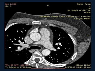
‫الرحي‬ ‫الرحمن‬ ‫هللا‬ ‫بسم‬‫م‬
27 years old male presented with
chest pain ,patient is not smoker
,not
hypertensive ,not diabetic
his TMT is not conclusive
CTA done for this patient
Diagnosis
Myocardial bridge
Myocardial bridge is abnormality in the course
of coronary artery .Muscle overlying the
intramyocardial segment of an epicardial
coronary artery, first mentioned by Reyman1
in 1737, is termed a myocardial bridge, and
the artery coursing within the myocardium is
called a tunneled artery
• For the most part, a myocardial bridge is harmless.
Patients with a myocardial bridge have it from birth,
and most never know they have the condition.
• However, some patients can develop myocardial
ischemia (lack of oxygen) because of a myocardial
bridge. When the heart "squeezes" (contracts) during
the heartbeat, the bridge of heart muscle can
"tighten down" on the artery, pinching it and
decreasing the blood flow. If there is less than 50%
blockage, then the condition is probably benign. A
result of at least 70% usually causes some pain
• Luckily, most of the blood flow through the
heart happens during the "rest" phase of the
heartbeat, not during the "squeezing" phase.
Even so, the tightening of the bridge on the
artery can decrease blood flow enough that
myocardial ischemia develops, especially
during exersise or when the heart is beating
quickly.
• Although all major epicardial coronary arteries
can be affected, involvement of the left anterior
descending coronary artery (LAD) is the most
common(the mid segment is commonly affected
).
• In the presence of two parallel LAD branches, one
frequently takes an intramural course. Diagonal
and marginal branches may be involved in 18%
and 40% of cases, respectively.
.
• Angiographically, myocardial bridges are almost
exclusively spotted in the LAD. They are located
at a depth of 1 to 10 mm with a typical length of
≈10 to 30 mm
• The rate of angiographic bridging is <5%,
attributable to thin bridges causing little
compression. In subjects with angiographically
normal coronary arteries, the use of provocation
tests may enhance systolic myocardial
compression and thereby reveal myocardial
bridges in ≤40% of cases
• A high prevalence has also been reported in
heart transplant recipients and in patients
with hypertrophic obstructive cardiomyopathy
(HOCM).In the latter, more rigorous
contraction may unmask otherwise
undetectable bridges. Myocardial bridging
may be found at multiple sites in HOCM, but
also in patients without
• DIAGNOSIS:
1.CORONARY CTA
2.CONVENTIONAL ANGIOGRAPHY
3.IVUS(INTRAVASCULAR US) AND ICD(INTRA-
CORONARY DOPPLER )
4. MRI
Typical systolic compression (arrows) of the mid
LAD at two sites in series.
IVUS-images of the myocardial bridge during diastole (left) and
systole (right). A “half-moon”–like area surrounding the tunneled
segment is present during the entire cardiac cycle
The “half-moon phenomenon”is a characteristic IVUS observation, but its physiology
and anatomy are not fully understood
• ICD studies, frequently reveals a characteristic
flow pattern, the “fingertip phenomenon” or
“spike-and-dome pattern.” This flow pattern
consists of a sharp acceleration of flow in early
diastole followed by immediate marked
deceleration and a mid-diastolic pressure
plateau. It can frequently be observed within
and just proximal to the tunneled segment
ICD-images of the myocardial bridge showing retrograde flow
during systole (double arrows) in the proximal segment of the
bridge after nitroglycerin provocation.
How is it treated?
• In most patients, a myocardial bridge is not treated if it is
not causing any symptoms. In patients with symptoms,
medicines such as beta blocker and calcium channel
blocker are usually the first line of treatment.
• In rare cases, patients need surgery to relieve their
symptoms. Surgery involves removing the bridge that is
pressing on the coronary artery surgery should be limited
to patients with severe angina and evidence for clinically
relevant ischemia. In bridges that take a deep
subendocardial course, the right ventricle may accidentally
be opened during surgery,and a case of aneurysm at the
site of myocardial cleavage has been reported
Myocardial bridge..case

Myocardial bridge..case

  • 1.
  • 2.
    27 years oldmale presented with chest pain ,patient is not smoker ,not hypertensive ,not diabetic his TMT is not conclusive
  • 3.
    CTA done forthis patient
  • 5.
  • 6.
  • 8.
    Myocardial bridge isabnormality in the course of coronary artery .Muscle overlying the intramyocardial segment of an epicardial coronary artery, first mentioned by Reyman1 in 1737, is termed a myocardial bridge, and the artery coursing within the myocardium is called a tunneled artery
  • 9.
    • For themost part, a myocardial bridge is harmless. Patients with a myocardial bridge have it from birth, and most never know they have the condition. • However, some patients can develop myocardial ischemia (lack of oxygen) because of a myocardial bridge. When the heart "squeezes" (contracts) during the heartbeat, the bridge of heart muscle can "tighten down" on the artery, pinching it and decreasing the blood flow. If there is less than 50% blockage, then the condition is probably benign. A result of at least 70% usually causes some pain
  • 10.
    • Luckily, mostof the blood flow through the heart happens during the "rest" phase of the heartbeat, not during the "squeezing" phase. Even so, the tightening of the bridge on the artery can decrease blood flow enough that myocardial ischemia develops, especially during exersise or when the heart is beating quickly.
  • 11.
    • Although allmajor epicardial coronary arteries can be affected, involvement of the left anterior descending coronary artery (LAD) is the most common(the mid segment is commonly affected ). • In the presence of two parallel LAD branches, one frequently takes an intramural course. Diagonal and marginal branches may be involved in 18% and 40% of cases, respectively. .
  • 12.
    • Angiographically, myocardialbridges are almost exclusively spotted in the LAD. They are located at a depth of 1 to 10 mm with a typical length of ≈10 to 30 mm • The rate of angiographic bridging is <5%, attributable to thin bridges causing little compression. In subjects with angiographically normal coronary arteries, the use of provocation tests may enhance systolic myocardial compression and thereby reveal myocardial bridges in ≤40% of cases
  • 13.
    • A highprevalence has also been reported in heart transplant recipients and in patients with hypertrophic obstructive cardiomyopathy (HOCM).In the latter, more rigorous contraction may unmask otherwise undetectable bridges. Myocardial bridging may be found at multiple sites in HOCM, but also in patients without
  • 14.
    • DIAGNOSIS: 1.CORONARY CTA 2.CONVENTIONALANGIOGRAPHY 3.IVUS(INTRAVASCULAR US) AND ICD(INTRA- CORONARY DOPPLER ) 4. MRI
  • 17.
    Typical systolic compression(arrows) of the mid LAD at two sites in series.
  • 18.
    IVUS-images of themyocardial bridge during diastole (left) and systole (right). A “half-moon”–like area surrounding the tunneled segment is present during the entire cardiac cycle The “half-moon phenomenon”is a characteristic IVUS observation, but its physiology and anatomy are not fully understood
  • 19.
    • ICD studies,frequently reveals a characteristic flow pattern, the “fingertip phenomenon” or “spike-and-dome pattern.” This flow pattern consists of a sharp acceleration of flow in early diastole followed by immediate marked deceleration and a mid-diastolic pressure plateau. It can frequently be observed within and just proximal to the tunneled segment
  • 20.
    ICD-images of themyocardial bridge showing retrograde flow during systole (double arrows) in the proximal segment of the bridge after nitroglycerin provocation.
  • 21.
    How is ittreated? • In most patients, a myocardial bridge is not treated if it is not causing any symptoms. In patients with symptoms, medicines such as beta blocker and calcium channel blocker are usually the first line of treatment. • In rare cases, patients need surgery to relieve their symptoms. Surgery involves removing the bridge that is pressing on the coronary artery surgery should be limited to patients with severe angina and evidence for clinically relevant ischemia. In bridges that take a deep subendocardial course, the right ventricle may accidentally be opened during surgery,and a case of aneurysm at the site of myocardial cleavage has been reported