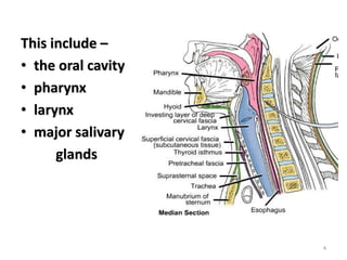
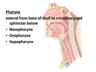
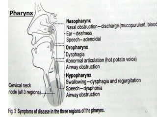
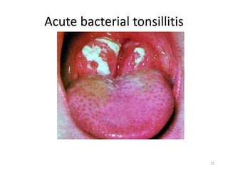
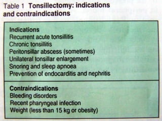
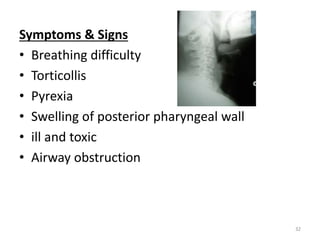
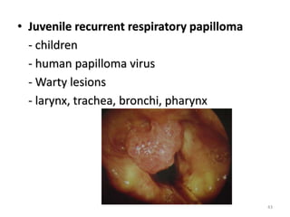
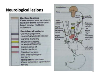
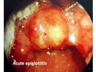
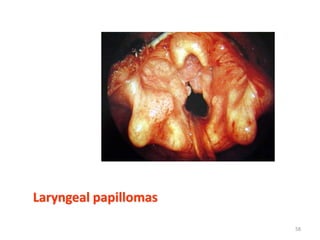
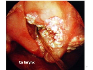
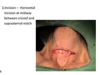
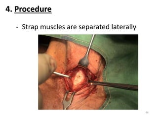
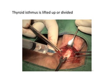
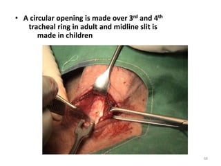
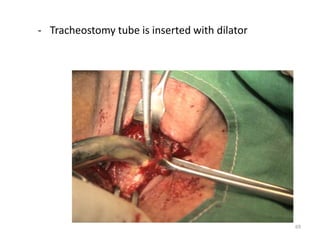
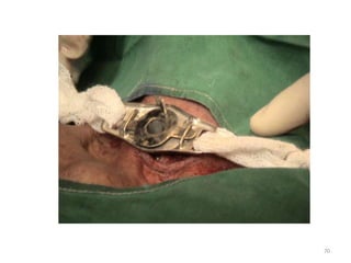
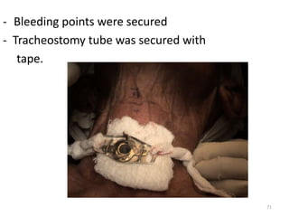
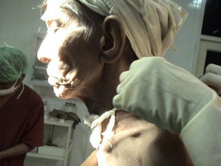
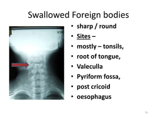
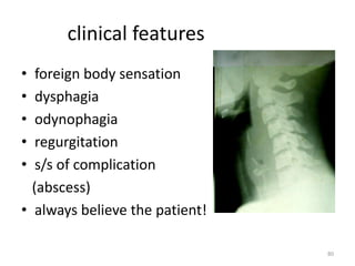
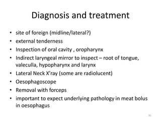
Throat
1
Contents
• Anatomy & physiology
• S/S, investigations
• Sore throat
• Abscess
• Dysphonia
• Stridor
2
3
This include –
• the oral cavity
• pharynx
• larynx
• major salivary
glands
4
Pharynx
extend from base of skull to cricopharyngel
sphincter below
• Nasopharynx
• Oropharynx
• Hypopharynx
5
Deglutition
3 stages –
(1) Oral phase
(2) Pharyngeal phase
(3) Oesophageal phase
6
7
8
Functions of larynx:
1.Protect tracheobronchial tree
2.Voice production
3.Respiratory passage
9
10
Symptoms, Signs & Examination
Oral cavity
• Pain
• masses
• ulceration
• haemorrhage
• halitosis
• ageusia
• discolouration
11
Pharynx
12
Larynx
• Change of voice
• respiratory difficulty / Stridor
• pain
• aspiration
Neck lumps
13
Examination
• Oral examination
• ENT examination
14
• Rigid Scope
15
• Flexible scope
16
• Direct or Micro Laryngoscope
17
• Imagings
18
SORE THROAT
19
Sore throats
• Acute pharyngitis
• Acute tonsilitis
• Peritonsilar abscess
• blood disorders
• candidiasis
• glandular fever
20
Viral infection
• Pharyngotonsilitis – common
• associated with running nose and cough
• conservative management
Bacterial tonsilitis
• secondary to viral infection
• streptococcus,H influzae etc
• dysphagia, odynophagia, fever , cervical L/N
• antibiotic, analgesic, antipyretic
21
Acute bacterial tonsillitis
22
23
Complications of tonsillectomy
• Haemorrhage
• Pain
• Infection
• Trauma
24
ABSCESS AROUND THE PHARYNX
25
Abscess around the Pharynx
Peritonsillar abscess (Quinsy)
Definition - an abscess between the tonsil and
the adjacent lateral pharyngeal wall (superior
constrictor muscle)
Pathogenesis - follow an acute attack of
tonsillitis usually unilateral and lies above the
tonsil near the soft palate preceded by
cellulitis.
26
Symptoms
• Severe pain
• Odynophagia
• Fever
• Otalgia
• Salivation + dribbling
• Thickened speech
• Trismus
27
Signs
Marked hyperaemia and edema of tonsils,
palate and uvula, pushing the latter to the
unaffected side
Management consist of - rest, fluid, I&D ,
tonsillectomy ( 6 weeks )
28
Ludwig’s Angina
• Definition – cellulitis of the floor of mouth
and submandibular space of neck
• Pathology – usually due to infection with
Hemolytic streptococcus as complication of
pharyngitis or oral sepsis.
• Symptoms – pain in floor of mouth ( tongue)
- drolling of saliva
- swelling neck
- difficulty in breathing
29
Signs
• Hard tender brawny swelling between the
chin and neck detected on bimanual exam
• Fluctuation may be negative
• Fever and toxemia present
• edema of glottis ( sudden death )
Management
full dose of antibiotics with I & D ( Hilton’s
method )
30
Retropharyngeal abscess
Definition – It is an abscess in the fascial space
behind the pharyngeal muscles
causes - suppuration of retropharyngeal
lymph nodes
- penetration of pharyngeal wall by
sharp FB
- caries of bodies of cervical
vertebra.
31
Symptoms & Signs
• Breathing difficulty
• Torticollis
• Pyrexia
• Swelling of posterior pharyngeal wall
• ill and toxic
• Airway obstruction
32
Management
• Medical – Antibiotics
• Surgical – I&D through open mouth
33
Parapharyngeal abscess
Definition - suppurative infection of the
parapharyngeal space
Causes - suppuration of lymph node
- penetration of lateral pharyngeal
wall by sharp FB
- spread of infection from tonsils,
peritonsillar space, lower wisdom tooth ,its
surrounding gums and bones
34
Symptoms
• Pain in throat especially on swallowing
• pyrexia
Signs
• trismus
• torticollis
• tender red fluctuant swelling of neck
• pharyngeal wall and tonsil are pushed
medially
35
Management
• Medical – antibiotics, analgesics
• Surgical – I&D via external approach
Complications
• acute edema of the larynx
• thrombophlebitis of jugular vein
• Septicaemia
• direct spread to mediastinum
36
HOARSENESS OF VOICE
37
Dysphonia (Organic causes)
“An alteration in the quality of voice”
• Inflammatory - acute laryngitis
- chronic laryngitis
• Neoplasia - Ca larynx, Papillomata
• Neurological - Myasthenia gravis, Ca lung
/breast, post thyroidectomy
• Systemic - Hypothyroidism,Rheumatoid
arthritis
38
Inflammatory lesion
Acute laryngitis
• Infection ~ very common
~ upper respiratory tract infection
~ associated with pain in throat
~ spontaneous resolution
~ Steam inhalation
~ referral if symptoms persist
39
• Non-infected ~ shouting
~ foreign bodies
~ fumes, tobacco, smokes or
chemicals
~ may produce edema and
respiratory embarrassment
40
• Inflammatory polyps
~ not uncommon
~ history similar to acute laryngitis
~ removal under microlaryngoscope
41
Neoplastic lesions
• Ca larynx
- Old age, male
- Smoking, alcohol
- Hoarseness of voice, stridor
- Direct laryngoscopy and biopsy
- Surgery and Radiotherapy
42
• Juvenile recurrent respiratory papilloma
- children
- human papilloma virus
- Warty lesions
- larynx, trachea, bronchi, pharynx
43
Neurological lesions
44
Systemic causes
• Hypothyroidism – chronic edema of vocal
cord
• Angioneurotic edema – type I allergic
response
• Rheumatoid arthritis – fixation of
cricoarytenoid joint
Management of dysphonia
• treat the underlying causes
45
46
It is a noisy and difficult
breathing due to partial
obstruction of upper airway.
47
A case of stridor due to
malignant infiltration to
trachea from Ca thyroid
48
Causes of stridor
1.Congenital
(a) Laryngeal stenosis
(b) Laryngomalacia – congenital
softness of larynx
(c) Laryngeal web
2.Traumatic
(a) External – blow on larynx
e g. dash-board injury
49
(b) Internal – foreign body
corrosive substances
fumes
3.Infection
Acute laryngotracheobronchitis
Acute epiglottitis
Ludwig’s angina
Diphtheria
50
4.Tumour
(a) Benign – Papillomas
(Juvenile recurrent
respiratory papillomatosis)
(b) Malignant – Ca larynx
5.Neurological
(a) Bilateral recurrent laryngeal nerve palsy
(b) Bulbar palsy
6. Miscellaneous
Angioneurotic edema 51
Laryngomalacia
52
Laryngeal web
53
Subglottis hemangioma
54
Acute laryngotracheobronchitis
55
Acute epiglottitis
56
Ludwig’s angina
57
Laryngeal papillomas
58
Ca larynx
59
Emergency Management
• Important to realize that relief of stridor is
more important than knowing causes of
stridor.
• Oxygen inhalation
Tracheostomy – don’t wait for obvious
cyanosis
60
Definitive Management
1. Confirmation of diagnosis
2. Remove the cause when possible, or
timely referral when removal of cause is
not possible.
3. Regular follow-up
61
Tracheostomy
An operation where by an opening made in
the anterior wall of trachea and converted
it to stoma
62
Indications
1. Relief of upper airway obstruction
2. Protection of tracheo-bronchial tree & to
facilitate tracheal toilet
3.Treatment of respiratory insufficiency
- to reduce dead space
- to institute IPPR
63
Standard Method of Tracheostomy
1. Anaesthesia – LA or GA
2. Position – Supine position
Extended neck (pillow under
the shoulder)
1
2
64
3.Incision – Horizontal
incision at midway
between cricoid and
suprasternal notch
3
4
5 65
- Strap muscles are separated laterally
4. Procedure
66
Thyroid isthmus is lifted up or divided
67
• A circular opening is made over 3rd and 4th
tracheal ring in adult and midline slit is
made in children
68
- Tracheostomy tube is inserted with dilator
69
70
- Bleeding points were secured
- Tracheostomy tube was secured with
tape.
71
72
Complications
1.Haemorrhage and infection
2.Dislodgement of tube (proper applying of
tape)
3.Injury to surrounding structures
73
4.Surgical emphysema, pneumothorax
5.Tube blockage – by dry mucous forming crusts
6.Subglottic stenosis – of 1st ring was cut
7.Decannulation problem
74
75
Foreign Bodies
Inhaled foreign bodies(airpassage)
• under 4 years (75%)
• features depend on types and location in
laryngotracheobronchial tree
• vegetable materials – severe mucosal react:
• may be rapidly fetal
• first aids
76
77
Clinical features
• Child previously healthy
with sudden onset
• aphonia,
wheezing,stridor,choking,
coughing
• chest and neck X’ray
• Scopy
Treatment
Scopy and removal
78
Swallowed Foreign bodies
• sharp / round
• Sites –
• mostly – tonsils,
• root of tongue,
• Valeculla
• Pyriform fossa,
• post cricoid
• oesophagus
79
clinical features
• foreign body sensation
• dysphagia
• odynophagia
• regurgitation
• s/s of complication
(abscess)
• always believe the patient!
80
Diagnosis and treatment
• site of foreign (midline/lateral?)
• external tenderness
• Inspection of oral cavity , oropharynx
• Indirect laryngeal mirror to inspect – root of tongue,
valeculla, hypopharynx and larynx
• Lateral Neck X’ray (some are radiolucent)
• Oesophagoscope
• Removal with forceps
• important to expect underlying pathology in meat bolus
in oesophagus
81

Throat.pptx

  • 1.
  • 2.
    Contents • Anatomy &physiology • S/S, investigations • Sore throat • Abscess • Dysphonia • Stridor 2
  • 3.
  • 4.
    This include – •the oral cavity • pharynx • larynx • major salivary glands 4
  • 5.
    Pharynx extend from baseof skull to cricopharyngel sphincter below • Nasopharynx • Oropharynx • Hypopharynx 5
  • 6.
    Deglutition 3 stages – (1)Oral phase (2) Pharyngeal phase (3) Oesophageal phase 6
  • 7.
  • 8.
  • 9.
    Functions of larynx: 1.Protecttracheobronchial tree 2.Voice production 3.Respiratory passage 9
  • 10.
  • 11.
    Symptoms, Signs &Examination Oral cavity • Pain • masses • ulceration • haemorrhage • halitosis • ageusia • discolouration 11
  • 12.
  • 13.
    Larynx • Change ofvoice • respiratory difficulty / Stridor • pain • aspiration Neck lumps 13
  • 14.
  • 15.
  • 16.
  • 17.
    • Direct orMicro Laryngoscope 17
  • 18.
  • 19.
  • 20.
    Sore throats • Acutepharyngitis • Acute tonsilitis • Peritonsilar abscess • blood disorders • candidiasis • glandular fever 20
  • 21.
    Viral infection • Pharyngotonsilitis– common • associated with running nose and cough • conservative management Bacterial tonsilitis • secondary to viral infection • streptococcus,H influzae etc • dysphagia, odynophagia, fever , cervical L/N • antibiotic, analgesic, antipyretic 21
  • 22.
  • 23.
  • 24.
    Complications of tonsillectomy •Haemorrhage • Pain • Infection • Trauma 24
  • 25.
  • 26.
    Abscess around thePharynx Peritonsillar abscess (Quinsy) Definition - an abscess between the tonsil and the adjacent lateral pharyngeal wall (superior constrictor muscle) Pathogenesis - follow an acute attack of tonsillitis usually unilateral and lies above the tonsil near the soft palate preceded by cellulitis. 26
  • 27.
    Symptoms • Severe pain •Odynophagia • Fever • Otalgia • Salivation + dribbling • Thickened speech • Trismus 27
  • 28.
    Signs Marked hyperaemia andedema of tonsils, palate and uvula, pushing the latter to the unaffected side Management consist of - rest, fluid, I&D , tonsillectomy ( 6 weeks ) 28
  • 29.
    Ludwig’s Angina • Definition– cellulitis of the floor of mouth and submandibular space of neck • Pathology – usually due to infection with Hemolytic streptococcus as complication of pharyngitis or oral sepsis. • Symptoms – pain in floor of mouth ( tongue) - drolling of saliva - swelling neck - difficulty in breathing 29
  • 30.
    Signs • Hard tenderbrawny swelling between the chin and neck detected on bimanual exam • Fluctuation may be negative • Fever and toxemia present • edema of glottis ( sudden death ) Management full dose of antibiotics with I & D ( Hilton’s method ) 30
  • 31.
    Retropharyngeal abscess Definition –It is an abscess in the fascial space behind the pharyngeal muscles causes - suppuration of retropharyngeal lymph nodes - penetration of pharyngeal wall by sharp FB - caries of bodies of cervical vertebra. 31
  • 32.
    Symptoms & Signs •Breathing difficulty • Torticollis • Pyrexia • Swelling of posterior pharyngeal wall • ill and toxic • Airway obstruction 32
  • 33.
    Management • Medical –Antibiotics • Surgical – I&D through open mouth 33
  • 34.
    Parapharyngeal abscess Definition -suppurative infection of the parapharyngeal space Causes - suppuration of lymph node - penetration of lateral pharyngeal wall by sharp FB - spread of infection from tonsils, peritonsillar space, lower wisdom tooth ,its surrounding gums and bones 34
  • 35.
    Symptoms • Pain inthroat especially on swallowing • pyrexia Signs • trismus • torticollis • tender red fluctuant swelling of neck • pharyngeal wall and tonsil are pushed medially 35
  • 36.
    Management • Medical –antibiotics, analgesics • Surgical – I&D via external approach Complications • acute edema of the larynx • thrombophlebitis of jugular vein • Septicaemia • direct spread to mediastinum 36
  • 37.
  • 38.
    Dysphonia (Organic causes) “Analteration in the quality of voice” • Inflammatory - acute laryngitis - chronic laryngitis • Neoplasia - Ca larynx, Papillomata • Neurological - Myasthenia gravis, Ca lung /breast, post thyroidectomy • Systemic - Hypothyroidism,Rheumatoid arthritis 38
  • 39.
    Inflammatory lesion Acute laryngitis •Infection ~ very common ~ upper respiratory tract infection ~ associated with pain in throat ~ spontaneous resolution ~ Steam inhalation ~ referral if symptoms persist 39
  • 40.
    • Non-infected ~shouting ~ foreign bodies ~ fumes, tobacco, smokes or chemicals ~ may produce edema and respiratory embarrassment 40
  • 41.
    • Inflammatory polyps ~not uncommon ~ history similar to acute laryngitis ~ removal under microlaryngoscope 41
  • 42.
    Neoplastic lesions • Calarynx - Old age, male - Smoking, alcohol - Hoarseness of voice, stridor - Direct laryngoscopy and biopsy - Surgery and Radiotherapy 42
  • 43.
    • Juvenile recurrentrespiratory papilloma - children - human papilloma virus - Warty lesions - larynx, trachea, bronchi, pharynx 43
  • 44.
  • 45.
    Systemic causes • Hypothyroidism– chronic edema of vocal cord • Angioneurotic edema – type I allergic response • Rheumatoid arthritis – fixation of cricoarytenoid joint Management of dysphonia • treat the underlying causes 45
  • 46.
  • 47.
    It is anoisy and difficult breathing due to partial obstruction of upper airway. 47
  • 48.
    A case ofstridor due to malignant infiltration to trachea from Ca thyroid 48
  • 49.
    Causes of stridor 1.Congenital (a)Laryngeal stenosis (b) Laryngomalacia – congenital softness of larynx (c) Laryngeal web 2.Traumatic (a) External – blow on larynx e g. dash-board injury 49
  • 50.
    (b) Internal –foreign body corrosive substances fumes 3.Infection Acute laryngotracheobronchitis Acute epiglottitis Ludwig’s angina Diphtheria 50
  • 51.
    4.Tumour (a) Benign –Papillomas (Juvenile recurrent respiratory papillomatosis) (b) Malignant – Ca larynx 5.Neurological (a) Bilateral recurrent laryngeal nerve palsy (b) Bulbar palsy 6. Miscellaneous Angioneurotic edema 51
  • 52.
  • 53.
  • 54.
  • 55.
  • 56.
  • 57.
  • 58.
  • 59.
  • 60.
    Emergency Management • Importantto realize that relief of stridor is more important than knowing causes of stridor. • Oxygen inhalation Tracheostomy – don’t wait for obvious cyanosis 60
  • 61.
    Definitive Management 1. Confirmationof diagnosis 2. Remove the cause when possible, or timely referral when removal of cause is not possible. 3. Regular follow-up 61
  • 62.
    Tracheostomy An operation whereby an opening made in the anterior wall of trachea and converted it to stoma 62
  • 63.
    Indications 1. Relief ofupper airway obstruction 2. Protection of tracheo-bronchial tree & to facilitate tracheal toilet 3.Treatment of respiratory insufficiency - to reduce dead space - to institute IPPR 63
  • 64.
    Standard Method ofTracheostomy 1. Anaesthesia – LA or GA 2. Position – Supine position Extended neck (pillow under the shoulder) 1 2 64
  • 65.
    3.Incision – Horizontal incisionat midway between cricoid and suprasternal notch 3 4 5 65
  • 66.
    - Strap musclesare separated laterally 4. Procedure 66
  • 67.
    Thyroid isthmus islifted up or divided 67
  • 68.
    • A circularopening is made over 3rd and 4th tracheal ring in adult and midline slit is made in children 68
  • 69.
    - Tracheostomy tubeis inserted with dilator 69
  • 70.
  • 71.
    - Bleeding pointswere secured - Tracheostomy tube was secured with tape. 71
  • 72.
  • 73.
    Complications 1.Haemorrhage and infection 2.Dislodgementof tube (proper applying of tape) 3.Injury to surrounding structures 73
  • 74.
    4.Surgical emphysema, pneumothorax 5.Tubeblockage – by dry mucous forming crusts 6.Subglottic stenosis – of 1st ring was cut 7.Decannulation problem 74
  • 75.
  • 76.
    Foreign Bodies Inhaled foreignbodies(airpassage) • under 4 years (75%) • features depend on types and location in laryngotracheobronchial tree • vegetable materials – severe mucosal react: • may be rapidly fetal • first aids 76
  • 77.
  • 78.
    Clinical features • Childpreviously healthy with sudden onset • aphonia, wheezing,stridor,choking, coughing • chest and neck X’ray • Scopy Treatment Scopy and removal 78
  • 79.
    Swallowed Foreign bodies •sharp / round • Sites – • mostly – tonsils, • root of tongue, • Valeculla • Pyriform fossa, • post cricoid • oesophagus 79
  • 80.
    clinical features • foreignbody sensation • dysphagia • odynophagia • regurgitation • s/s of complication (abscess) • always believe the patient! 80
  • 81.
    Diagnosis and treatment •site of foreign (midline/lateral?) • external tenderness • Inspection of oral cavity , oropharynx • Indirect laryngeal mirror to inspect – root of tongue, valeculla, hypopharynx and larynx • Lateral Neck X’ray (some are radiolucent) • Oesophagoscope • Removal with forceps • important to expect underlying pathology in meat bolus in oesophagus 81