Downloaded 400 times

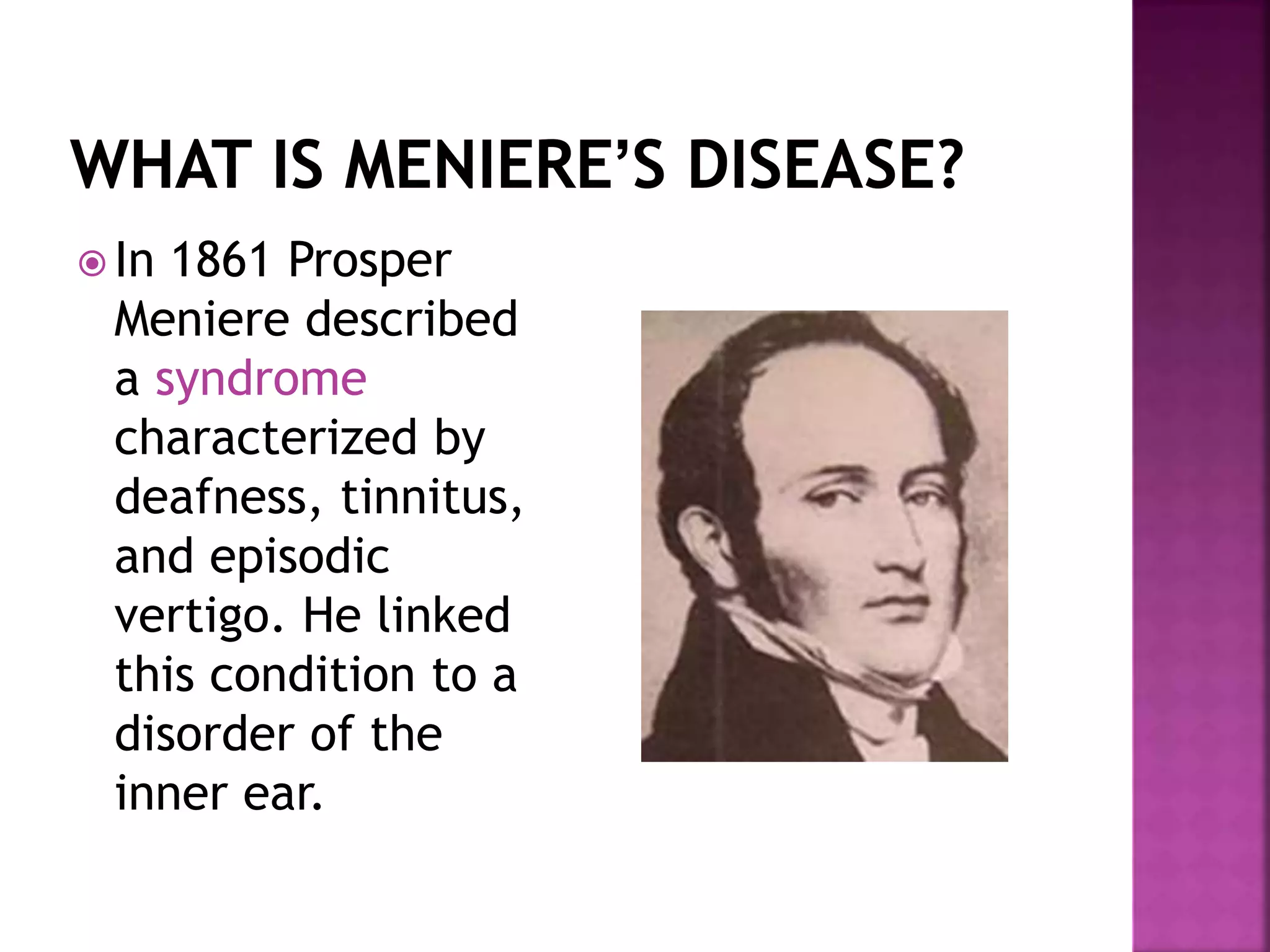


This document discusses the history, presentation, diagnosis and management of Meniere's disease. Some key points: - Meniere's disease was first described in 1861 and is characterized by hearing loss, tinnitus, and vertigo due to endolymphatic hydrops (fluid buildup) in the inner ear. - Diagnosis is based on recurrent vertigo spells lasting 20 minutes to 24 hours, fluctuating hearing loss, tinnitus and aural fullness. Tests like electrocochleography and VEMPs can provide supportive evidence. - Treatment includes dietary sodium restriction, diuretics, medications and surgical options like intratympanic injections if conservative measures fail. The