1. Parts & Branches of an Aorta
a) The Descending aorta
b) The veins system of the thorax
c) The lymphatics of the thorax
2. The nervous system of the thorax
a) anterior and posterior branches of the thoracic nerves
b) the phrenic nerve
c) the thoracic part of the sympathetic trunk
d) the thoracic part of the vagus (10th) nerve
Slideshow: Hip Joint and Pelvic Gateways
The Funky Professor videos can be viewed here;
http://publishing.rcseng.ac.uk/journal/video?videoTaxonomy=FUNK
Slideshow: Hip Joint and Pelvic Gateways
The Funky Professor videos can be viewed here;
http://publishing.rcseng.ac.uk/journal/video?videoTaxonomy=FUNK
Mediastinum is the space present in the thoracic cavity
.Two divisions of the mediastinum -- Superior and inferior are shown with relations, contents and boundaries in three dimension.
Inferior mediastinum has three divisions - anterior, middle and posterior,Relations and contents and boundaries are shown
.Applied aspects like - mediatinities and mediasinal syndrome. is written.
This is useful for undergraduate students
Anatomy of Blood vessels of abdomen pelvic cavities. Portacaval & Cavacaval A...Eneutron
1. The abdominal aorta
a. the parietal branches
b. the visceral branches
2. The common iliac arteries and veins
3. The external iliac artery and veins
4. The internal iliac artery and veins
5. The inferior vena cava
6. The portal vein
7. The cavacacal Anastomoses
8. The portacaval Anastomoses
9. The Fetal Circulation
1. Blood supply of the upper limb. Major arterial anastomoses of the upper extremity.
2. The veins of the upper limb.
3. Innervation of the upper limb. Schematic representation of the innervation of the skin of the upper limb.
4. Lymphatic vessels of the upper extremity.
Blood supply of head & neck. Arterial & venous anastomosesEneutron
1. The coomon carotid artery
a) topography
- carotid sinus
- carotid body
2. Neurovascular bundles of the neck
3. The external carotid artery
4. The internal carotid artery
- arterial supply of the brain
5. Arterial anastomoses head and neck
6. Veins of the head and neck
USMLE MSK L020 Upper 09 Anatomical regions anatomy.pdfAHMED ASHOUR
The upper limb is divided into several anatomical regions, each with distinct structures and functions.
Understanding these anatomical regions is essential for healthcare professionals, anatomists, and individuals studying the upper limb for medical or educational purposes. Each region plays a specific role in the overall function and movement of the upper limb.
NVBDCP.pptx Nation vector borne disease control programSapna Thakur
NVBDCP was launched in 2003-2004 . Vector-Borne Disease: Disease that results from an infection transmitted to humans and other animals by blood-feeding arthropods, such as mosquitoes, ticks, and fleas. Examples of vector-borne diseases include Dengue fever, West Nile Virus, Lyme disease, and malaria.
These simplified slides by Dr. Sidra Arshad present an overview of the non-respiratory functions of the respiratory tract.
Learning objectives:
1. Enlist the non-respiratory functions of the respiratory tract
2. Briefly explain how these functions are carried out
3. Discuss the significance of dead space
4. Differentiate between minute ventilation and alveolar ventilation
5. Describe the cough and sneeze reflexes
Study Resources:
1. Chapter 39, Guyton and Hall Textbook of Medical Physiology, 14th edition
2. Chapter 34, Ganong’s Review of Medical Physiology, 26th edition
3. Chapter 17, Human Physiology by Lauralee Sherwood, 9th edition
4. Non-respiratory functions of the lungs https://academic.oup.com/bjaed/article/13/3/98/278874
Pulmonary Thromboembolism - etilogy, types, medical- Surgical and nursing man...VarunMahajani
Disruption of blood supply to lung alveoli due to blockage of one or more pulmonary blood vessels is called as Pulmonary thromboembolism. In this presentation we will discuss its causes, types and its management in depth.
Acute scrotum is a general term referring to an emergency condition affecting the contents or the wall of the scrotum.
There are a number of conditions that present acutely, predominantly with pain and/or swelling
A careful and detailed history and examination, and in some cases, investigations allow differentiation between these diagnoses. A prompt diagnosis is essential as the patient may require urgent surgical intervention
Testicular torsion refers to twisting of the spermatic cord, causing ischaemia of the testicle.
Testicular torsion results from inadequate fixation of the testis to the tunica vaginalis producing ischemia from reduced arterial inflow and venous outflow obstruction.
The prevalence of testicular torsion in adult patients hospitalized with acute scrotal pain is approximately 25 to 50 percent
These lecture slides, by Dr Sidra Arshad, offer a quick overview of physiological basis of a normal electrocardiogram.
Learning objectives:
1. Define an electrocardiogram (ECG) and electrocardiography
2. Describe how dipoles generated by the heart produce the waveforms of the ECG
3. Describe the components of a normal electrocardiogram of a typical bipolar leads (limb II)
4. Differentiate between intervals and segments
5. Enlist some common indications for obtaining an ECG
Study Resources:
1. Chapter 11, Guyton and Hall Textbook of Medical Physiology, 14th edition
2. Chapter 9, Human Physiology - From Cells to Systems, Lauralee Sherwood, 9th edition
3. Chapter 29, Ganong’s Review of Medical Physiology, 26th edition
4. Electrocardiogram, StatPearls - https://www.ncbi.nlm.nih.gov/books/NBK549803/
5. ECG in Medical Practice by ABM Abdullah, 4th edition
6. ECG Basics, http://www.nataliescasebook.com/tag/e-c-g-basics
The prostate is an exocrine gland of the male mammalian reproductive system
It is a walnut-sized gland that forms part of the male reproductive system and is located in front of the rectum and just below the urinary bladder
Function is to store and secrete a clear, slightly alkaline fluid that constitutes 10-30% of the volume of the seminal fluid that along with the spermatozoa, constitutes semen
A healthy human prostate measures (4cm-vertical, by 3cm-horizontal, 2cm ant-post ).
It surrounds the urethra just below the urinary bladder. It has anterior, median, posterior and two lateral lobes
It’s work is regulated by androgens which are responsible for male sex characteristics
Generalised disease of the prostate due to hormonal derangement which leads to non malignant enlargement of the gland (increase in the number of epithelial cells and stromal tissue)to cause compression of the urethra leading to symptoms (LUTS
Report Back from SGO 2024: What’s the Latest in Cervical Cancer?bkling
Are you curious about what’s new in cervical cancer research or unsure what the findings mean? Join Dr. Emily Ko, a gynecologic oncologist at Penn Medicine, to learn about the latest updates from the Society of Gynecologic Oncology (SGO) 2024 Annual Meeting on Women’s Cancer. Dr. Ko will discuss what the research presented at the conference means for you and answer your questions about the new developments.
Explore natural remedies for syphilis treatment in Singapore. Discover alternative therapies, herbal remedies, and lifestyle changes that may complement conventional treatments. Learn about holistic approaches to managing syphilis symptoms and supporting overall health.
Title: Sense of Taste
Presenter: Dr. Faiza, Assistant Professor of Physiology
Qualifications:
MBBS (Best Graduate, AIMC Lahore)
FCPS Physiology
ICMT, CHPE, DHPE (STMU)
MPH (GC University, Faisalabad)
MBA (Virtual University of Pakistan)
Learning Objectives:
Describe the structure and function of taste buds.
Describe the relationship between the taste threshold and taste index of common substances.
Explain the chemical basis and signal transduction of taste perception for each type of primary taste sensation.
Recognize different abnormalities of taste perception and their causes.
Key Topics:
Significance of Taste Sensation:
Differentiation between pleasant and harmful food
Influence on behavior
Selection of food based on metabolic needs
Receptors of Taste:
Taste buds on the tongue
Influence of sense of smell, texture of food, and pain stimulation (e.g., by pepper)
Primary and Secondary Taste Sensations:
Primary taste sensations: Sweet, Sour, Salty, Bitter, Umami
Chemical basis and signal transduction mechanisms for each taste
Taste Threshold and Index:
Taste threshold values for Sweet (sucrose), Salty (NaCl), Sour (HCl), and Bitter (Quinine)
Taste index relationship: Inversely proportional to taste threshold
Taste Blindness:
Inability to taste certain substances, particularly thiourea compounds
Example: Phenylthiocarbamide
Structure and Function of Taste Buds:
Composition: Epithelial cells, Sustentacular/Supporting cells, Taste cells, Basal cells
Features: Taste pores, Taste hairs/microvilli, and Taste nerve fibers
Location of Taste Buds:
Found in papillae of the tongue (Fungiform, Circumvallate, Foliate)
Also present on the palate, tonsillar pillars, epiglottis, and proximal esophagus
Mechanism of Taste Stimulation:
Interaction of taste substances with receptors on microvilli
Signal transduction pathways for Umami, Sweet, Bitter, Sour, and Salty tastes
Taste Sensitivity and Adaptation:
Decrease in sensitivity with age
Rapid adaptation of taste sensation
Role of Saliva in Taste:
Dissolution of tastants to reach receptors
Washing away the stimulus
Taste Preferences and Aversions:
Mechanisms behind taste preference and aversion
Influence of receptors and neural pathways
Impact of Sensory Nerve Damage:
Degeneration of taste buds if the sensory nerve fiber is cut
Abnormalities of Taste Detection:
Conditions: Ageusia, Hypogeusia, Dysgeusia (parageusia)
Causes: Nerve damage, neurological disorders, infections, poor oral hygiene, adverse drug effects, deficiencies, aging, tobacco use, altered neurotransmitter levels
Neurotransmitters and Taste Threshold:
Effects of serotonin (5-HT) and norepinephrine (NE) on taste sensitivity
Supertasters:
25% of the population with heightened sensitivity to taste, especially bitterness
Increased number of fungiform papillae
New Drug Discovery and Development .....NEHA GUPTA
The "New Drug Discovery and Development" process involves the identification, design, testing, and manufacturing of novel pharmaceutical compounds with the aim of introducing new and improved treatments for various medical conditions. This comprehensive endeavor encompasses various stages, including target identification, preclinical studies, clinical trials, regulatory approval, and post-market surveillance. It involves multidisciplinary collaboration among scientists, researchers, clinicians, regulatory experts, and pharmaceutical companies to bring innovative therapies to market and address unmet medical needs.
New Directions in Targeted Therapeutic Approaches for Older Adults With Mantl...i3 Health
i3 Health is pleased to make the speaker slides from this activity available for use as a non-accredited self-study or teaching resource.
This slide deck presented by Dr. Kami Maddocks, Professor-Clinical in the Division of Hematology and
Associate Division Director for Ambulatory Operations
The Ohio State University Comprehensive Cancer Center, will provide insight into new directions in targeted therapeutic approaches for older adults with mantle cell lymphoma.
STATEMENT OF NEED
Mantle cell lymphoma (MCL) is a rare, aggressive B-cell non-Hodgkin lymphoma (NHL) accounting for 5% to 7% of all lymphomas. Its prognosis ranges from indolent disease that does not require treatment for years to very aggressive disease, which is associated with poor survival (Silkenstedt et al, 2021). Typically, MCL is diagnosed at advanced stage and in older patients who cannot tolerate intensive therapy (NCCN, 2022). Although recent advances have slightly increased remission rates, recurrence and relapse remain very common, leading to a median overall survival between 3 and 6 years (LLS, 2021). Though there are several effective options, progress is still needed towards establishing an accepted frontline approach for MCL (Castellino et al, 2022). Treatment selection and management of MCL are complicated by the heterogeneity of prognosis, advanced age and comorbidities of patients, and lack of an established standard approach for treatment, making it vital that clinicians be familiar with the latest research and advances in this area. In this activity chaired by Michael Wang, MD, Professor in the Department of Lymphoma & Myeloma at MD Anderson Cancer Center, expert faculty will discuss prognostic factors informing treatment, the promising results of recent trials in new therapeutic approaches, and the implications of treatment resistance in therapeutic selection for MCL.
Target Audience
Hematology/oncology fellows, attending faculty, and other health care professionals involved in the treatment of patients with mantle cell lymphoma (MCL).
Learning Objectives
1.) Identify clinical and biological prognostic factors that can guide treatment decision making for older adults with MCL
2.) Evaluate emerging data on targeted therapeutic approaches for treatment-naive and relapsed/refractory MCL and their applicability to older adults
3.) Assess mechanisms of resistance to targeted therapies for MCL and their implications for treatment selection
- Video recording of this lecture in English language: https://youtu.be/lK81BzxMqdo
- Video recording of this lecture in Arabic language: https://youtu.be/Ve4P0COk9OI
- Link to download the book free: https://nephrotube.blogspot.com/p/nephrotube-nephrology-books.html
- Link to NephroTube website: www.NephroTube.com
- Link to NephroTube social media accounts: https://nephrotube.blogspot.com/p/join-nephrotube-on-social-media.html
Flu Vaccine Alert in Bangalore Karnatakaaddon Scans
As flu season approaches, health officials in Bangalore, Karnataka, are urging residents to get their flu vaccinations. The seasonal flu, while common, can lead to severe health complications, particularly for vulnerable populations such as young children, the elderly, and those with underlying health conditions.
Dr. Vidisha Kumari, a leading epidemiologist in Bangalore, emphasizes the importance of getting vaccinated. "The flu vaccine is our best defense against the influenza virus. It not only protects individuals but also helps prevent the spread of the virus in our communities," he says.
This year, the flu season is expected to coincide with a potential increase in other respiratory illnesses. The Karnataka Health Department has launched an awareness campaign highlighting the significance of flu vaccinations. They have set up multiple vaccination centers across Bangalore, making it convenient for residents to receive their shots.
To encourage widespread vaccination, the government is also collaborating with local schools, workplaces, and community centers to facilitate vaccination drives. Special attention is being given to ensuring that the vaccine is accessible to all, including marginalized communities who may have limited access to healthcare.
Residents are reminded that the flu vaccine is safe and effective. Common side effects are mild and may include soreness at the injection site, mild fever, or muscle aches. These side effects are generally short-lived and far less severe than the flu itself.
Healthcare providers are also stressing the importance of continuing COVID-19 precautions. Wearing masks, practicing good hand hygiene, and maintaining social distancing are still crucial, especially in crowded places.
Protect yourself and your loved ones by getting vaccinated. Together, we can help keep Bangalore healthy and safe this flu season. For more information on vaccination centers and schedules, residents can visit the Karnataka Health Department’s official website or follow their social media pages.
Stay informed, stay safe, and get your flu shot today!
Anatomy of Blood vessels & Nerves of pectoral cavity
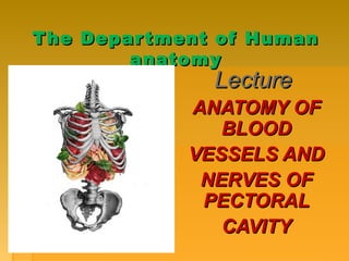
1. The Department of HumanThe Department of Human
anatomyanatomy
LectureLecture
ANATOMY OFANATOMY OF
BLOODBLOOD
VESSELS ANDVESSELS AND
NERVES OFNERVES OF
PECTORALPECTORAL
CAVITYCAVITY
2. PLANPLAN
I. PARTS AND BRANCHES OF AN AORTAI. PARTS AND BRANCHES OF AN AORTA
a) THE DESCENDING AORTAa) THE DESCENDING AORTA
b) THE VEINS SYSTEM OF THE THORAXb) THE VEINS SYSTEM OF THE THORAX
c) THE LYMPHATICS OF THE THORAXc) THE LYMPHATICS OF THE THORAX
II.II.THE NERVOUS SYSTEM OF THE THORAXTHE NERVOUS SYSTEM OF THE THORAX
a) anterior and posterior branches of the thoracica) anterior and posterior branches of the thoracic
nervesnerves
b) the phrenic nerveb) the phrenic nerve
c) the thoracic part of the sympathetic trunkc) the thoracic part of the sympathetic trunk
d) the thoracic part of the vagusd) the thoracic part of the vagus (10(10thth
) nerve) nerve
Topicality: normal functioning of the walls
and the chest cavity depends on their correct
blood supply and innervation.
3. PARTS AND BRANCHESPARTS AND BRANCHES
OF ANOF AN AORTAAORTA
AortaAorta isis contents of:contents of:
1. ASCENDING PART OF1. ASCENDING PART OF
AN AORTAAN AORTA
22.. ARCH ORARCH OR
AORTIC ARCHAORTIC ARCH
3. DESCENDING PART OF3. DESCENDING PART OF
AN AORTAAN AORTA
All of them have own branches.All of them have own branches.
The arch of aorta, arcus
aortae begins at the level of
sternal angle. It arches
superiorly, posteriorly and
to the left, and then
inferiorly.
On passing over the left
main bronchus, the arch of
aorta descends to the
posterior mediastinum and
reaches the body of Th4 on
the left. Here it becomes
continuous with the
descending part of thoracic
aorta.
4. 1.1. AN ASCENDING AORTAAN ASCENDING AORTA
The ascending aortaThe ascending aorta
begins between the left atriumbegins between the left atrium
and the infundibulum. Thisand the infundibulum. This
the first part of the aorta. Thethe first part of the aorta. The
ascendinq aorta has threeascendinq aorta has three
aortic sinusesaortic sinuses and bulb.and bulb. ItIt
contains of two branches:contains of two branches:
a) the left coronary arterya) the left coronary artery
(arteria coronaria cordis(arteria coronaria cordis
sinistra)sinistra)
b) the right coronary arteryb) the right coronary artery
(arteria coronaria cordis(arteria coronaria cordis
dextra).dextra).
6. Branches of arch ofBranches of arch of
aortaaorta
1)1) BrachiocephalicBrachiocephalic
trunc (artery)trunc (artery) ::
Right subclavianRight subclavian
Right common carotidRight common carotid
2)2) Left commonLeft common
carotidcarotid
External carotidExternal carotid
Internal carotidInternal carotid
3)3) Left subclavianLeft subclavian
7. TheThe descending aortadescending aorta
TheThe descending aortadescending aorta,, parspars
descendens aortae runs alongdescendens aortae runs along
the vertebral column on the left.the vertebral column on the left.
Its upper portion resides withinIts upper portion resides within
the posterior mediastinum (thethe posterior mediastinum (the
thoracic aorta),thoracic aorta), on passing theon passing the
aortic hiatus,aortic hiatus, the thoracic aortathe thoracic aorta
becomes continuous with thebecomes continuous with the
abdominal aorta.abdominal aorta. The abdominalThe abdominal
aortaaorta ends by splitting intoends by splitting into
common iliac arteries at thecommon iliac arteries at the
level oflevel of L4L4 (the(the aorticaortic
bifurcation,bifurcation, bifurcatio aortae).bifurcatio aortae).
8. THE THORACIC AORTATHE THORACIC AORTA (PARS THORACICA(PARS THORACICA
AORTAE)AORTAE)
is a continuation of the aortic arch. Itis a continuation of the aortic arch. It
resides within the posterior mediastinumresides within the posterior mediastinum
next to the vertebral column. The aortanext to the vertebral column. The aorta
resides to the left and then posteriorlyresides to the left and then posteriorly
from the esophagus. The thoracic aortafrom the esophagus. The thoracic aorta
passes through the aortic hiatus topasses through the aortic hiatus to
become continuous with the abdominalbecome continuous with the abdominal
aorta. Other neighboring organs are theaorta. Other neighboring organs are the
thoracic duct (found on the left) thethoracic duct (found on the left) the
azygos and hemiazygos veins and the leftazygos and hemiazygos veins and the left
sympathetic trunk. The branches ofsympathetic trunk. The branches of
thoracic aorta are subdivided into thethoracic aorta are subdivided into the
parietal and the visceral.parietal and the visceral.
9. The parietal branches are only the
posterior intercostal and the superior
phrenic arteries:
- the posterior intercostal arteries,
arteriae intercostales posteriores
(10 pairs) run along intercostal spaces 3
through 11. The branches running below
the 12 rib are the subcostal arteries
(arteriae subcostales).
- the superior phrenic arteries, arteriae
phrenicae arise from the lower portion of
the thoracic aorta. They supply the lumbar
part of diaphragm.
10. The visceral branchesThe visceral branches supplysupply
the thoracic viscera:the thoracic viscera:
-- thethe bronchial branches,bronchial branches, ramirami
bronchiales accompany thebronchiales accompany the
bronchi on their way to the lungsbronchi on their way to the lungs
- the- the esophageal branches,esophageal branches,
rami oesophagcalcs arise fromrami oesophagcalcs arise from
the anterior surface of thethe anterior surface of the
thoracic aorta. The upperthoracic aorta. The upper
esophageal branchesesophageal branches
anastomose with the inferioranastomose with the inferior
thyroid artery and the lower withthyroid artery and the lower with
the left gastric artery;the left gastric artery;
- the- the pericardial branchespericardial branches,, ramirami
pericardiac are the smallpericardiac are the small
branches that reach the posteriorbranches that reach the posterior
surface of pericardium;surface of pericardium;
- the- the mediastinal branches,mediastinal branches, ramirami
mediastinals are the smallmediastinals are the small
branches that supply mediastinalbranches that supply mediastinal
fat.fat.
11. THE VEINS SYSTEM OF THE THORAXTHE VEINS SYSTEM OF THE THORAX
The superior vena cavaThe superior vena cava
the great vein draining bloodthe great vein draining blood
from the head and neck isfrom the head and neck is
located in the superiorlocated in the superior
mediastinum. About 7cmmediastinum. About 7cm
long, this large vessel enterslong, this large vessel enters
the right atrium of the heartthe right atrium of the heart
vertically from its superiorvertically from its superior
aspect.aspect.
The brachiocephalic veinsThe brachiocephalic veins
are located in the superiorare located in the superior
mediastinum.mediastinum.
a)a) THETHE INTERNAL JUGULAR VEININTERNAL JUGULAR VEIN
b) THEb) THE SUBCLAVIAN VEINSUBCLAVIAN VEIN
12. The thoracic wall andThe thoracic wall and
upper lumbar regionupper lumbar region
are drained by theare drained by the
posterior intercostalposterior intercostal
and lumbar veins intoand lumbar veins into
thethe azygous veinazygous vein..
These consist ofThese consist of
longitudinal trunkslongitudinal trunks
on right and lefton right and left
sides. There is asides. There is a
single trunk on thesingle trunk on the
right. On the left sideright. On the left side
thethe hemiazygoushemiazygous
13. The lymphatic glandsThe lymphatic glands ((nodulesnodules)) of theof the
viscera of the thorax are:viscera of the thorax are:
1)The bronchial lymphatic glands-are situated1)The bronchial lymphatic glands-are situated
round the bifurcation of the trachea and rootsround the bifurcation of the trachea and roots
of the lungs. They are ten or twelve in number,of the lungs. They are ten or twelve in number,
the largest being placed opposite thethe largest being placed opposite the
bifurcation of the trachea. The smallest roundbifurcation of the trachea. The smallest round
the bronchi and their primary divisions forthe bronchi and their primary divisions for
some little distance within the substance of thesome little distance within the substance of the
lungs.lungs.
2) The superior mediastinal or cardiac glands2) The superior mediastinal or cardiac glands
- lie in front of the transverse aorta and left- lie in front of the transverse aorta and left
innominate vein.innominate vein.
:
14. The lymphatic glands of theThe lymphatic glands of the
thoracic wall are:thoracic wall are:
The intercostal lymphaticThe intercostal lymphatic
glands - are small, andglands - are small, and
situated on each side of thesituated on each side of the
spine near the costo-spine near the costo-
vertebral articulations. Theyvertebral articulations. They
vary from one to three invary from one to three in
each space.each space.
The sternal or internalThe sternal or internal
mammary lymphatic glands -mammary lymphatic glands -
are placed at the anteriorare placed at the anterior
extremity of each intercostalextremity of each intercostal
space, by the side of thespace, by the side of the
internal mammary vessels.internal mammary vessels.
15. The SUPERFICiAl LYMPHATIC VESSELSThe SUPERFICiAl LYMPHATIC VESSELS -- of theof the
front of the thorax run across the greatfront of the thorax run across the great
pectoral muscle, and those on the backpectoral muscle, and those on the back
part of this cavity lie upon the trapeziuspart of this cavity lie upon the trapezius
and latissimus dorsi.and latissimus dorsi.
THE DEEP LYMPHATIC VESSELS OF THETHE DEEP LYMPHATIC VESSELS OF THE
THORACIC WALLTHORACIC WALL areare
I) The intercostal lymphatic vesselsI) The intercostal lymphatic vessels
2) Tne internal mammary lymphatic2) Tne internal mammary lymphatic
vesselsvessels
3)The lymphatic vessels of the3)The lymphatic vessels of the
diaphragmdiaphragm
LYMPHATIC VESSELS OF THE THORAXLYMPHATIC VESSELS OF THE THORAX areare
1)The lymphatic vessels of the lung1)The lymphatic vessels of the lung
2)2) The cardiac lymphatic vesselsThe cardiac lymphatic vessels
3)3) The thymic lymphatic vesselsThe thymic lymphatic vessels
4)4) The lymphatic vessels of theThe lymphatic vessels of the
oesophagusoesophagus
16. THE INNERVATION OF THE WALLS OFTHE INNERVATION OF THE WALLS OF
THORATIC CAVITYTHORATIC CAVITY
The anterior branchesThe anterior branches (rami(rami
ventrales)ventrales) of the spinal nervesof the spinal nerves
innervate the skin and musclesinnervate the skin and muscles
of the ventral wall of the bodyof the ventral wall of the body
and both pairs of limbs. Theand both pairs of limbs. The
anterior branches of the spinalanterior branches of the spinal
nerves preserve their originalnerves preserve their original
metameric structure only in themetameric structure only in the
thoracic segment (nn.thoracic segment (nn.
intercostales). In the otherintercostales). In the other
segments connected with thesegments connected with the
limbs in whose development thelimbs in whose development the
segmentary character is lost, thesegmentary character is lost, the
nerves arising from the anteriornerves arising from the anterior
spinal branches intertwine.spinal branches intertwine.
17. THE POSTERIORTHE POSTERIOR BRANCHESBRANCHES
(rami dorsales)(rami dorsales) of the thoracicof the thoracic
nerves divide into medialnerves divide into medial
and lateral / branchesand lateral / branches
giving rise to branchesgiving rise to branches
running to therunning to the
autochthonous muscles;autochthonous muscles;
the skin branches of thethe skin branches of the
superior thoracic nervessuperior thoracic nerves
originate only from ramioriginate only from rami
mediales, while those ofmediales, while those of
the inferior thoracicthe inferior thoracic
nerves, from raminerves, from rami
laterales.laterales.
18. TheThe phrenic nervephrenic nerve
TheThe phrenic nervephrenic nerve is theis the
mixed branches of themixed branches of the
cervical plexus- its motorcervical plexus- its motor
branches innervate thebranches innervate the
diaphragm. It sendsdiaphragm. It sends
sensory nerves to the pleurasensory nerves to the pleura
and pericardium.and pericardium.
Some of the terminal branchesSome of the terminal branches
of the nerve pass throughof the nerve pass through
the diaphragm into thethe diaphragm into the
abdominal cavitiabdominal caviti
(NN.PHRENICOABDOMINALES(NN.PHRENICOABDOMINALES))
19. The thoracic part of the sympatheticThe thoracic part of the sympathetic
trunktrunk
The thoracic part of the sympathetic trunkThe thoracic part of the sympathetic trunk consists of 10 toconsists of 10 to
12 ganglia of a more or less triangular shape. The thoracic12 ganglia of a more or less triangular shape. The thoracic
part is characterized by the presence ofpart is characterized by the presence of whitewhite
communicating branches (rami communicantes albicommunicating branches (rami communicantes albi) which) which
connect the anterior roots of the spinal nerves with theconnect the anterior roots of the spinal nerves with the
sympathetic trunk ganglia.sympathetic trunk ganglia.
The branches of the thoracic part are as follows:The branches of the thoracic part are as follows:
- the cardiac branches- the cardiac branches (nervi cardiaci thoracici);(nervi cardiaci thoracici);
-- the grey communicating branchesthe grey communicating branches (rami communicantes grisei(rami communicantes grisei));;
-- the pulmonar branchesthe pulmonar branches (rami pulmonales);(rami pulmonales);
- the aortic branches- the aortic branches (rami aorlici)(rami aorlici) form a thoracic aortic plexusform a thoracic aortic plexus (plexus(plexus
aorticus thoracicus),aorticus thoracicus), partly on the oesophagus - oesophageal plexuspartly on the oesophagus - oesophageal plexus
(plexus esophageus(plexus esophageus), and on the thoracic duct (the vagus nerve also), and on the thoracic duct (the vagus nerve also
contributes to the formation of these plexuses);contributes to the formation of these plexuses);
- the greater and lesser splanchnic nerves- the greater and lesser splanchnic nerves (nervi splanchnici major(nervi splanchnici major andand
minor).minor).
20. The thoracic part of the sympathetic trunkThe thoracic part of the sympathetic trunk
21. The thoracic part of theThe thoracic part of the vagusvagus
nerve:nerve:
1. The recurrent laryngeal
nerve (n. laryngeus recurrens)
2.Cardiac branches (lower)
(ramus cardiaci cervicales
inferiores)
3.Pulmonary and tracheal
branches (rami bronchiales
and tracheales)
4.Thoracic cardiac branches
(rami cardiaci thoracici)
5.Oesophageal branches
(rami esophagei)