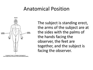
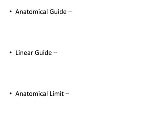
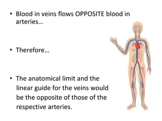
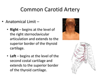
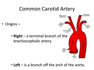
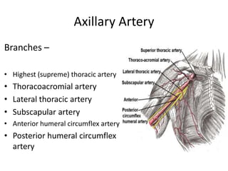
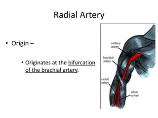
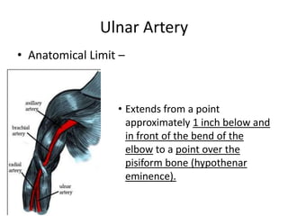
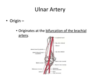
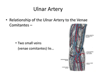
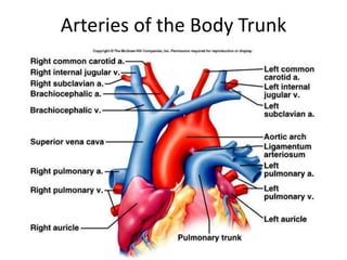
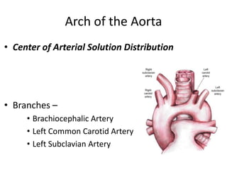
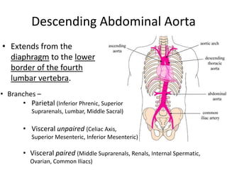
Munh me chhale hona ek aam bat si ho chuki hai jiske bahut se karan hai jaise mirch mashaledar bhojan, chay, bidi, paan, gutkha ityadi. Munh me hone wale chhale bahut jyada dard utpann karte hai jiski vajah se marij khana bhi theek se nahi kha pata. Munh ke chhalo ki angreji dawa se achha hai ki gharelu ilaj kiya jaye.
Cervical Arterial Dysfunction & Clinical Practice: Moving ForwardAlan J Taylor
A short description of current concepts in the understanding of vascular risk in the cervical spine, with specific reference to cervical arterial dysfunction (CAD). This presentation gives an overview of this 'umbrella term' and relates to the pathologies that fall under it.
Arterial Pulse: Radial
To assess cardiac function. To assess state of health.
NEW BLOGSITE ADDRESS:
"Nurses Information Site"
http://nursesinfosite.blogspot.com
In human anatomy, the thigh is the area between the hip (pelvis) and the knee. Anatomically, it is part of the lower limb. The single bone in the thigh is called the femur.
1. Blood supply of the upper limb. Major arterial anastomoses of the upper extremity.
2. The veins of the upper limb.
3. Innervation of the upper limb. Schematic representation of the innervation of the skin of the upper limb.
4. Lymphatic vessels of the upper extremity.
General Introduction of Cardiovascular System and Anatomy of Cardiovascular System.
In this slide, you will be able to find the general anatomy of the heart and Basic introduction of Cardiovascular Sstem
The femoral triangle is a wedge-shaped area located within the superomedial aspect of the anterior thigh. The femoral triangle is a hollow region located in the supero-medial part of the anterior thigh.
The female reproductive system provides several functions.
The ovaries produce the egg cells, called the ova or oocytes.
The oocytes are then transported to the fallopian tube where fertilization by a sperm may occur.
The fertilized egg then moves to the uterus, where the uterine lining has thickened in response to the normal hormones of the reproductive cycle.
Once in the uterus, the fertilized egg can implant into thickened uterine lining and continue to develop.
If implantation does not take place, the uterine lining is shed as menstrual flow.
In addition, the female reproductive system produces female sex hormones that maintain the reproductive cycle.
During menopause, the female reproductive system gradually stops making the female hormones necessary for the reproductive cycle to work. At this point, menstrual cycles can become irregular and eventually stop.
One year after menstrual cycles stop, the woman is considered to be menopausal.
The work of the peripheral vascular surgeon ranges widely through the anatomy of the whole body and he is required to command a wide range of anatomical knowledge.Both diseased and healthy arteries may be surprisingly friable, and rough dissection may cause severe damage and be sufficient to jeopardize the result of the vascular reconstruction. This power point presentation outlines the different exposure techniques to almost all vessels in the body which makes the surgeon familiar and confident when faced with a clinical situation that mandates exposure of certain vessel whether as an emergency or elective procedure.
Be the first to comment
Anatomy And Physiology of Human Heart
1. ANATOMY OF THE HEART By: Dr Mohammed Faez
2. The Heart The heart is a chambered muscular organ that pumps blood received from the veins into the arteries, thereby maintaining the flow of blood through the entire circulatory system.
3. The Heart • The heart is surrounded by membrane called Pericardium.
4. The Pericardium • The pericardium is a fibroserous sac that encloses the heart and the roots of the great vessels. • The pericardium lies within the middle mediastinum.
5. The Pericardium
6. The Pericardium • Its function is to restrict excessive movements of the heart as a whole and to serve as a lubricated container in which the different parts of the heart can contract.
The thoracic structure refers to the anatomical components of the thorax, which is the region of the body between the neck and the abdomen, also known as the chest. It includes the thoracic vertebrae, ribs, sternum (breastbone), and associated muscles, ligaments, and organs such as the heart and lungs. The thoracic structure plays a crucial role in protecting vital organs, supporting the upper body, and facilitating respiration.
The thoracic structure encompasses the anatomical elements of the chest region, including the thoracic vertebrae, ribs, sternum, associated muscles, and vital organs such as the heart and lungs. It provides protection, support, and facilitates respiration, highlighting its critical role in overall bodily function and health.The thoracic structure comprises the chest's anatomical components, including vertebrae, ribs, sternum, muscles, and vital organs like the heart and lungs, crucial for protection, support, and respiration..The thoracic structure includes the chest's bones, muscles, and organs, vital for breathing and protecting the heart and lungs.
1. **Thoracic Vertebrae:** The thoracic spine consists of twelve vertebrae (T1-T12) that form the posterior aspect of the thoracic structure. These vertebrae are larger than those in the cervical or lumbar regions and articulate with the ribs, providing stability and support for the upper body.
2. **Ribs:** There are twelve pairs of ribs that attach posteriorly to the thoracic vertebrae and curve anteriorly to meet the sternum. Ribs play a vital role in protecting the internal organs of the thoracic cavity, such as the heart and lungs. The upper seven pairs are true ribs, while the lower five pairs are false ribs (some of which do not directly attach to the sternum) or floating ribs.
3. **Sternum:** The sternum is a flat bone located in the center of the anterior thoracic wall. It consists of three parts: the manubrium, body, and xiphoid process. The sternum serves as an attachment point for the ribs and provides structural support to the chest.
4. **Muscles:** Several muscles surround the thoracic cavity, contributing to breathing and movement of the chest wall. These include the intercostal muscles (external, internal, and innermost), which are located between the ribs and aid in respiration, as well as muscles such as the diaphragm, which separates the thoracic and abdominal cavities and plays a primary role in breathing.
5. **Organs:** The thoracic cavity houses important organs such as the heart and lungs. The heart is situated in the mediastinum, a central compartment of the thoracic cavity, while the lungs occupy the lateral portions. The thoracic structure provides protection and support for these vital organs while allowing for their necessary movements during respiration and circulation.
Understanding the anatomy and function of the thoracic structure is essential for various medical disciplines, including anatomy, physiology, orthopedics, cardiology, and res
Lung Cancer: Artificial Intelligence, Synergetics, Complex System Analysis, S...Oleg Kshivets
RESULTS: Overall life span (LS) was 2252.1±1742.5 days and cumulative 5-year survival (5YS) reached 73.2%, 10 years – 64.8%, 20 years – 42.5%. 513 LCP lived more than 5 years (LS=3124.6±1525.6 days), 148 LCP – more than 10 years (LS=5054.4±1504.1 days).199 LCP died because of LC (LS=562.7±374.5 days). 5YS of LCP after bi/lobectomies was significantly superior in comparison with LCP after pneumonectomies (78.1% vs.63.7%, P=0.00001 by log-rank test). AT significantly improved 5YS (66.3% vs. 34.8%) (P=0.00000 by log-rank test) only for LCP with N1-2. Cox modeling displayed that 5YS of LCP significantly depended on: phase transition (PT) early-invasive LC in terms of synergetics, PT N0—N12, cell ratio factors (ratio between cancer cells- CC and blood cells subpopulations), G1-3, histology, glucose, AT, blood cell circuit, prothrombin index, heparin tolerance, recalcification time (P=0.000-0.038). Neural networks, genetic algorithm selection and bootstrap simulation revealed relationships between 5YS and PT early-invasive LC (rank=1), PT N0—N12 (rank=2), thrombocytes/CC (3), erythrocytes/CC (4), eosinophils/CC (5), healthy cells/CC (6), lymphocytes/CC (7), segmented neutrophils/CC (8), stick neutrophils/CC (9), monocytes/CC (10); leucocytes/CC (11). Correct prediction of 5YS was 100% by neural networks computing (area under ROC curve=1.0; error=0.0).
CONCLUSIONS: 5YS of LCP after radical procedures significantly depended on: 1) PT early-invasive cancer; 2) PT N0--N12; 3) cell ratio factors; 4) blood cell circuit; 5) biochemical factors; 6) hemostasis system; 7) AT; 8) LC characteristics; 9) LC cell dynamics; 10) surgery type: lobectomy/pneumonectomy; 11) anthropometric data. Optimal diagnosis and treatment strategies for LC are: 1) screening and early detection of LC; 2) availability of experienced thoracic surgeons because of complexity of radical procedures; 3) aggressive en block surgery and adequate lymph node dissection for completeness; 4) precise prediction; 5) adjuvant chemoimmunoradiotherapy for LCP with unfavorable prognosis.
Ozempic: Preoperative Management of Patients on GLP-1 Receptor Agonists Saeid Safari
Preoperative Management of Patients on GLP-1 Receptor Agonists like Ozempic and Semiglutide
ASA GUIDELINE
NYSORA Guideline
2 Case Reports of Gastric Ultrasound
- Video recording of this lecture in English language: https://youtu.be/lK81BzxMqdo
- Video recording of this lecture in Arabic language: https://youtu.be/Ve4P0COk9OI
- Link to download the book free: https://nephrotube.blogspot.com/p/nephrotube-nephrology-books.html
- Link to NephroTube website: www.NephroTube.com
- Link to NephroTube social media accounts: https://nephrotube.blogspot.com/p/join-nephrotube-on-social-media.html
These simplified slides by Dr. Sidra Arshad present an overview of the non-respiratory functions of the respiratory tract.
Learning objectives:
1. Enlist the non-respiratory functions of the respiratory tract
2. Briefly explain how these functions are carried out
3. Discuss the significance of dead space
4. Differentiate between minute ventilation and alveolar ventilation
5. Describe the cough and sneeze reflexes
Study Resources:
1. Chapter 39, Guyton and Hall Textbook of Medical Physiology, 14th edition
2. Chapter 34, Ganong’s Review of Medical Physiology, 26th edition
3. Chapter 17, Human Physiology by Lauralee Sherwood, 9th edition
4. Non-respiratory functions of the lungs https://academic.oup.com/bjaed/article/13/3/98/278874
Recomendações da OMS sobre cuidados maternos e neonatais para uma experiência pós-natal positiva.
Em consonância com os ODS – Objetivos do Desenvolvimento Sustentável e a Estratégia Global para a Saúde das Mulheres, Crianças e Adolescentes, e aplicando uma abordagem baseada nos direitos humanos, os esforços de cuidados pós-natais devem expandir-se para além da cobertura e da simples sobrevivência, de modo a incluir cuidados de qualidade.
Estas diretrizes visam melhorar a qualidade dos cuidados pós-natais essenciais e de rotina prestados às mulheres e aos recém-nascidos, com o objetivo final de melhorar a saúde e o bem-estar materno e neonatal.
Uma “experiência pós-natal positiva” é um resultado importante para todas as mulheres que dão à luz e para os seus recém-nascidos, estabelecendo as bases para a melhoria da saúde e do bem-estar a curto e longo prazo. Uma experiência pós-natal positiva é definida como aquela em que as mulheres, pessoas que gestam, os recém-nascidos, os casais, os pais, os cuidadores e as famílias recebem informação consistente, garantia e apoio de profissionais de saúde motivados; e onde um sistema de saúde flexível e com recursos reconheça as necessidades das mulheres e dos bebês e respeite o seu contexto cultural.
Estas diretrizes consolidadas apresentam algumas recomendações novas e já bem fundamentadas sobre cuidados pós-natais de rotina para mulheres e neonatos que recebem cuidados no pós-parto em unidades de saúde ou na comunidade, independentemente dos recursos disponíveis.
É fornecido um conjunto abrangente de recomendações para cuidados durante o período puerperal, com ênfase nos cuidados essenciais que todas as mulheres e recém-nascidos devem receber, e com a devida atenção à qualidade dos cuidados; isto é, a entrega e a experiência do cuidado recebido. Estas diretrizes atualizam e ampliam as recomendações da OMS de 2014 sobre cuidados pós-natais da mãe e do recém-nascido e complementam as atuais diretrizes da OMS sobre a gestão de complicações pós-natais.
O estabelecimento da amamentação e o manejo das principais intercorrências é contemplada.
Recomendamos muito.
Vamos discutir essas recomendações no nosso curso de pós-graduação em Aleitamento no Instituto Ciclos.
Esta publicação só está disponível em inglês até o momento.
Prof. Marcus Renato de Carvalho
www.agostodourado.com
Ethanol (CH3CH2OH), or beverage alcohol, is a two-carbon alcohol
that is rapidly distributed in the body and brain. Ethanol alters many
neurochemical systems and has rewarding and addictive properties. It
is the oldest recreational drug and likely contributes to more morbidity,
mortality, and public health costs than all illicit drugs combined. The
5th edition of the Diagnostic and Statistical Manual of Mental Disorders
(DSM-5) integrates alcohol abuse and alcohol dependence into a single
disorder called alcohol use disorder (AUD), with mild, moderate,
and severe subclassifications (American Psychiatric Association, 2013).
In the DSM-5, all types of substance abuse and dependence have been
combined into a single substance use disorder (SUD) on a continuum
from mild to severe. A diagnosis of AUD requires that at least two of
the 11 DSM-5 behaviors be present within a 12-month period (mild
AUD: 2–3 criteria; moderate AUD: 4–5 criteria; severe AUD: 6–11 criteria).
The four main behavioral effects of AUD are impaired control over
drinking, negative social consequences, risky use, and altered physiological
effects (tolerance, withdrawal). This chapter presents an overview
of the prevalence and harmful consequences of AUD in the U.S.,
the systemic nature of the disease, neurocircuitry and stages of AUD,
comorbidities, fetal alcohol spectrum disorders, genetic risk factors, and
pharmacotherapies for AUD.
Pulmonary Thromboembolism - etilogy, types, medical- Surgical and nursing man...VarunMahajani
Disruption of blood supply to lung alveoli due to blockage of one or more pulmonary blood vessels is called as Pulmonary thromboembolism. In this presentation we will discuss its causes, types and its management in depth.
TEST BANK for Operations Management, 14th Edition by William J. Stevenson, Ve...kevinkariuki227
TEST BANK for Operations Management, 14th Edition by William J. Stevenson, Verified Chapters 1 - 19, Complete Newest Version.pdf
New Directions in Targeted Therapeutic Approaches for Older Adults With Mantl...i3 Health
i3 Health is pleased to make the speaker slides from this activity available for use as a non-accredited self-study or teaching resource.
This slide deck presented by Dr. Kami Maddocks, Professor-Clinical in the Division of Hematology and
Associate Division Director for Ambulatory Operations
The Ohio State University Comprehensive Cancer Center, will provide insight into new directions in targeted therapeutic approaches for older adults with mantle cell lymphoma.
STATEMENT OF NEED
Mantle cell lymphoma (MCL) is a rare, aggressive B-cell non-Hodgkin lymphoma (NHL) accounting for 5% to 7% of all lymphomas. Its prognosis ranges from indolent disease that does not require treatment for years to very aggressive disease, which is associated with poor survival (Silkenstedt et al, 2021). Typically, MCL is diagnosed at advanced stage and in older patients who cannot tolerate intensive therapy (NCCN, 2022). Although recent advances have slightly increased remission rates, recurrence and relapse remain very common, leading to a median overall survival between 3 and 6 years (LLS, 2021). Though there are several effective options, progress is still needed towards establishing an accepted frontline approach for MCL (Castellino et al, 2022). Treatment selection and management of MCL are complicated by the heterogeneity of prognosis, advanced age and comorbidities of patients, and lack of an established standard approach for treatment, making it vital that clinicians be familiar with the latest research and advances in this area. In this activity chaired by Michael Wang, MD, Professor in the Department of Lymphoma & Myeloma at MD Anderson Cancer Center, expert faculty will discuss prognostic factors informing treatment, the promising results of recent trials in new therapeutic approaches, and the implications of treatment resistance in therapeutic selection for MCL.
Target Audience
Hematology/oncology fellows, attending faculty, and other health care professionals involved in the treatment of patients with mantle cell lymphoma (MCL).
Learning Objectives
1.) Identify clinical and biological prognostic factors that can guide treatment decision making for older adults with MCL
2.) Evaluate emerging data on targeted therapeutic approaches for treatment-naive and relapsed/refractory MCL and their applicability to older adults
3.) Assess mechanisms of resistance to targeted therapies for MCL and their implications for treatment selection
Acute scrotum is a general term referring to an emergency condition affecting the contents or the wall of the scrotum.
There are a number of conditions that present acutely, predominantly with pain and/or swelling
A careful and detailed history and examination, and in some cases, investigations allow differentiation between these diagnoses. A prompt diagnosis is essential as the patient may require urgent surgical intervention
Testicular torsion refers to twisting of the spermatic cord, causing ischaemia of the testicle.
Testicular torsion results from inadequate fixation of the testis to the tunica vaginalis producing ischemia from reduced arterial inflow and venous outflow obstruction.
The prevalence of testicular torsion in adult patients hospitalized with acute scrotal pain is approximately 25 to 50 percent
2. Anatomical Position
• The subject is standing erect,
the arms of the subject are at
the sides with the palms of
the hands facing the
observer, the feet are
together, and the subject is
facing the observer.
4. • Blood in veins flows OPPOSITE blood in
arteries…
• Therefore…
• The anatomical limit and the
linear guide for the veins would
be the opposite of those of the
respective arteries.
5. • The anatomical guides for arteries and veins
would be the same.
8. Common Carotid Artery
• Anatomical Limit –
• Right – begins at the level of
the right sternoclavicular
articulation and extends to the
superior border of the thyroid
cartilage.
• Left – begins at the level of the
second costal cartilage and
extends to the superior border
of the thyroid cartilage.
9. Common Carotid Artery
• Origins –
• Right – a terminal branch of the
brachiocephalic artery.
• Left – is a branch off the arch of the aorta.
10. Common Carotid Artery
• Branches –
• Right – no branches of the right common carotid,
except the terminal bifurcation into the right internal
and external carotid arteries.
• Left – no branches except the terminal bifurcation into
the left internal and external carotid arteries.
12. Common Carotid Artery
• Branches of the Right and Left External
Carotid Arteries –
• Ascending pharyngeal
• Superior thyroid
• Lingual
• Facial
• Occipital
• Posterior auricular
• Maxillary
• Superficial temporal
13. Common Carotid Artery
• Branches of the Right and Left Internal Carotid
Arteries –
• Branches arising
within the carotid canal
• Ophthalmic
• Anterior cerebral
• Middle cerebral
• Posterior communicating
• Choroidal branches
14. Common Carotid Artery
• Relationship of the Common Carotid to the
Internal Jugular Vein –
• The internal jugular vein lies…
15. Common Carotid Artery
• Contents of the Carotid Sheath –
• Internal jugular vein
• Vagus nerve
• Common carotid artery
What is the carotid sheath?
18. Axillary Artery
• Anatomical Limit –
• Extends from a point
beginning at the lateral
border of the first rib and
extends to the inferior
border of the tendon of the
teres major muscle.
19. Axillary Artery
• Origin –
• A continuation of the
subclavian artery.
25. Brachial Artery
• Anatomical Limit –
• Extends from a point beginning at the
inferior border of the tendon of the
teres major muscle and extends to a
point inferior to the antecubital fossa.
31. Radial Artery
• Anatomical Limit –
• Extends from a point approximately 1 inch below
an in front of the bend
of the elbow to a point
over the base of the
thumb (thenar
eminence).
36. Ulnar Artery
• Anatomical Limit –
• Extends from a point
approximately 1 inch below and
in front of the bend of the
elbow to a point over the
pisiform bone (hypothenar
eminence).
38. Ulnar Artery
• Relationship of the Ulnar Artery to the Venae
Comitantes –
• Two small veins
(venae comitantes) lie…
39. Arteries of the Body Trunk
• Ascending Aorta
• Arch of the Aorta
• Right Subclavian
• Left Subclavian
• Descending Thoracic Aorta
• Descending Abdominal Aorta
42. Arch of the Aorta
• Center of Arterial Solution Distribution
• Branches –
• Brachiocephalic Artery
• Left Common Carotid Artery
• Left Subclavian Artery
43. Right Subclavian
• Begins at the right sternoclavicular articulation
and extends to the lateral border of the first
rib.
• For full autopsy (neck organs removed) –
branches need to be clamped.
• Braches –
• Vertebral Artery
• Internal Thoracic artery
• Inferior Thyroid
44. Left Subclavian
• Begins at the level of the
left second costal cartilage
and extends to the lateral
border of the first rib.
46. Descending Abdominal Aorta
• Extends from the
diaphragm to the lower
border of the fourth
lumbar vertebra.
• Branches –
• Parietal (Inferior Phrenic, Superior
Suprarenals, Lumbar, Middle Sacral)
• Visceral unpaired (Celiac Axis,
Superior Mesenteric, Inferior Mesenteric)
• Visceral paired (Middle Suprarenals, Renals, Internal Spermatic,
Ovarian, Common Iliacs)
47. External Iliac Artery and Vein
• Continuation of the common iliac artery.
• Common iliac is a terminal branch of the
abdominal aorta.
• Extends to a point under the center of the
inguinal ligament.
• Lies exactly at this ligament lateral to the external
iliac vein.
48. External Iliac Artery and Vein
• Used in autopsied bodies for the injection of the
lower extremities.
• In unautopsied bodies, it
can be used for the
morbidly obese.
• Iliofemoral –