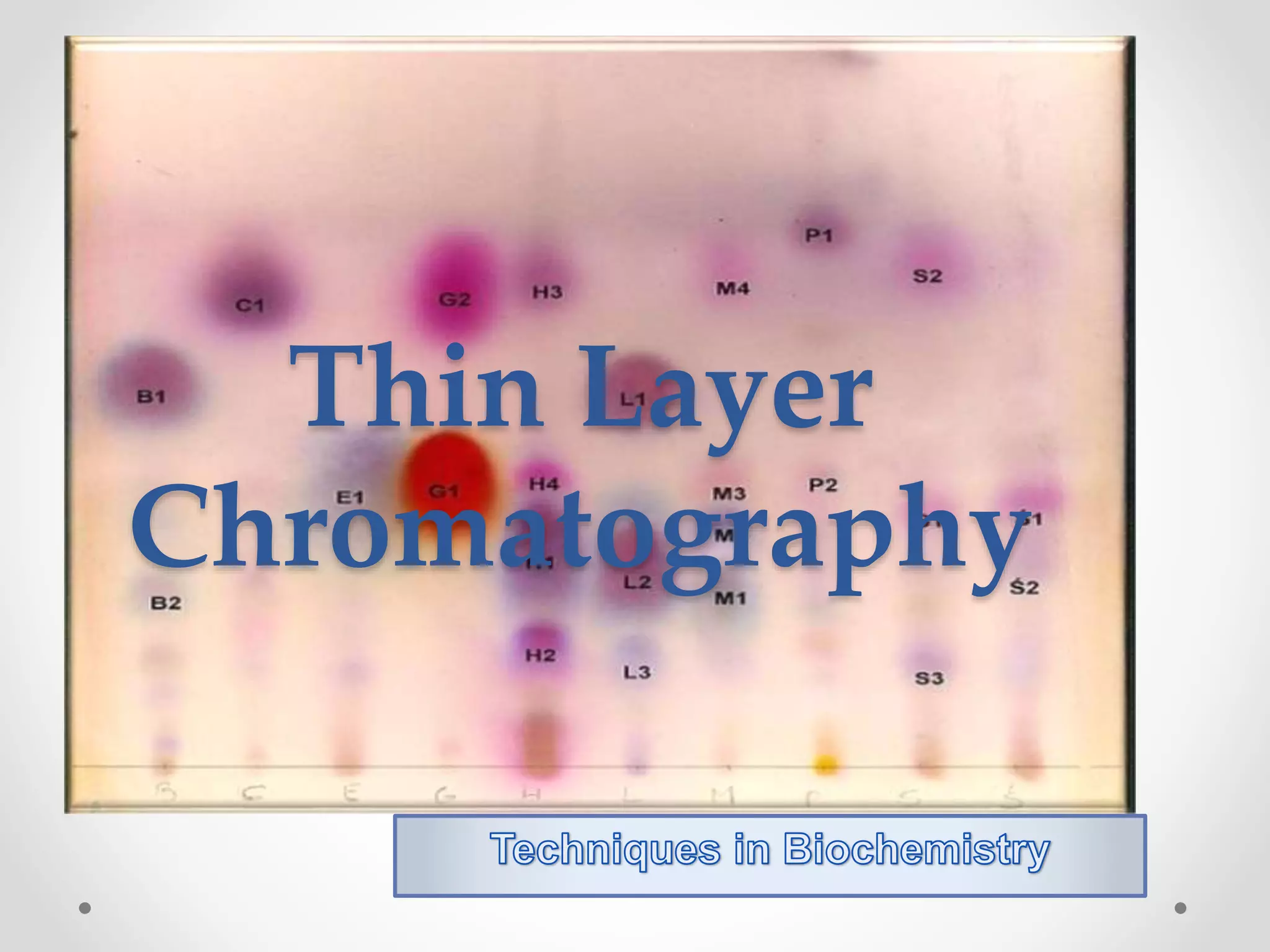
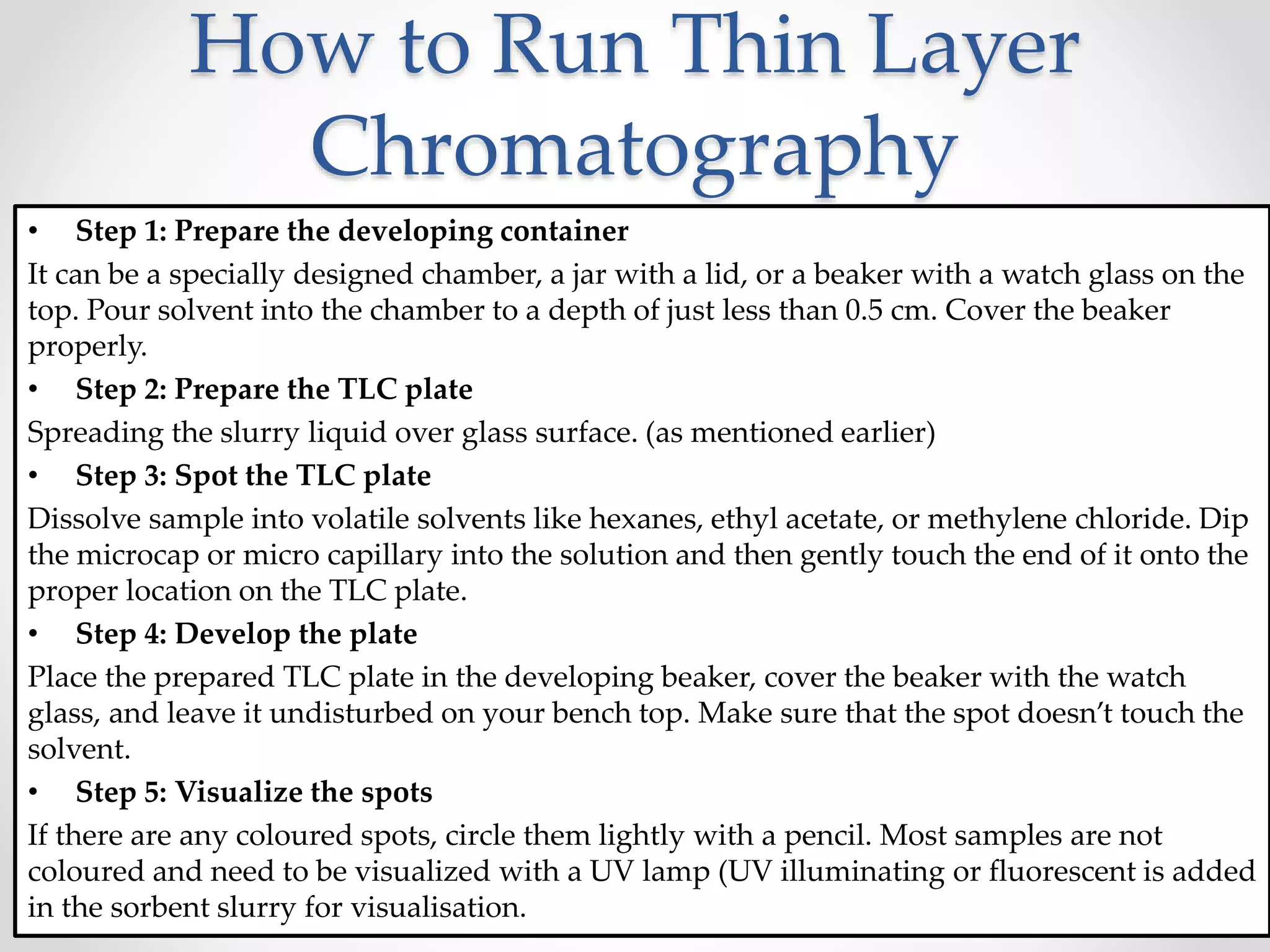
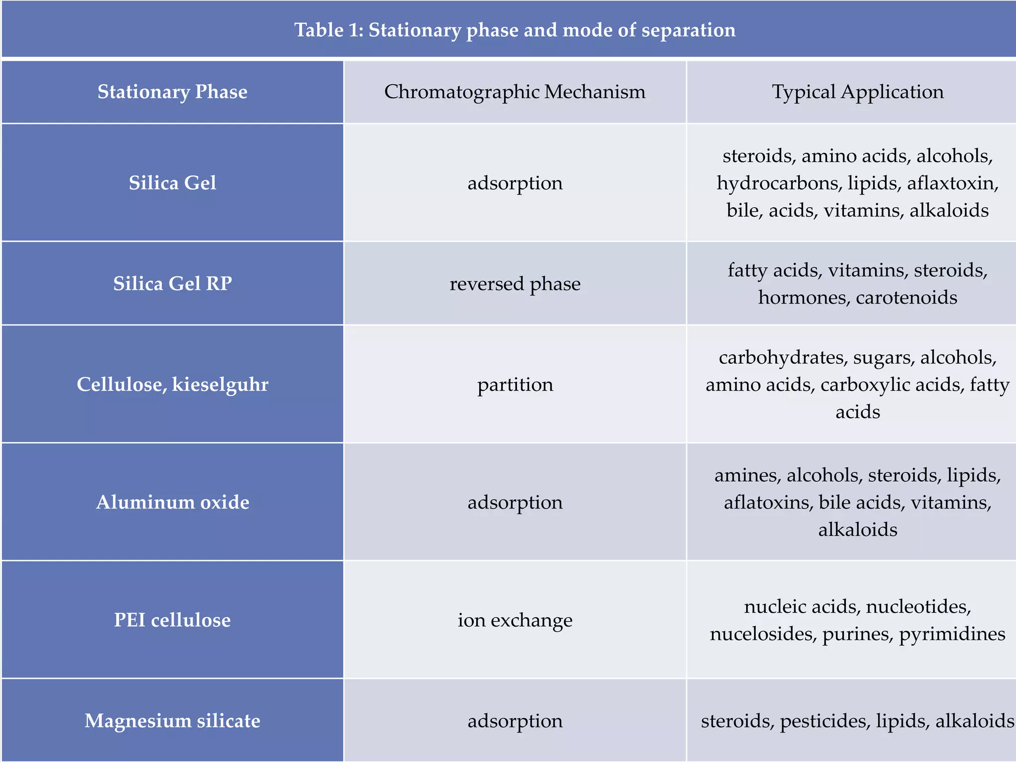
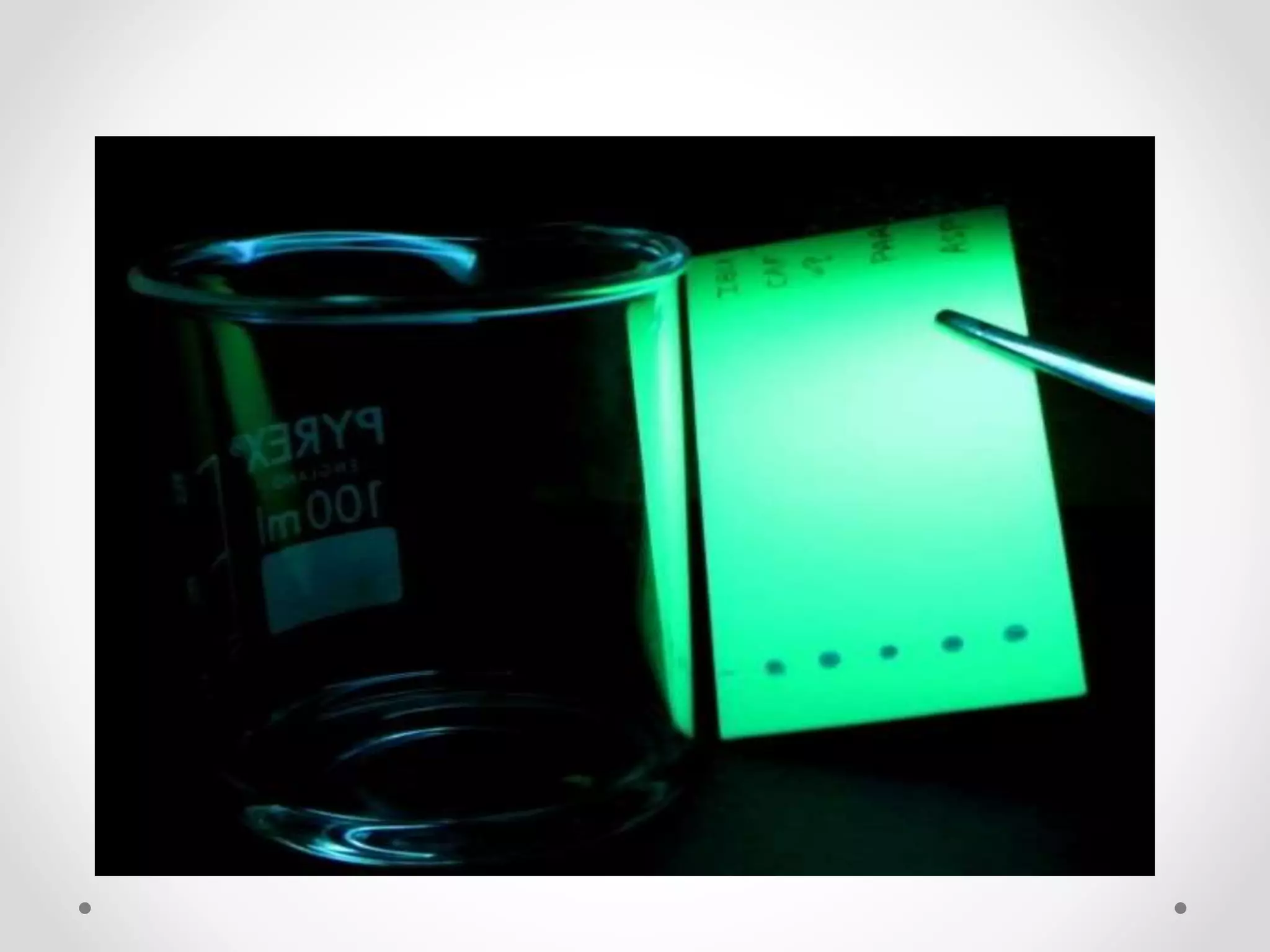
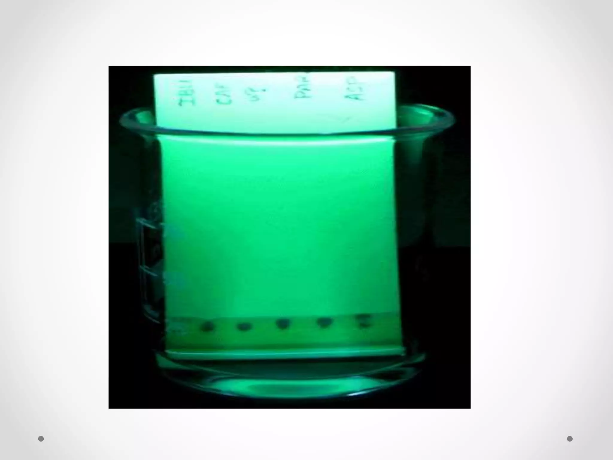
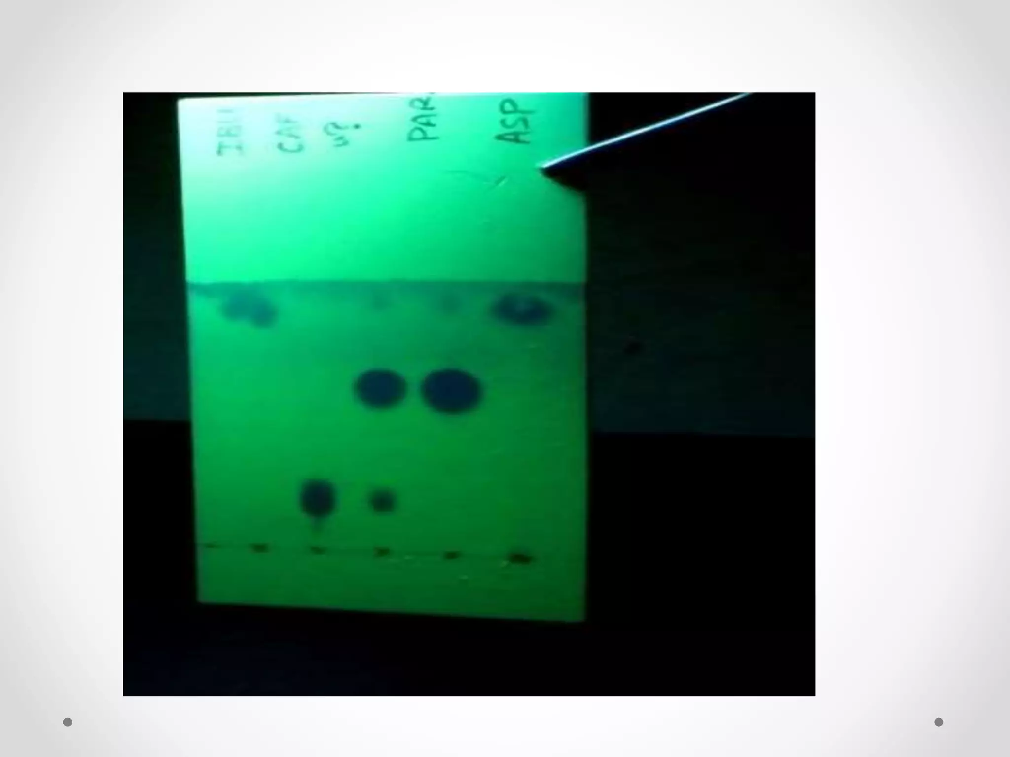
X-Ray Crystallography is a technique used to determine the atomic and molecular structure of a crystal, in which the crystalline atoms cause a beam of incident X-rays to diffract into many specific directions.
Atomic Absorption Spectroscopy is a very common technique for detecting metals and metalloids in samples.
It is very reliable and simple to use.
It can analyze over 62 elements.
It also measures the concentration of metals in the sample.
Dynamic light scattering (DLS) or Quasi-Elastic Light Scattering (QELS), is a non-invasive, well-established technique for measuring the size and size distribution of molecules and particles typically in the submicron region, and with the latest technology lower than 1nm.
In This slide the working principle and the function of DLS is Explained in brief and precise way.
GAS CHROMATOGRAPHY-MASS SPECTROSCOPY [GC-MS]Shikha Popali
Â
THIS PRESENTATION GIVES A DETAIL ACCOUNT ON THE GC-MS WITH ITS INTRODUCTION, BASIC PRINCIPLE OF BOTH COMBINED AND INDIVIDUALLY WITH ITS INSTRUMENTATION, APPLICATION AND EXAMPLES, MAKES EASY TO COLLECT ALL THE DATA AT A PLACE ACCORDING TO THE M.PHARM SYLLABUS S PER PCI
SEM is a type of electron microscope designed for directly studying the surfaces of solid objects, that utilizes a beam of focused electron of relatively low energy as an electron probe that is scanned in a regular manner over the specimen.
Atomic Absorption spectrometer is an instrument used for quantitative analysis of most of the metals in nano grams. This is highly sensitive technique used for analysis.
A scanning electron microscope is a type of electron microscope that produces images of a sample by scanning the surface with a focused beam of electrons. The electrons interact with atoms in the sample, producing various signals that contain information about the sample's surface topography and composition.
SEMs can magnify an object from about 10 times up to 300,000 times. A scale bar is often provided on an SEM image. From this the actual size of structures in the image can be calculated.
X-ray crystallography is a technique used for determining the atomic and molecular structure of a crystal, in which the crystalline atoms cause a beam of incident X-rays to diffract into many specific directions.
introduction, history, principle, experimental techniques, evaluation on chromatogram, adv. & dis-adv., common problems, comparision, applications and analysis of drugs through TLC(2000-2017)
This is about on TLC. and I hope it will helpful for you.
In this describe about their introduction, principle, application, procedure, methodology, RF value, and their advantage, disadvantage
Thank youđ
Comparing Evolved Extractive Text Summary Scores of Bidirectional Encoder Rep...University of Maribor
Â
Slides from:
11th International Conference on Electrical, Electronics and Computer Engineering (IcETRAN), NiĹĄ, 3-6 June 2024
Track: Artificial Intelligence
https://www.etran.rs/2024/en/home-english/
A brief information about the SCOP protein database used in bioinformatics.
The Structural Classification of Proteins (SCOP) database is a comprehensive and authoritative resource for the structural and evolutionary relationships of proteins. It provides a detailed and curated classification of protein structures, grouping them into families, superfamilies, and folds based on their structural and sequence similarities.
Slide 1: Title Slide
Extrachromosomal Inheritance
Slide 2: Introduction to Extrachromosomal Inheritance
Definition: Extrachromosomal inheritance refers to the transmission of genetic material that is not found within the nucleus.
Key Components: Involves genes located in mitochondria, chloroplasts, and plasmids.
Slide 3: Mitochondrial Inheritance
Mitochondria: Organelles responsible for energy production.
Mitochondrial DNA (mtDNA): Circular DNA molecule found in mitochondria.
Inheritance Pattern: Maternally inherited, meaning it is passed from mothers to all their offspring.
Diseases: Examples include Leberâs hereditary optic neuropathy (LHON) and mitochondrial myopathy.
Slide 4: Chloroplast Inheritance
Chloroplasts: Organelles responsible for photosynthesis in plants.
Chloroplast DNA (cpDNA): Circular DNA molecule found in chloroplasts.
Inheritance Pattern: Often maternally inherited in most plants, but can vary in some species.
Examples: Variegation in plants, where leaf color patterns are determined by chloroplast DNA.
Slide 5: Plasmid Inheritance
Plasmids: Small, circular DNA molecules found in bacteria and some eukaryotes.
Features: Can carry antibiotic resistance genes and can be transferred between cells through processes like conjugation.
Significance: Important in biotechnology for gene cloning and genetic engineering.
Slide 6: Mechanisms of Extrachromosomal Inheritance
Non-Mendelian Patterns: Do not follow Mendelâs laws of inheritance.
Cytoplasmic Segregation: During cell division, organelles like mitochondria and chloroplasts are randomly distributed to daughter cells.
Heteroplasmy: Presence of more than one type of organellar genome within a cell, leading to variation in expression.
Slide 7: Examples of Extrachromosomal Inheritance
Four Oâclock Plant (Mirabilis jalapa): Shows variegated leaves due to different cpDNA in leaf cells.
Petite Mutants in Yeast: Result from mutations in mitochondrial DNA affecting respiration.
Slide 8: Importance of Extrachromosomal Inheritance
Evolution: Provides insight into the evolution of eukaryotic cells.
Medicine: Understanding mitochondrial inheritance helps in diagnosing and treating mitochondrial diseases.
Agriculture: Chloroplast inheritance can be used in plant breeding and genetic modification.
Slide 9: Recent Research and Advances
Gene Editing: Techniques like CRISPR-Cas9 are being used to edit mitochondrial and chloroplast DNA.
Therapies: Development of mitochondrial replacement therapy (MRT) for preventing mitochondrial diseases.
Slide 10: Conclusion
Summary: Extrachromosomal inheritance involves the transmission of genetic material outside the nucleus and plays a crucial role in genetics, medicine, and biotechnology.
Future Directions: Continued research and technological advancements hold promise for new treatments and applications.
Slide 11: Questions and Discussion
Invite Audience: Open the floor for any questions or further discussion on the topic.
Observation of Ioâs Resurfacing via Plume Deposition Using Ground-based Adapt...SĂŠrgio Sacani
Â
Since volcanic activity was first discovered on Io from Voyager images in 1979, changes
on Ioâs surface have been monitored from both spacecraft and ground-based telescopes.
Here, we present the highest spatial resolution images of Io ever obtained from a groundbased telescope. These images, acquired by the SHARK-VIS instrument on the Large
Binocular Telescope, show evidence of a major resurfacing event on Ioâs trailing hemisphere. When compared to the most recent spacecraft images, the SHARK-VIS images
show that a plume deposit from a powerful eruption at Pillan Patera has covered part
of the long-lived Pele plume deposit. Although this type of resurfacing event may be common on Io, few have been detected due to the rarity of spacecraft visits and the previously low spatial resolution available from Earth-based telescopes. The SHARK-VIS instrument ushers in a new era of high resolution imaging of Ioâs surface using adaptive
optics at visible wavelengths.
Multi-source connectivity as the driver of solar wind variability in the heli...SĂŠrgio Sacani
Â
The ambient solar wind that flls the heliosphere originates from multiple
sources in the solar corona and is highly structured. It is often described
as high-speed, relatively homogeneous, plasma streams from coronal
holes and slow-speed, highly variable, streams whose source regions are
under debate. A key goal of ESA/NASAâs Solar Orbiter mission is to identify
solar wind sources and understand what drives the complexity seen in the
heliosphere. By combining magnetic feld modelling and spectroscopic
techniques with high-resolution observations and measurements, we show
that the solar wind variability detected in situ by Solar Orbiter in March
2022 is driven by spatio-temporal changes in the magnetic connectivity to
multiple sources in the solar atmosphere. The magnetic feld footpoints
connected to the spacecraft moved from the boundaries of a coronal hole
to one active region (12961) and then across to another region (12957). This
is refected in the in situ measurements, which show the transition from fast
to highly AlfvĂŠnic then to slow solar wind that is disrupted by the arrival of
a coronal mass ejection. Our results describe solar wind variability at 0.5âau
but are applicable to near-Earth observatories.
What is greenhouse gasses and how many gasses are there to affect the Earth.moosaasad1975
Â
What are greenhouse gasses how they affect the earth and its environment what is the future of the environment and earth how the weather and the climate effects.
THE IMPORTANCE OF MARTIAN ATMOSPHERE SAMPLE RETURN.SĂŠrgio Sacani
Â
The return of a sample of near-surface atmosphere from Mars would facilitate answers to several first-order science questions surrounding the formation and evolution of the planet. One of the important aspects of terrestrial planet formation in general is the role that primary atmospheres played in influencing the chemistry and structure of the planets and their antecedents. Studies of the martian atmosphere can be used to investigate the role of a primary atmosphere in its history. Atmosphere samples would also inform our understanding of the near-surface chemistry of the planet, and ultimately the prospects for life. High-precision isotopic analyses of constituent gases are needed to address these questions, requiring that the analyses are made on returned samples rather than in situ.
(May 29th, 2024) Advancements in Intravital Microscopy- Insights for Preclini...Scintica Instrumentation
Â
Intravital microscopy (IVM) is a powerful tool utilized to study cellular behavior over time and space in vivo. Much of our understanding of cell biology has been accomplished using various in vitro and ex vivo methods; however, these studies do not necessarily reflect the natural dynamics of biological processes. Unlike traditional cell culture or fixed tissue imaging, IVM allows for the ultra-fast high-resolution imaging of cellular processes over time and space and were studied in its natural environment. Real-time visualization of biological processes in the context of an intact organism helps maintain physiological relevance and provide insights into the progression of disease, response to treatments or developmental processes.
In this webinar we give an overview of advanced applications of the IVM system in preclinical research. IVIM technology is a provider of all-in-one intravital microscopy systems and solutions optimized for in vivo imaging of live animal models at sub-micron resolution. The systemâs unique features and user-friendly software enables researchers to probe fast dynamic biological processes such as immune cell tracking, cell-cell interaction as well as vascularization and tumor metastasis with exceptional detail. This webinar will also give an overview of IVM being utilized in drug development, offering a view into the intricate interaction between drugs/nanoparticles and tissues in vivo and allows for the evaluation of therapeutic intervention in a variety of tissues and organs. This interdisciplinary collaboration continues to drive the advancements of novel therapeutic strategies.
This pdf is about the Schizophrenia.
For more details visit on YouTube; @SELF-EXPLANATORY;
https://www.youtube.com/channel/UCAiarMZDNhe1A3Rnpr_WkzA/videos
Thanks...!
2. Introduction
⢠TLC is one of the simplest, speed of separation, highly
sensitive and low cost of several chromatographic
techniques used in qualitative and quantitative analysis
to separate organic compounds and to test the purity of
compounds.
⢠Thin Layer Chromatography is a method of separation
or identification of a mixture/substance into individual
components by using finely divided adsorbent solid /
(liquid) spread over a glass plate and liquid as a mobile
phase.
3. ⢠TLC is a form of liquid chromatography consisting of:
⢠A mobile phase (developing solvent)
⢠A stationary phase (a plate or strip coated with a form of
silica gel or Aluminium)
⢠Analysis is performed on a flat surface under
atmospheric pressure and room temperature
4. Principle
⢠The principle TLC is based on adsorption
chromatography or partition chromatography or
combination of both and depends upon:
⢠Adsorbent and its treatment
⢠nature of the solvents
⢠Affinity of the components for stationary and mobile
phases
5. Mechanism
⢠In TLC, a solid phase, the adsorbent, is coated onto a solid
support (thin sheet of glass, plastic, and aluminium ) as a thin
layer (about 0.25 mm thick).
⢠In many cases, a small amount of a binder such as plaster of
Paris is mixed with the absorbent to facilitate the coating.
⢠The mixture to be separated is dissolved in a solvent and the
resulting solution is spotted onto the thin layer plate near the
bottom.
⢠A solvent, or mixture of solvents, called the elutant, is allowed
to flow up the plate by capillary action.
⢠At all times, the solid will adsorb a certain fraction of each
component of the mixture and the remainder will be in
solution.
6. Separation of Mixtures
Components of the samples will separate on the stationary
phase according to:
⢠How much they adsorb on the stationary phase versus
how much they dissolve in the mobile phase.
⢠Movement of a solvent across a flat surface
⢠Components migrate at different rates due to differences
in solubility, adsorption, size or charge.
⢠Elution is halted when or before the solvent front
reaches the opposite side of the surface and the
components examined in situ or removed for further
analysis
7. Factors affecting Rf value
It depends on following factors:
⢠Nature adsorbent
⢠Mobile phase
⢠Activity
⢠Thickness of layer
⢠Temperature
⢠Equilibrium
⢠Loading
⢠Dipping zone
⢠Chromatographic techniques
8. Selection of Stationary
Phase
The chose of stationary Phase in following characters are
considered:
⢠The chemical composition of the stationary phase and in
particular that of its surface, must be suitable for the
task.
⢠To obtain satisfactory separation efficiency, the mean
particle size, the particle size distribution and the
morphology of the particle must be considered.
9. Selection of Mobile Phase
⢠A mixture of an organic solvent and water with the addition of
acid, base or complexing agent to optimize the solubility of the
components of a mixture can be used e.g., good separations of
polar or ionic solutes can be achieved with a mixture of water
and n-butanol.
⢠Addition of Acetic acid allows more water to be incorporated
and increases the solubility of basic materials.
⢠Addition of ammonia increases the solubility of acidic materials.
⢠If the stationary phase is hydrophobic, various mixtures of
benzene, cyclohexane and chloroform provide satisfactory
mobile phases.
⢠For TLC on silica gel, a mobile phase with as low a polarity as
possible should be used consistent with achieving a satisfactory
separation. Polar solvents can themselves become strongly
adsorbed thereby producing a partition system, a situation
which may not be as desirable
10. PREPARATION OF
CHROMATOPLATES
⢠Glass plates or flexible plates are commonly used for
adsorbent.
⢠Size used depends on type of separation to be carried
out, the type of chromatographic tank and spreading
apparatus available.
⢠The standard sizes are 20 x 5 cm, 20 x 10 cm or 20 x 20
cm .
⢠The surface should be flat without irregularities.
⢠The standard film thickness is 250um
11. Methods for application
of adsorbent
⢠Pouring:
The adsorbent of finely divided and homogeneous particle size is made
into slurry and is poured on a plate and allowed to flow over it so that it is
evenly covered.
⢠Dipping:
This technique is used for small plates by dipping the two plates at a time,
back to back in a slurry of adsorbent in chloroform or other volatile
solvents. Exact thickness of layer is not known and evenness of layer may
not be good.
⢠Spraying:
Slurry is diluted further for the operation of sprayer. But this technique is
not used now a days as it is difficult to get uniform layer.
⢠Spreading :
All the above methods fail to give thin and uniform layers. Modern
methods utilize the spreading devices for preparation of uniform thin
layers on glass plates. Commercial spreaders are of two types Moving
spreader and Moving plate type. It gives layer thickness from 0.2 to 2.0 mm.
12. ACTIVATION OF
PLATES
⢠After spreading plates are allowed to dry in air and
further dried and activated by heating at about 1000 ď°c
for 30 mins.
⢠By removing the liquids associated with layer
completely, the adsorbent layer is activated.
13. How to Run Thin Layer
Chromatography
⢠Step 1: Prepare the developing container
It can be a specially designed chamber, a jar with a lid, or a beaker with a watch glass on the
top. Pour solvent into the chamber to a depth of just less than 0.5 cm. Cover the beaker
properly.
⢠Step 2: Prepare the TLC plate
Spreading the slurry liquid over glass surface. (as mentioned earlier)
⢠Step 3: Spot the TLC plate
Dissolve sample into volatile solvents like hexanes, ethyl acetate, or methylene chloride. Dip
the microcap or micro capillary into the solution and then gently touch the end of it onto the
proper location on the TLC plate.
⢠Step 4: Develop the plate
Place the prepared TLC plate in the developing beaker, cover the beaker with the watch
glass, and leave it undisturbed on your bench top. Make sure that the spot doesnât touch the
solvent.
⢠Step 5: Visualize the spots
If there are any coloured spots, circle them lightly with a pencil. Most samples are not
coloured and need to be visualized with a UV lamp (UV illuminating or fluorescent is added
in the sorbent slurry for visualisation.
19. When TLC is used?
⢠The substances are non-volatile or low volatility
⢠The substances are strongly polar, medium polar or non-
polar
⢠A large number of samples can be analysed
⢠The samples to analysed which can damage column in
liquid and gas column chromatography
20. Where TLC is used
⢠Pharmaceutical and Drugs
Identification, purity testing and determination of active ingredients,
drugs preparation and quality check for the drugs manufacturing.
⢠Clinical Chemistry, Forensic Chemistry and Biochemistry
Determination of active substances, metabolites, proteins
⢠Cosmetology
Dye raw materials and end products, preservatives, surfactants , fatty
acids and perfumeâs constituents.
⢠Food Analysis
Determination of pesticides and fungicides in drinking water, residues in
vegetables, meat, vitamins in soft drinks
⢠Environmental Analysis
Determination of pollutants in water and soil, decomposition of azo dyes
⢠Analysis of inorganic substance
Determination of metals and ions.
21. Assignment# 01
Explain the Development of Thin-Layer Chromatograms.
Hints:
⢠One-dimensional development
⢠Radial development
⢠Multiple development