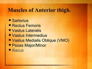
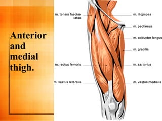
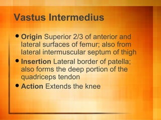
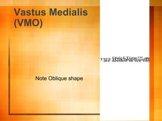
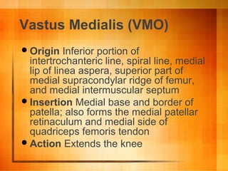
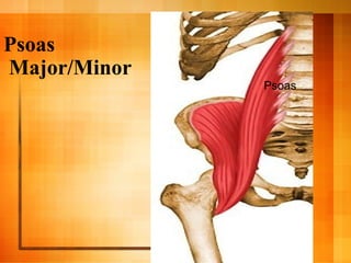
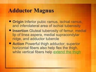
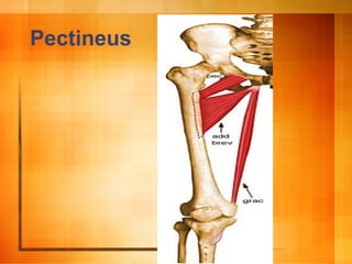
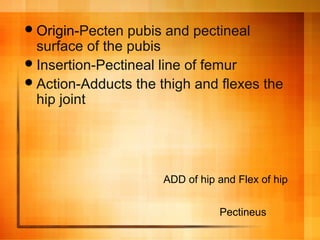
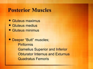
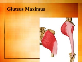
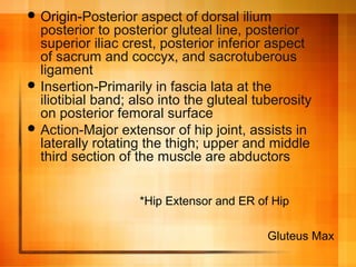
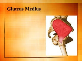
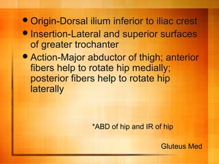
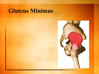
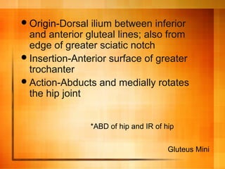
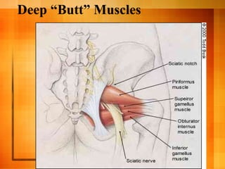
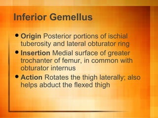
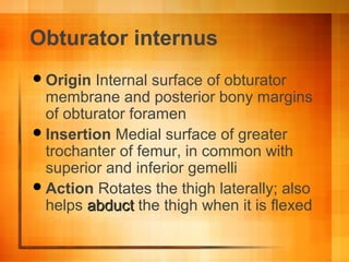
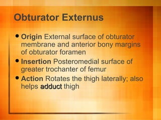
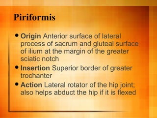
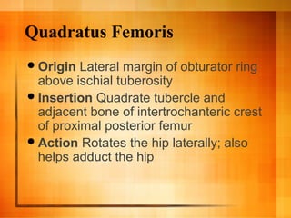
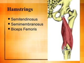
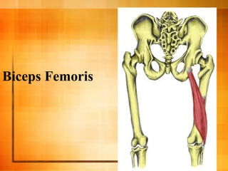
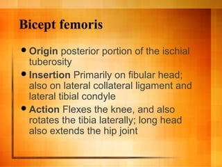
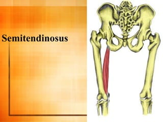
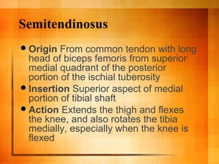
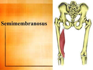
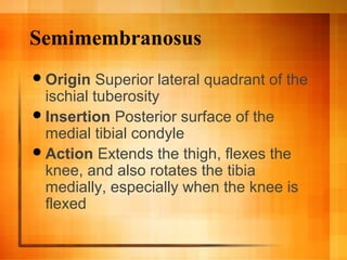
Thigh and Pelvic Muscles
"Obstacles are those frightful things
you see when you take your eyes off
your goal."
- Henry Ford (1863-1947)
Ok Here’s a Joke…
Q. Why are fish easy to weigh?
A. They have their own scales.
Q. What did the fish say when it ran
into the concrete wall?
Dam!
Goals and Objectives
 To identify vast majority of leg and pelvic
muscles to class
 For students to be able to identify muscles or
know where they are at end of lesson
 For students to be able to understand
function of muscles in this powerpoint
 To be able to link body movements with
certain muscles for strength training, injury
recognition and for just better understanding
of human movement and function
Muscles of Anterior thigh.
Sartorius
Rectus Femoris
Vastus Lateralis
Vastus Intermedius
Vastus Medialis Oblique (VMO)
Psoas Major/Minor
Iliacus
Anterior
and
medial
thigh.
Sartorius
QuickTime™ and aTIFF (Uncompressed) decompressorare needed to see this picture.
Sartorius
Origin Anterior superior iliac spine
Insertion Superior aspect of the
medial surface of the tibial shaft near
the tibial tuberosity
Action Flexes and laterally rotates the
hip joint and flexes the knee
Rectus Femoris
QuickTime™ and aTIFF (Uncompressed) decompare needed to see this picture
Rectus Femoris
Origin Straight head from anterior
inferior iliac spine; reflected head from
groove just above acetabulum
Insertion Base of patella to form the
more central portion of the quadriceps
femoris tendon
Action Hip Flexion and maybe some
Knee extension.
Vastus Lateralis
QuickTime™ and aTIFF (Uncompressed) decompresare needed to see this picture.
Note all Three
Quad muscles
Here.
Vastus Lateralis
 Origin Superior portion of intertrochanteric
line, anterior and inferior borders of greater
trochanter, superior portion of lateral lip of
linea aspera, and lateral portion of gluteal
tuberosity of femur
 Insertion Lateral base and border of patella;
also forms the lateral patellar retinaculum
and lateral side of quadriceps femoris
tendon
 Action Extends the knee
Vastus Intermedius
QuickTime™ and aTIFF (Uncompressed) decompressorare needed to see this picture.
Vastus Intermedius
Origin Superior 2/3 of anterior and
lateral surfaces of femur; also from
lateral intermuscular septum of thigh
Insertion Lateral border of patella;
also forms the deep portion of the
quadriceps tendon
Action Extends the knee
Vastus Medialis
(VMO)
QuickTime™ and aTIFF (Uncompressed) decomare needed to see this pictu
Note Oblique shape
Vastus Medialis (VMO)
Origin Inferior portion of
intertrochanteric line, spiral line, medial
lip of linea aspera, superior part of
medial supracondylar ridge of femur,
and medial intermuscular septum
Insertion Medial base and border of
patella; also forms the medial patellar
retinaculum and medial side of
quadriceps femoris tendon
Action Extends the knee
Psoas
Major/Minor
Psoas
Psoas Major/Minor
Origin Anterior surfaces and lower
borders of transverse processes of L1 -
L5 and bodies and discs of T12 - L5
Insertion Lesser trochanter
Action Flex the torso and thigh with
respect to each other
Iliacus
Origin Upper 2/3 of iliac fossa of ilium,
internal lip of iliac crest, lateral aspect
of sacrum, ventral sacroiliac ligament,
and lower portion of iliolumbar ligament
Insertion Lesser trochanter
Action Flex the torso and thigh with
respect to each other
Iliacus
QuickTime™ and a
TIFF (Uncompressed) decompressor
are needed to see this picture.
Iliacus
Take Two……..
 Why don't skeletons like parties?
 They have no body to dance with.
 Why didn't the skeleton cross the road?
 He didn't have the guts.
Anterior-Medial, to more
Medial
 Gracilis
 Adductor Brevis
 Adductor Magnus
 Adductor Longus
 Pectineus
Anterior Hip (Flexors / rotators)
– Psoas Major / Minor
– Iliopsoas
– Iliacus
QuickTime™ and a
TIFF (Uncompressed) decompressor
are needed to see this picture.
Deep medial / Medial muscles
Gracilis
Adductor Brevis
Adductor Magnus
Adductor Longus
Pectineus
Gracilis
QuickTime™ and aTIFF (Uncompressed) decompresare needed to see this picture.
Pectineus--->
Gracilis---->
Gracilis
Origin Inferior margin of pubic
symphysis, inferior ramus of pubis, and
adjacent ramus of ischium
Insertion Medial surface of tibial shaft,
just posterior to sartorius
Action Flexes the knee, adducts the
thigh, and helps to medially rotate the
tibia on the femur
Adductor Brevis and Longus
QuickTime™ and aTIFF (Uncompressed) decompressorare needed to see this picture.
<---Brevis
Longus--->
Adductor Brevis and Longus
 Origin Anterior surface of body of pubis, just
lateral to pubic symphysis
 Insertion Middle third of linea aspera,
between the more medial adductor magnus
and brevis insertions and the more lateral
origin of the vastus medialis
 Action Adducts and flexes the thigh, and
helps to laterally rotate the hip joint
***what other muscle is similar???? Hint:
“LONG”
Adductor Magnus
QuickTime™ and aTIFF (Uncompressed) decompreare needed to see this picture.
Adductor Magnus
 Origin Inferior pubic ramus, ischial ramus,
and inferolateral area of ischial tuberosity
 Insertion Gluteal tuberosity of femur, medial
lip of linea aspera, medial supracondylar
ridge, and adductor tubercle
 Action Powerful thigh adductor; superior
horizontal fibers also help flex the thigh,
while vertical fibers help extend the thigh
QuickTime™ and aTIFF (Uncompressed) decompressorare needed to see this picture.
QuickTime™ and aTIFF (Uncompressed) decare needed to see this pic
magnus Brevis Longus
A
D
D
U
C
T
O
R
S
Pectineus
Origin-Pecten pubis and pectineal
surface of the pubis
Insertion-Pectineal line of femur
Action-Adducts the thigh and flexes the
hip joint
ADD of hip and Flex of hip
Pectineus
QuickTime™ and a
TIFF (Uncompressed) decompressor
are needed to see this picture.
Quick review
Quick Break…..
QuickTime™ and a
TIFF (Uncompressed) decompressor
are needed to see this picture.
Hum??
Lateral Muscles
IT-band (Iliotibial)
Tensor Fasciae Latae
IT-band (Iliotibial)
 iliotibial band is a longitudinal fibrous
reinforcement of the Fascia lata.
 It is attached to the midpoint of the
external lip of the Iliac crest and to the
lateral condyle of the tibia.
The part of the iliotibial band which lies
beneath the Tensor fasciæ latæ is
prolonged upward to join the lateral
part of the capsule of the hip-joint.
IT-band (Iliotibial)
QuickTime™ and a
TIFF (Uncompressed) decompressor
are needed to see this picture.
White Band
QuickTime™ and a
TIFF (Uncompressed) decompressor
are needed to see this picture.
Tensor
Fascia Lata
QuickTime™ and a
TIFF (Uncompressed) decompressor
are needed to see this picture.
Tensor Fascia Lata
Origin Anterior superior iliac spine,
outer lip of anterior iliac crest and
fascia lata
Insertion Iliotibial band
Action Helps stabilize and steady the
hip and knee joints by putting tension
on the iliotibial band of fascia
Posterior Muscles
 Gluteus maximus
 Gluteus medius
 Gluteus minimus
 Deeper “Butt” muscles;
Piriformis
Gamelius Superior and Inferior
Obturator Internus and Exturnus
Quadratus Femoris
Take two……….
QuickTime™ and a
TIFF (Uncompressed) decompressor
are needed to see this picture.
Power House “The Glutes”
Gluteus Maximus
Gluteus Medius
Gluteus Minimus
Gluteus Maximus
 Origin-Posterior aspect of dorsal ilium
posterior to posterior gluteal line, posterior
superior iliac crest, posterior inferior aspect
of sacrum and coccyx, and sacrotuberous
ligament
 Insertion-Primarily in fascia lata at the
iliotibial band; also into the gluteal tuberosity
on posterior femoral surface
 Action-Major extensor of hip joint, assists in
laterally rotating the thigh; upper and middle
third section of the muscle are abductors
*Hip Extensor and ER of Hip
Gluteus Max
Gluteus Medius
Origin-Dorsal ilium inferior to iliac crest
Insertion-Lateral and superior surfaces
of greater trochanter
Action-Major abductor of thigh; anterior
fibers help to rotate hip medially;
posterior fibers help to rotate hip
laterally
*ABD of hip and IR of hip
Gluteus Med
Gluteus Minimus
Origin-Dorsal ilium between inferior
and anterior gluteal lines; also from
edge of greater sciatic notch
Insertion-Anterior surface of greater
trochanter
Action-Abducts and medially rotates
the hip joint
*ABD of hip and IR of hip
Gluteus Mini
Deep “Butt” Muscles
Inferior
Gemellus QuickTime™ and a
TIFF (Uncompressed) decompressor
are needed to see this picture.
Inferior Gemellus
Origin Posterior portions of ischial
tuberosity and lateral obturator ring
Insertion Medial surface of greater
trochanter of femur, in common with
obturator internus
Action Rotates the thigh laterally; also
helps abduct the flexed thigh
Obturator Internus /
Externus
QuickTime™ and a
TIFF (Uncompressed) decompressor
are needed to see this picture.QuickTime™ and a
TIFF (Uncompressed) decompressor
are needed to see this picture.
Externus
Obturator internus
Origin Internal surface of obturator
membrane and posterior bony margins
of obturator foramen
Insertion Medial surface of greater
trochanter of femur, in common with
superior and inferior gemelli
Action Rotates the thigh laterally; also
helps abductabduct the thigh when it is flexed
Obturator Externus
Origin External surface of obturator
membrane and anterior bony margins
of obturator foramen
Insertion Posteromedial surface of
greater trochanter of femur
Action Rotates the thigh laterally; also
helps adductadduct thigh
Piriformis
QuickTime™ and a
TIFF (Uncompressed) decompressor
are needed to see this picture.
Piriformis
Origin Anterior surface of lateral
process of sacrum and gluteal surface
of ilium at the margin of the greater
sciatic notch
Insertion Superior border of greater
trochanter
Action Lateral rotator of the hip joint;
also helps abduct the hip if it is flexed
Quadratus Femoris
QuickTime™ and a
TIFF (Uncompressed) decompressor
are needed to see this picture.
<--Here
Quadratus Femoris
Origin Lateral margin of obturator ring
above ischial tuberosity
Insertion Quadrate tubercle and
adjacent bone of intertrochanteric crest
of proximal posterior femur
Action Rotates the hip laterally; also
helps adduct the hip
Getting
cold…
QuickTime™ and a
TIFF (Uncompressed) decompressor
are needed to see this picture.
“Hammies”
QuickTime™ and a
TIFF (Uncompressed) decompressor
are needed to see this picture.
QuickTime™ and a
TIFF (Uncompressed) decompressor
are needed to see this picture.
Hamstrings
Semitendinosus
Semimembranosus
Biceps Femoris
Biceps Femoris
Biceps Femoris
QuickTime™ andTIFF (Uncompressed)are needed to see this
QuickTime™ and aTIFF (Uncompressed) decompressorare needed to see this picture.
Long Head
Short Head
Bicept femoris
Origin posterior portion of the ischial
tuberosity
Insertion Primarily on fibular head;
also on lateral collateral ligament and
lateral tibial condyle
Action Flexes the knee, and also
rotates the tibia laterally; long head
also extends the hip joint
Semitendinosus
Semitendinosus
Origin From common tendon with long
head of biceps femoris from superior
medial quadrant of the posterior
portion of the ischial tuberosity
Insertion Superior aspect of medial
portion of tibial shaft
Action Extends the thigh and flexes
the knee, and also rotates the tibia
medially, especially when the knee is
flexed
Semimembranosus
Semimembranosus
Origin Superior lateral quadrant of the
ischial tuberosity
Insertion Posterior surface of the
medial tibial condyle
Action Extends the thigh, flexes the
knee, and also rotates the tibia
medially, especially when the knee is
flexed
We… Are… Doneee….
Back to Work!!

Thigh and hip muscles

  • 1.
    Thigh and PelvicMuscles "Obstacles are those frightful things you see when you take your eyes off your goal." - Henry Ford (1863-1947)
  • 2.
    Ok Here’s aJoke… Q. Why are fish easy to weigh?
  • 3.
    A. They havetheir own scales.
  • 4.
    Q. What didthe fish say when it ran into the concrete wall?
  • 5.
  • 6.
    Goals and Objectives To identify vast majority of leg and pelvic muscles to class  For students to be able to identify muscles or know where they are at end of lesson  For students to be able to understand function of muscles in this powerpoint  To be able to link body movements with certain muscles for strength training, injury recognition and for just better understanding of human movement and function
  • 7.
    Muscles of Anteriorthigh. Sartorius Rectus Femoris Vastus Lateralis Vastus Intermedius Vastus Medialis Oblique (VMO) Psoas Major/Minor Iliacus
  • 8.
  • 9.
    Sartorius QuickTime™ and aTIFF(Uncompressed) decompressorare needed to see this picture.
  • 10.
    Sartorius Origin Anterior superioriliac spine Insertion Superior aspect of the medial surface of the tibial shaft near the tibial tuberosity Action Flexes and laterally rotates the hip joint and flexes the knee
  • 11.
    Rectus Femoris QuickTime™ andaTIFF (Uncompressed) decompare needed to see this picture
  • 12.
    Rectus Femoris Origin Straighthead from anterior inferior iliac spine; reflected head from groove just above acetabulum Insertion Base of patella to form the more central portion of the quadriceps femoris tendon Action Hip Flexion and maybe some Knee extension.
  • 13.
    Vastus Lateralis QuickTime™ andaTIFF (Uncompressed) decompresare needed to see this picture. Note all Three Quad muscles Here.
  • 14.
    Vastus Lateralis  OriginSuperior portion of intertrochanteric line, anterior and inferior borders of greater trochanter, superior portion of lateral lip of linea aspera, and lateral portion of gluteal tuberosity of femur  Insertion Lateral base and border of patella; also forms the lateral patellar retinaculum and lateral side of quadriceps femoris tendon  Action Extends the knee
  • 15.
    Vastus Intermedius QuickTime™ andaTIFF (Uncompressed) decompressorare needed to see this picture.
  • 16.
    Vastus Intermedius Origin Superior2/3 of anterior and lateral surfaces of femur; also from lateral intermuscular septum of thigh Insertion Lateral border of patella; also forms the deep portion of the quadriceps tendon Action Extends the knee
  • 17.
    Vastus Medialis (VMO) QuickTime™ andaTIFF (Uncompressed) decomare needed to see this pictu Note Oblique shape
  • 18.
    Vastus Medialis (VMO) OriginInferior portion of intertrochanteric line, spiral line, medial lip of linea aspera, superior part of medial supracondylar ridge of femur, and medial intermuscular septum Insertion Medial base and border of patella; also forms the medial patellar retinaculum and medial side of quadriceps femoris tendon Action Extends the knee
  • 19.
  • 20.
    Psoas Major/Minor Origin Anteriorsurfaces and lower borders of transverse processes of L1 - L5 and bodies and discs of T12 - L5 Insertion Lesser trochanter Action Flex the torso and thigh with respect to each other
  • 21.
    Iliacus Origin Upper 2/3of iliac fossa of ilium, internal lip of iliac crest, lateral aspect of sacrum, ventral sacroiliac ligament, and lower portion of iliolumbar ligament Insertion Lesser trochanter Action Flex the torso and thigh with respect to each other
  • 22.
    Iliacus QuickTime™ and a TIFF(Uncompressed) decompressor are needed to see this picture. Iliacus
  • 23.
    Take Two……..  Whydon't skeletons like parties?  They have no body to dance with.  Why didn't the skeleton cross the road?  He didn't have the guts.
  • 24.
    Anterior-Medial, to more Medial Gracilis  Adductor Brevis  Adductor Magnus  Adductor Longus  Pectineus Anterior Hip (Flexors / rotators) – Psoas Major / Minor – Iliopsoas – Iliacus QuickTime™ and a TIFF (Uncompressed) decompressor are needed to see this picture.
  • 25.
    Deep medial /Medial muscles Gracilis Adductor Brevis Adductor Magnus Adductor Longus Pectineus
  • 26.
    Gracilis QuickTime™ and aTIFF(Uncompressed) decompresare needed to see this picture. Pectineus---> Gracilis---->
  • 27.
    Gracilis Origin Inferior marginof pubic symphysis, inferior ramus of pubis, and adjacent ramus of ischium Insertion Medial surface of tibial shaft, just posterior to sartorius Action Flexes the knee, adducts the thigh, and helps to medially rotate the tibia on the femur
  • 28.
    Adductor Brevis andLongus QuickTime™ and aTIFF (Uncompressed) decompressorare needed to see this picture. QuickTime™ and aTIFF (Uncompressed) decompressorare needed to see this picture. <---Brevis Longus--->
  • 29.
    Adductor Brevis andLongus  Origin Anterior surface of body of pubis, just lateral to pubic symphysis  Insertion Middle third of linea aspera, between the more medial adductor magnus and brevis insertions and the more lateral origin of the vastus medialis  Action Adducts and flexes the thigh, and helps to laterally rotate the hip joint ***what other muscle is similar???? Hint: “LONG”
  • 30.
    Adductor Magnus QuickTime™ andaTIFF (Uncompressed) decompreare needed to see this picture.
  • 31.
    Adductor Magnus  OriginInferior pubic ramus, ischial ramus, and inferolateral area of ischial tuberosity  Insertion Gluteal tuberosity of femur, medial lip of linea aspera, medial supracondylar ridge, and adductor tubercle  Action Powerful thigh adductor; superior horizontal fibers also help flex the thigh, while vertical fibers help extend the thigh
  • 32.
    QuickTime™ and aTIFF(Uncompressed) decompressorare needed to see this picture. QuickTime™ and aTIFF (Uncompressed) decompressorare needed to see this picture. QuickTime™ and aTIFF (Uncompressed) decare needed to see this pic magnus Brevis Longus A D D U C T O R S
  • 33.
  • 34.
    Origin-Pecten pubis andpectineal surface of the pubis Insertion-Pectineal line of femur Action-Adducts the thigh and flexes the hip joint ADD of hip and Flex of hip Pectineus
  • 35.
    QuickTime™ and a TIFF(Uncompressed) decompressor are needed to see this picture. Quick review
  • 36.
    Quick Break….. QuickTime™ anda TIFF (Uncompressed) decompressor are needed to see this picture. Hum??
  • 37.
  • 38.
    IT-band (Iliotibial)  iliotibialband is a longitudinal fibrous reinforcement of the Fascia lata.  It is attached to the midpoint of the external lip of the Iliac crest and to the lateral condyle of the tibia. The part of the iliotibial band which lies beneath the Tensor fasciæ latæ is prolonged upward to join the lateral part of the capsule of the hip-joint.
  • 39.
    IT-band (Iliotibial) QuickTime™ anda TIFF (Uncompressed) decompressor are needed to see this picture. White Band
  • 40.
    QuickTime™ and a TIFF(Uncompressed) decompressor are needed to see this picture.
  • 41.
    Tensor Fascia Lata QuickTime™ anda TIFF (Uncompressed) decompressor are needed to see this picture.
  • 42.
    Tensor Fascia Lata OriginAnterior superior iliac spine, outer lip of anterior iliac crest and fascia lata Insertion Iliotibial band Action Helps stabilize and steady the hip and knee joints by putting tension on the iliotibial band of fascia
  • 43.
    Posterior Muscles  Gluteusmaximus  Gluteus medius  Gluteus minimus  Deeper “Butt” muscles; Piriformis Gamelius Superior and Inferior Obturator Internus and Exturnus Quadratus Femoris
  • 44.
    Take two………. QuickTime™ anda TIFF (Uncompressed) decompressor are needed to see this picture.
  • 45.
    Power House “TheGlutes” Gluteus Maximus Gluteus Medius Gluteus Minimus
  • 46.
  • 47.
     Origin-Posterior aspectof dorsal ilium posterior to posterior gluteal line, posterior superior iliac crest, posterior inferior aspect of sacrum and coccyx, and sacrotuberous ligament  Insertion-Primarily in fascia lata at the iliotibial band; also into the gluteal tuberosity on posterior femoral surface  Action-Major extensor of hip joint, assists in laterally rotating the thigh; upper and middle third section of the muscle are abductors *Hip Extensor and ER of Hip Gluteus Max
  • 48.
  • 49.
    Origin-Dorsal ilium inferiorto iliac crest Insertion-Lateral and superior surfaces of greater trochanter Action-Major abductor of thigh; anterior fibers help to rotate hip medially; posterior fibers help to rotate hip laterally *ABD of hip and IR of hip Gluteus Med
  • 50.
  • 51.
    Origin-Dorsal ilium betweeninferior and anterior gluteal lines; also from edge of greater sciatic notch Insertion-Anterior surface of greater trochanter Action-Abducts and medially rotates the hip joint *ABD of hip and IR of hip Gluteus Mini
  • 52.
  • 53.
    Inferior Gemellus QuickTime™ anda TIFF (Uncompressed) decompressor are needed to see this picture.
  • 54.
    Inferior Gemellus Origin Posteriorportions of ischial tuberosity and lateral obturator ring Insertion Medial surface of greater trochanter of femur, in common with obturator internus Action Rotates the thigh laterally; also helps abduct the flexed thigh
  • 55.
    Obturator Internus / Externus QuickTime™and a TIFF (Uncompressed) decompressor are needed to see this picture.QuickTime™ and a TIFF (Uncompressed) decompressor are needed to see this picture. Externus
  • 56.
    Obturator internus Origin Internalsurface of obturator membrane and posterior bony margins of obturator foramen Insertion Medial surface of greater trochanter of femur, in common with superior and inferior gemelli Action Rotates the thigh laterally; also helps abductabduct the thigh when it is flexed
  • 57.
    Obturator Externus Origin Externalsurface of obturator membrane and anterior bony margins of obturator foramen Insertion Posteromedial surface of greater trochanter of femur Action Rotates the thigh laterally; also helps adductadduct thigh
  • 58.
    Piriformis QuickTime™ and a TIFF(Uncompressed) decompressor are needed to see this picture.
  • 59.
    Piriformis Origin Anterior surfaceof lateral process of sacrum and gluteal surface of ilium at the margin of the greater sciatic notch Insertion Superior border of greater trochanter Action Lateral rotator of the hip joint; also helps abduct the hip if it is flexed
  • 60.
    Quadratus Femoris QuickTime™ anda TIFF (Uncompressed) decompressor are needed to see this picture. <--Here
  • 61.
    Quadratus Femoris Origin Lateralmargin of obturator ring above ischial tuberosity Insertion Quadrate tubercle and adjacent bone of intertrochanteric crest of proximal posterior femur Action Rotates the hip laterally; also helps adduct the hip
  • 62.
    Getting cold… QuickTime™ and a TIFF(Uncompressed) decompressor are needed to see this picture.
  • 63.
    “Hammies” QuickTime™ and a TIFF(Uncompressed) decompressor are needed to see this picture. QuickTime™ and a TIFF (Uncompressed) decompressor are needed to see this picture.
  • 64.
  • 65.
  • 66.
    Biceps Femoris QuickTime™ andTIFF(Uncompressed)are needed to see this QuickTime™ and aTIFF (Uncompressed) decompressorare needed to see this picture. Long Head Short Head
  • 67.
    Bicept femoris Origin posteriorportion of the ischial tuberosity Insertion Primarily on fibular head; also on lateral collateral ligament and lateral tibial condyle Action Flexes the knee, and also rotates the tibia laterally; long head also extends the hip joint
  • 68.
  • 69.
    Semitendinosus Origin From commontendon with long head of biceps femoris from superior medial quadrant of the posterior portion of the ischial tuberosity Insertion Superior aspect of medial portion of tibial shaft Action Extends the thigh and flexes the knee, and also rotates the tibia medially, especially when the knee is flexed
  • 70.
  • 71.
    Semimembranosus Origin Superior lateralquadrant of the ischial tuberosity Insertion Posterior surface of the medial tibial condyle Action Extends the thigh, flexes the knee, and also rotates the tibia medially, especially when the knee is flexed
  • 72.

Editor's Notes

  • #11 InnervationFemoral nerve (L2, L3, L4)
  • #13 Innervation Muscular branches of femoral nerve (L2, L3, L4)
  • #15 Innervation Muscular branches of femoral nerve (L2, L3, L4)
  • #17 Innervation Muscular branches of femoral nerve (L2, L3, L4)
  • #19 InnervationMuscular branches of femoral nerve (L2, L3, L4)
  • #21 Innervation Direct fibers of L1 - L3 of lumbar plexus (L1, L2, L3)
  • #22 Innervation Muscular branch of femoral nerve (L1, L2, L3)
  • #28 Innervation Anterior division of obturator nerve (L2, L3)
  • #30 Innervation Anterior division of obturator nerve (L2, L3, L4)
  • #32 InnervationPosterior division of obturator nerve innervates most of the adductor magnus; vertical or hamstring portion innervated by tibial nerve (L2, L3, L4)
  • #43 InnervationSuperior gluteal nerve (L4, L5, S1) (L4, L5, S1)
  • #48 Inferior gluteal nerve (L5, S1, S2) (L5, S1, S2)
  • #50 Superior gluteal nerve (L4, L5, S1) (L4, L5, S1)
  • #52 Superior gluteal nerve (L4, L5, S1) (L4, L5, S1)
  • #55 InnervationNerve to the obturator internus and inferior gemellus -- a branch of the sacral plexus (L5, S1) (L5, S1)
  • #57 Innervation Nerve to the obturator internus and superior gemellus -- a branch of the sacral plexus (L5, S1) (L5, S1)
  • #58 InnervationPosterior division of obturator nerve innervates most of the adductor magnus; vertical or hamstring portion innervated by tibial nerve (L3, L4)
  • #60 InnervationPiriformis nerve (L5, S1, S2) (L5, S1, S2)
  • #62 Innervation Quadratus femoris branch of nerve to the quadratus femoris and inferior gemellus (L5, S1) (L5, S1)
  • #68 Innervation Tibial nerve (L5, S1, S2)
  • #70 InnervationTibial nerve (L5, S1, S2)
  • #72 Innervation Tibial nerve (L5, S1, S2)