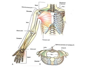
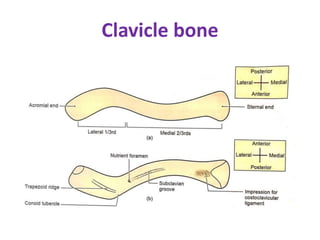
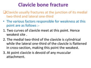
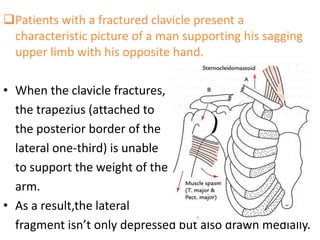
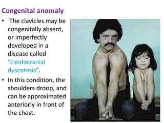
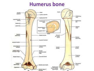
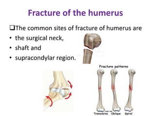
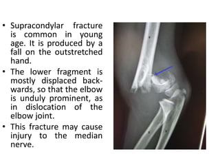
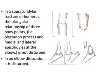
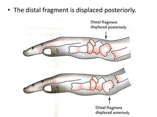
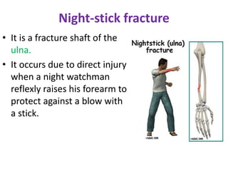
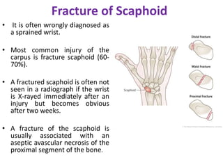
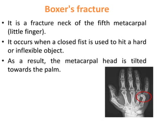
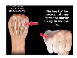
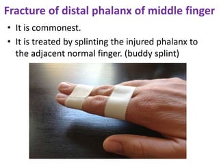
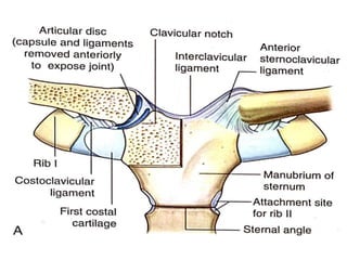
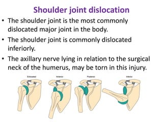
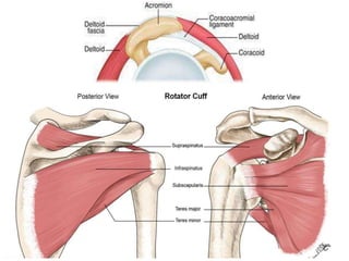
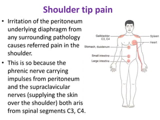
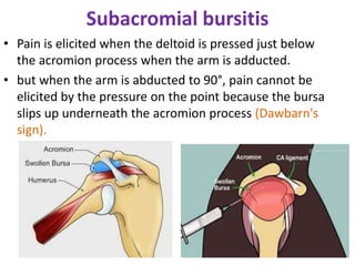
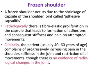
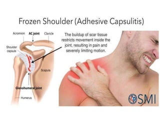
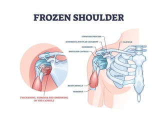
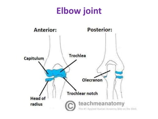
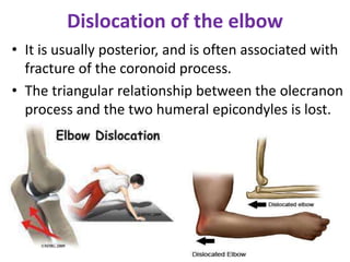
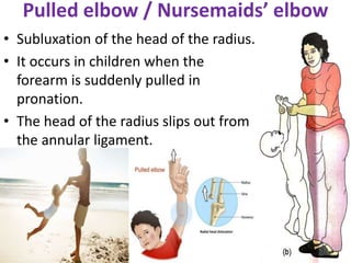
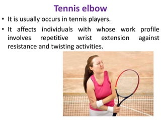
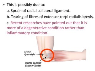
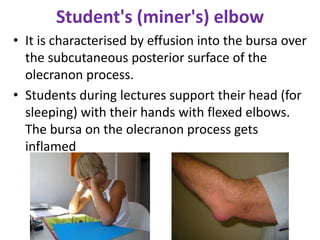
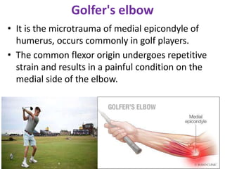
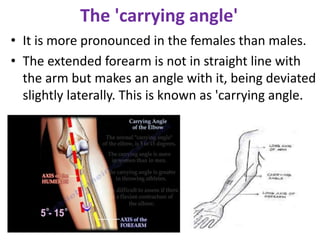
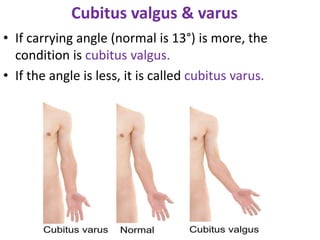
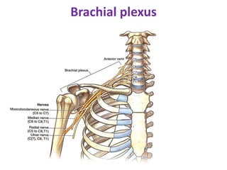
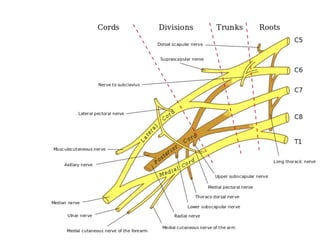
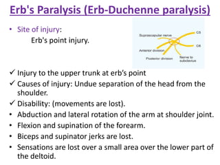
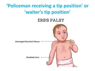
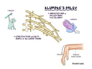
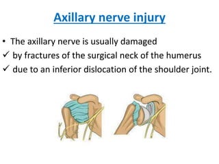
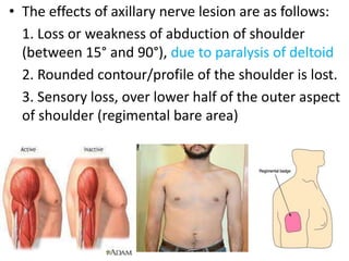
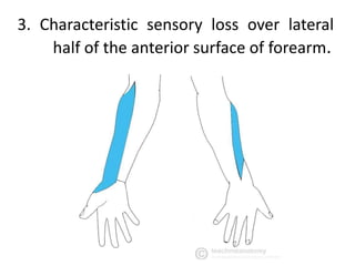
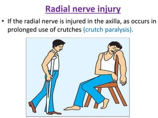
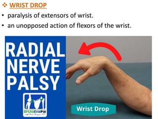
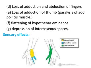
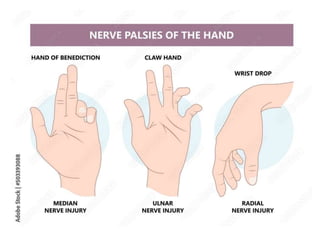
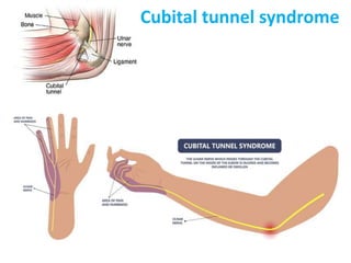
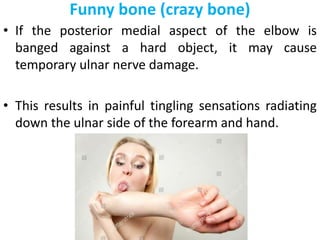
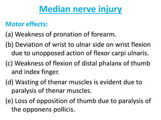
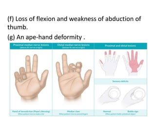
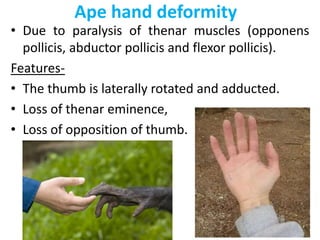
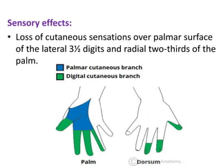
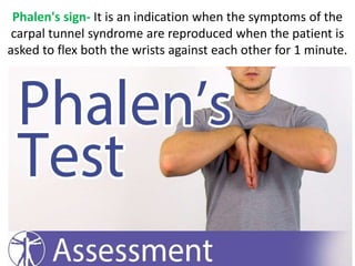
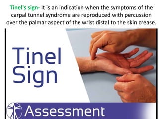
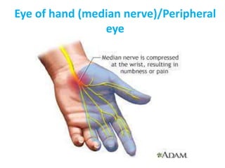
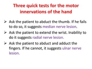
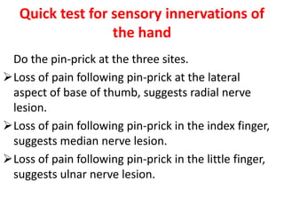
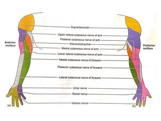
“Clinical anatomy (upper limb)”
By
Dr. Neeraj Tiwari
Lecturer
Clavicle bone
Clavicle bone fracture
Clavicle usually fractures at the junction of its medial
two-third and lateral one-third
• The various factors responsible for weakness at this
point are as follows :-
1. Two curves of clavicle meet at this point. Hence
weakest site .
2. The medial two-third of the clavicle is cylindrical
while the lateral one-third of the clavicle is flattened
in cross-section, making this point the weakest.
3. At point clavicle is devoid of any muscular
attachment.
4. Two primary centres of ossification appear quite
close to each other at this junction. They soon
unite.
5. A fissure of small or great extent may divide the
lateral one-third from medial two-third of clavicle.
5 It pierced by a nutrient foramen.
6. Supraclavicular nerves may pierce the clavicle at
the junction.
“Clavicle is the most commonly fractured bone
in the body”
Patients with a fractured clavicle present a
characteristic picture of a man supporting his sagging
upper limb with his opposite hand.
• When the clavicle fractures,
the trapezius (attached to
the posterior border of the
lateral one-third) is unable
to support the weight of the
arm.
• As a result,the lateral
fragment isn’t only depressed but also drawn medially.
Congenital anomaly
• The clavicles may be
congenitally absent,
or imperfectly
developed in a
disease called
“cleidocranial
dysostosis”.
• In this condition, the
shoulders droop, and
can be approximated
anteriorly in front of
the chest.
Winging of scapula
• Paralysis of the serratus
anterior. (injury to long
thoracic nerve)
• the medial border of
the bone becomes unduly
prominent.
• the arm can not be
abducted beyond 90 degree.
• Swimmers palsy
Humerus bone
Fracture of the humerus
The common sites of fracture of humerus are
• the surgical neck,
• shaft and
• supracondylar region.
• Supracondylar fracture
is common in young
age. It is produced by a
fall on the outstretched
hand.
• The lower fragment is
mostly displaced back-
wards, so that the elbow
is unduly prominent, as
in dislocation of the
elbow joint.
• This fracture may cause
injury to the median
nerve.
• In a supracondylar
fracture of humerus,
the triangular
relationship of three
bony points, (i.e.
olecranon process and
medial and lateral
epicondyles at the
elbow,) is not disturbed.
• In an elbow dislocation,
it is disturbed.
• The axillary, radial
and ulnar nerves
may be damaged in
fractures of the
humerus.
'Colles' fracture'
• It presents a characteristic 'dinner fork
deformity.
• The radius fractures about one inch proximal to
the wrist joint following a fall on an
outstretched hand.
• The distal fragment is displaced posteriorly.
• As a result, the wrist shows a bend that is
known as the 'dinner fork deformity because
the forearm and wrist resemble the shape of a
dinner fork.
Smith’s fracture
• Smith's fracture is a fracture of the distal end of
radius and occurs from a fall on the back of the
hand.
• It is termed as a reverse Colles, fracture because
the distal fragment is displaced anteriorly.
Monteggia fracture
• The Monteggia fracture is a fracture of the
proximal ulna associated with dislocation of the
radial head at the superior radio-ulnar joint.
• It occurs due to fall on an outstretched hand with
extended elbow.
Greenstick Fracture
• It is a partial thickness fracture of long bone of
children, where only the cortex and periosteum are
interrupted on one side of the bone but remain
uninterrupted on the other.
Night-stick fracture
• It is a fracture shaft of the
ulna.
• It occurs due to direct injury
when a night watchman
reflexly raises his forearm to
protect against a blow with
a stick.
Fracture of Scaphoid
• It is often wrongly diagnosed as
a sprained wrist.
• Most common injury of the
carpus is fracture scaphoid (60-
70%).
• A fractured scaphoid is often not
seen in a radiograph if the wrist
is X-rayed immediately after an
injury but becomes obvious
after two weeks.
• A fracture of the scaphoid is
usually associated with an
aseptic avascular necrosis of the
proximal segment of the bone.
Boxer's fracture
• It is a fracture neck of the fifth metacarpal
(little finger).
• It occurs when a closed fist is used to hit a hard
or inflexible object.
• As a result, the metacarpal head is tilted
towards the palm.
Bennett's fracture
• Fracture of the base of
the first metacarpal is
called.
• It involves the anterior
part of the base, and is
caused by a force along
its long axis.
• The thumb is forced into
a semiflexed position
and cannot be opposed.
• The fist cannot be
clenched.
Polydactyly
• It is an abnormality of fingers or toes.
• There are extra digits which are not formed fully.
• poor muscular development and are useless.
• either medial or lateral to the hand or foot.
• Polydactyly is inherited as a dominant characterstic.
Fracture of distal phalanx of middle finger
• It is commonest.
• It is treated by splinting the injured phalanx to
the adjacent normal finger. (buddy splint)
Dislocation Of Upper Limbs
 Clavicular displacement
• The clavicle may be dislocated at either of its
ends.
• At the medial end, it is usually dislocated
forwards.
• Backward dislocation is rare as it is prevented by
the costo clavicular ligament.
• The clavicle dislocates upwards at the acromio-
clavicular joint, because the clavicle overrides the
acromion process.
Shoulder joint
Shoulder joint dislocation
• The shoulder joint is the most commonly
dislocated major joint in the body.
• The shoulder joint is commonly dislocated
inferiorly.
• The axillary nerve lying in relation to the surgical
neck of the humerus, may be torn in this injury.
Shoulder tip pain
• Irritation of the peritoneum
underlying diaphragm from
any surrounding pathology
causes referred pain in the
shoulder.
• This is so because the
phrenic nerve carrying
impulses from peritoneum
and the supraclavicular
nerves (supplying the skin
over the shoulder) both arise
from spinal segments C3, C4.
Subacromial bursitis
• Pain is elicited when the deltoid is pressed just below
the acromion process when the arm is adducted.
• but when the arm is abducted to 90°, pain cannot be
elicited by the pressure on the point because the bursa
slips up underneath the acromion process (Dawbarn's
sign).
Frozen shoulder
• A frozen shoulder occurs due to the shrinkage of
capsule of the shoulder joint called ‘adhesive
capsulitis’.
• Pathologically there is fibro-elastic proliferation in
the capsule that leads to formation of adhesions
and consequent stiffness and pain on attempted
movements.
• Clinically, the patient (usually 40- 60 years of age)
complains of progressively increasing pain in the
shoulder, stiffness in the joint and restriction of all
movements. though there is no evidence of radio-
logical changes in the joint.
Exclusion of shoulder joint disease
Elbow joint
Dislocation of the elbow
• It is usually posterior, and is often associated with
fracture of the coronoid process.
• The triangular relationship between the olecranon
process and the two humeral epicondyles is lost.
Pulled elbow / Nursemaids’ elbow
• Subluxation of the head of the radius.
• It occurs in children when the
forearm is suddenly pulled in
pronation.
• The head of the radius slips out from
the annular ligament.
Tennis elbow
• It is usually occurs in tennis players.
• It affects individuals with whose work profile
involves repetitive wrist extension against
resistance and twisting activities.
• This is possibly due to:
a. Sprain of radial collateral ligament.
b. Tearing of fibres of extensor carpi radialis brevis.
c. Recent researches have pointed out that it is
more of a degenerative condition rather than
inflammatory condition.
Student's (miner's) elbow
• It is characterised by effusion into the bursa over
the subcutaneous posterior surface of the
olecranon process.
• Students during lectures support their head (for
sleeping) with their hands with flexed elbows.
The bursa on the olecranon process gets
inflamed
Golfer's elbow
• It is the microtrauma of medial epicondyle of
humerus, occurs commonly in golf players.
• The common flexor origin undergoes repetitive
strain and results in a painful condition on the
medial side of the elbow.
The 'carrying angle'
• It is more pronounced in the females than males.
• The extended forearm is not in straight line with
the arm but makes an angle with it, being deviated
slightly laterally. This is known as 'carrying angle.
• The anatomical factors responsible for producing
carrying angle are:
1. Medial flange of trochlea is 6 mm below than the
lateral flange.
2. Superior articular surface of the coronoid process
of the ulna is placed obliquely to the long axis of the
ulna.
Cubitus valgus & varus
• If carrying angle (normal is 13°) is more, the
condition is cubitus valgus.
• If the angle is less, it is called cubitus varus.
Site of injection
• Deltoid muscle is a
preferred site for I/M
injection.
• well-developed
muscle
• easily accessible
muscle.
• Site - middle or lower
half of the muscle
• To avoid injury to the
axillary nerve.
Clinical anatomy related with
brachial plexus & related
nerves
Brachial plexus
Erb's Paralysis (Erb-Duchenne paralysis)
• Site of injury:
Erb's point injury.
 Injury to the upper trunk at erb’s point
 Causes of injury: Undue separation of the head from the
shoulder.
 Disability: (movements are lost).
• Abduction and lateral rotation of the arm at shoulder joint.
• Flexion and supination of the forearm.
• Biceps and supinator jerks are lost.
• Sensations are lost over a small area over the lower part of
the deltoid.
‘Policeman receiving a tip position' or
'waiter's tip position'
Klumpke's Paralysis
Site of injury: Lower trunk of the brachial plexus.
Cause of injury:
1.Undue abduction of the arm, as in clutching
something with the hands after a fall from a height,
2. birth injury.
Disability :-
• Claw hand.
• Horner's syndrome: Ptosis, miosis, anhydrosis,
enophthalmos.
Axillary nerve injury
• The axillary nerve is usually damaged
 by fractures of the surgical neck of the humerus
 due to an inferior dislocation of the shoulder joint.
• The effects of axillary nerve lesion are as follows:
1. Loss or weakness of abduction of shoulder
(between 15° and 90°), due to paralysis of deltoid
2. Rounded contour/profile of the shoulder is lost.
3. Sensory loss, over lower half of the outer aspect
of shoulder (regimental bare area)
Musculocutaneous nerve lesion
• It is sometimes damaged in a fracture of the humerus.
• The effects will be as follows:
1. Weakness of elbow flexion (due to paralysis of biceps
brachii, and medial two-third of brachialis.)
2. Weak supination of forearm with the elbow flexed at
90° (due to paralysis of biceps brachii).
3. Characteristic sensory loss over lateral
half of the anterior surface of forearm.
Radial nerve injury
• If the radial nerve is injured in the axilla, as occurs in
prolonged use of crutches (crutch paralysis).
• The effects are as follows:
1. Loss of extension of elbow due to paralysis of
triceps.
2. Loss of extension of wrist due to paralysis of
extensor muscles of the forearm.(wrist drop)
3.
 WRIST DROP
• paralysis of extensors of wrist.
• an unopposed action of flexors of the wrist.
3. Supination of the extended elbow is not possible
due to paralysis of supinator.
4. Loss of triceps and supinator reflexes.
5. Loss of sensation over
Saturday night paralysis & honeymoon
palsy
Ulnar nerve injury
• It is injured at the elbow.
Motor effects:
(a) Loss of flexion of the terminal phalanges of the
ring and little fingers. (FDP paralysis)
(b)Weakness of flexion and adduction of wrist (FCU
paralysis)
(c) Ulnar claw hand- due to paralysis
of all interossei and medial two
lumbricals.
(d) Loss of adduction and abduction of fingers
(e) Loss of adduction of thumb (paralysis of add.
pollicis muscle.)
(f) flattening of hypothenar eminence
(g) depression of interosseous spaces.
Sensory effects:
Cubital tunnel syndrome
Funny bone (crazy bone)
• If the posterior medial aspect of the elbow is
banged against a hard object, it may cause
temporary ulnar nerve damage.
• This results in painful tingling sensations radiating
down the ulnar side of the forearm and hand.
Median nerve injury
Motor effects:
(a) Weakness of pronation of forearm.
(b) Deviation of wrist to ulnar side on wrist flexion
due to unopposed action of flexor carpi ulnaris.
(c) Weakness of flexion of distal phalanx of thumb
and index finger.
(d) Wasting of thenar muscles is evident due to
paralysis of thenar muscles.
(e) Loss of opposition of thumb due to paralysis of
the opponens pollicis.
(f) Loss of flexion and weakness of abduction of
thumb.
(g) An ape-hand deformity .
Ape hand deformity
• Due to paralysis of thenar muscles (opponens
pollicis, abductor pollicis and flexor pollicis).
Features-
• The thumb is laterally rotated and adducted.
• Loss of thenar eminence,
• Loss of opposition of thumb.
Sensory effects:
• Loss of cutaneous sensations over palmar surface
of the lateral 3½ digits and radial two-thirds of the
palm.
CARPAL TUNNEL SYNDROME
Phalen's sign- It is an indication when the symptoms of the
carpal tunnel syndrome are reproduced when the patient is
asked to flex both the wrists against each other for 1 minute.
Tinel's sign- It is an indication when the symptoms of the
carpal tunnel syndrome are reproduced with percussion
over the palmar aspect of the wrist distal to the skin crease.
Eye of hand (median nerve)/Peripheral
eye
Three quick tests for the motor
innervations of the hand
 Ask the patient to abduct the thumb. If he fails
to do so, it suggests median nerve lesion.
 Ask the patient to extend the wrist. Inability to
do it suggests radial nerve lesion.
 Ask the patient to abduct and adduct the
fingers. If he cannot, it suggests ulnar nerve
lesion.
Quick test for sensory innervations of
the hand
Do the pin-prick at the three sites.
Loss of pain following pin-prick at the lateral
aspect of base of thumb, suggests radial nerve
lesion.
Loss of pain following pin-prick in the index finger,
suggests median nerve lesion.
Loss of pain following pin-prick in the little finger,
suggests ulnar nerve lesion.
Thank You for your attention…

clinical anatomy (upper limb)

  • 1.
    “Clinical anatomy (upperlimb)” By Dr. Neeraj Tiwari Lecturer
  • 3.
  • 5.
    Clavicle bone fracture Clavicleusually fractures at the junction of its medial two-third and lateral one-third • The various factors responsible for weakness at this point are as follows :- 1. Two curves of clavicle meet at this point. Hence weakest site . 2. The medial two-third of the clavicle is cylindrical while the lateral one-third of the clavicle is flattened in cross-section, making this point the weakest. 3. At point clavicle is devoid of any muscular attachment.
  • 7.
    4. Two primarycentres of ossification appear quite close to each other at this junction. They soon unite. 5. A fissure of small or great extent may divide the lateral one-third from medial two-third of clavicle. 5 It pierced by a nutrient foramen. 6. Supraclavicular nerves may pierce the clavicle at the junction. “Clavicle is the most commonly fractured bone in the body”
  • 8.
    Patients with afractured clavicle present a characteristic picture of a man supporting his sagging upper limb with his opposite hand. • When the clavicle fractures, the trapezius (attached to the posterior border of the lateral one-third) is unable to support the weight of the arm. • As a result,the lateral fragment isn’t only depressed but also drawn medially.
  • 9.
    Congenital anomaly • Theclavicles may be congenitally absent, or imperfectly developed in a disease called “cleidocranial dysostosis”. • In this condition, the shoulders droop, and can be approximated anteriorly in front of the chest.
  • 10.
    Winging of scapula •Paralysis of the serratus anterior. (injury to long thoracic nerve) • the medial border of the bone becomes unduly prominent. • the arm can not be abducted beyond 90 degree. • Swimmers palsy
  • 11.
  • 12.
    Fracture of thehumerus The common sites of fracture of humerus are • the surgical neck, • shaft and • supracondylar region.
  • 13.
    • Supracondylar fracture iscommon in young age. It is produced by a fall on the outstretched hand. • The lower fragment is mostly displaced back- wards, so that the elbow is unduly prominent, as in dislocation of the elbow joint. • This fracture may cause injury to the median nerve.
  • 14.
    • In asupracondylar fracture of humerus, the triangular relationship of three bony points, (i.e. olecranon process and medial and lateral epicondyles at the elbow,) is not disturbed. • In an elbow dislocation, it is disturbed.
  • 15.
    • The axillary,radial and ulnar nerves may be damaged in fractures of the humerus.
  • 16.
    'Colles' fracture' • Itpresents a characteristic 'dinner fork deformity. • The radius fractures about one inch proximal to the wrist joint following a fall on an outstretched hand.
  • 17.
    • The distalfragment is displaced posteriorly.
  • 18.
    • As aresult, the wrist shows a bend that is known as the 'dinner fork deformity because the forearm and wrist resemble the shape of a dinner fork.
  • 19.
    Smith’s fracture • Smith'sfracture is a fracture of the distal end of radius and occurs from a fall on the back of the hand. • It is termed as a reverse Colles, fracture because the distal fragment is displaced anteriorly.
  • 20.
    Monteggia fracture • TheMonteggia fracture is a fracture of the proximal ulna associated with dislocation of the radial head at the superior radio-ulnar joint. • It occurs due to fall on an outstretched hand with extended elbow.
  • 21.
    Greenstick Fracture • Itis a partial thickness fracture of long bone of children, where only the cortex and periosteum are interrupted on one side of the bone but remain uninterrupted on the other.
  • 22.
    Night-stick fracture • Itis a fracture shaft of the ulna. • It occurs due to direct injury when a night watchman reflexly raises his forearm to protect against a blow with a stick.
  • 23.
    Fracture of Scaphoid •It is often wrongly diagnosed as a sprained wrist. • Most common injury of the carpus is fracture scaphoid (60- 70%). • A fractured scaphoid is often not seen in a radiograph if the wrist is X-rayed immediately after an injury but becomes obvious after two weeks. • A fracture of the scaphoid is usually associated with an aseptic avascular necrosis of the proximal segment of the bone.
  • 25.
    Boxer's fracture • Itis a fracture neck of the fifth metacarpal (little finger). • It occurs when a closed fist is used to hit a hard or inflexible object. • As a result, the metacarpal head is tilted towards the palm.
  • 27.
    Bennett's fracture • Fractureof the base of the first metacarpal is called. • It involves the anterior part of the base, and is caused by a force along its long axis. • The thumb is forced into a semiflexed position and cannot be opposed. • The fist cannot be clenched.
  • 28.
    Polydactyly • It isan abnormality of fingers or toes. • There are extra digits which are not formed fully. • poor muscular development and are useless. • either medial or lateral to the hand or foot. • Polydactyly is inherited as a dominant characterstic.
  • 29.
    Fracture of distalphalanx of middle finger • It is commonest. • It is treated by splinting the injured phalanx to the adjacent normal finger. (buddy splint)
  • 30.
    Dislocation Of UpperLimbs  Clavicular displacement • The clavicle may be dislocated at either of its ends. • At the medial end, it is usually dislocated forwards. • Backward dislocation is rare as it is prevented by the costo clavicular ligament. • The clavicle dislocates upwards at the acromio- clavicular joint, because the clavicle overrides the acromion process.
  • 34.
  • 36.
    Shoulder joint dislocation •The shoulder joint is the most commonly dislocated major joint in the body. • The shoulder joint is commonly dislocated inferiorly. • The axillary nerve lying in relation to the surgical neck of the humerus, may be torn in this injury.
  • 38.
    Shoulder tip pain •Irritation of the peritoneum underlying diaphragm from any surrounding pathology causes referred pain in the shoulder. • This is so because the phrenic nerve carrying impulses from peritoneum and the supraclavicular nerves (supplying the skin over the shoulder) both arise from spinal segments C3, C4.
  • 39.
    Subacromial bursitis • Painis elicited when the deltoid is pressed just below the acromion process when the arm is adducted. • but when the arm is abducted to 90°, pain cannot be elicited by the pressure on the point because the bursa slips up underneath the acromion process (Dawbarn's sign).
  • 40.
    Frozen shoulder • Afrozen shoulder occurs due to the shrinkage of capsule of the shoulder joint called ‘adhesive capsulitis’. • Pathologically there is fibro-elastic proliferation in the capsule that leads to formation of adhesions and consequent stiffness and pain on attempted movements. • Clinically, the patient (usually 40- 60 years of age) complains of progressively increasing pain in the shoulder, stiffness in the joint and restriction of all movements. though there is no evidence of radio- logical changes in the joint.
  • 43.
    Exclusion of shoulderjoint disease
  • 44.
  • 45.
    Dislocation of theelbow • It is usually posterior, and is often associated with fracture of the coronoid process. • The triangular relationship between the olecranon process and the two humeral epicondyles is lost.
  • 46.
    Pulled elbow /Nursemaids’ elbow • Subluxation of the head of the radius. • It occurs in children when the forearm is suddenly pulled in pronation. • The head of the radius slips out from the annular ligament.
  • 48.
    Tennis elbow • Itis usually occurs in tennis players. • It affects individuals with whose work profile involves repetitive wrist extension against resistance and twisting activities.
  • 49.
    • This ispossibly due to: a. Sprain of radial collateral ligament. b. Tearing of fibres of extensor carpi radialis brevis. c. Recent researches have pointed out that it is more of a degenerative condition rather than inflammatory condition.
  • 50.
    Student's (miner's) elbow •It is characterised by effusion into the bursa over the subcutaneous posterior surface of the olecranon process. • Students during lectures support their head (for sleeping) with their hands with flexed elbows. The bursa on the olecranon process gets inflamed
  • 51.
    Golfer's elbow • Itis the microtrauma of medial epicondyle of humerus, occurs commonly in golf players. • The common flexor origin undergoes repetitive strain and results in a painful condition on the medial side of the elbow.
  • 52.
    The 'carrying angle' •It is more pronounced in the females than males. • The extended forearm is not in straight line with the arm but makes an angle with it, being deviated slightly laterally. This is known as 'carrying angle.
  • 53.
    • The anatomicalfactors responsible for producing carrying angle are: 1. Medial flange of trochlea is 6 mm below than the lateral flange. 2. Superior articular surface of the coronoid process of the ulna is placed obliquely to the long axis of the ulna.
  • 54.
    Cubitus valgus &varus • If carrying angle (normal is 13°) is more, the condition is cubitus valgus. • If the angle is less, it is called cubitus varus.
  • 55.
    Site of injection •Deltoid muscle is a preferred site for I/M injection. • well-developed muscle • easily accessible muscle. • Site - middle or lower half of the muscle • To avoid injury to the axillary nerve.
  • 56.
    Clinical anatomy relatedwith brachial plexus & related nerves
  • 57.
  • 59.
    Erb's Paralysis (Erb-Duchenneparalysis) • Site of injury: Erb's point injury.  Injury to the upper trunk at erb’s point  Causes of injury: Undue separation of the head from the shoulder.  Disability: (movements are lost). • Abduction and lateral rotation of the arm at shoulder joint. • Flexion and supination of the forearm. • Biceps and supinator jerks are lost. • Sensations are lost over a small area over the lower part of the deltoid.
  • 60.
    ‘Policeman receiving atip position' or 'waiter's tip position'
  • 61.
    Klumpke's Paralysis Site ofinjury: Lower trunk of the brachial plexus. Cause of injury: 1.Undue abduction of the arm, as in clutching something with the hands after a fall from a height, 2. birth injury. Disability :- • Claw hand. • Horner's syndrome: Ptosis, miosis, anhydrosis, enophthalmos.
  • 63.
    Axillary nerve injury •The axillary nerve is usually damaged  by fractures of the surgical neck of the humerus  due to an inferior dislocation of the shoulder joint.
  • 64.
    • The effectsof axillary nerve lesion are as follows: 1. Loss or weakness of abduction of shoulder (between 15° and 90°), due to paralysis of deltoid 2. Rounded contour/profile of the shoulder is lost. 3. Sensory loss, over lower half of the outer aspect of shoulder (regimental bare area)
  • 65.
    Musculocutaneous nerve lesion •It is sometimes damaged in a fracture of the humerus. • The effects will be as follows: 1. Weakness of elbow flexion (due to paralysis of biceps brachii, and medial two-third of brachialis.) 2. Weak supination of forearm with the elbow flexed at 90° (due to paralysis of biceps brachii).
  • 66.
    3. Characteristic sensoryloss over lateral half of the anterior surface of forearm.
  • 67.
    Radial nerve injury •If the radial nerve is injured in the axilla, as occurs in prolonged use of crutches (crutch paralysis).
  • 68.
    • The effectsare as follows: 1. Loss of extension of elbow due to paralysis of triceps. 2. Loss of extension of wrist due to paralysis of extensor muscles of the forearm.(wrist drop) 3.
  • 69.
     WRIST DROP •paralysis of extensors of wrist. • an unopposed action of flexors of the wrist.
  • 70.
    3. Supination ofthe extended elbow is not possible due to paralysis of supinator. 4. Loss of triceps and supinator reflexes. 5. Loss of sensation over
  • 71.
    Saturday night paralysis& honeymoon palsy
  • 72.
    Ulnar nerve injury •It is injured at the elbow. Motor effects: (a) Loss of flexion of the terminal phalanges of the ring and little fingers. (FDP paralysis) (b)Weakness of flexion and adduction of wrist (FCU paralysis) (c) Ulnar claw hand- due to paralysis of all interossei and medial two lumbricals.
  • 73.
    (d) Loss ofadduction and abduction of fingers (e) Loss of adduction of thumb (paralysis of add. pollicis muscle.) (f) flattening of hypothenar eminence (g) depression of interosseous spaces. Sensory effects:
  • 75.
  • 76.
    Funny bone (crazybone) • If the posterior medial aspect of the elbow is banged against a hard object, it may cause temporary ulnar nerve damage. • This results in painful tingling sensations radiating down the ulnar side of the forearm and hand.
  • 77.
    Median nerve injury Motoreffects: (a) Weakness of pronation of forearm. (b) Deviation of wrist to ulnar side on wrist flexion due to unopposed action of flexor carpi ulnaris. (c) Weakness of flexion of distal phalanx of thumb and index finger. (d) Wasting of thenar muscles is evident due to paralysis of thenar muscles. (e) Loss of opposition of thumb due to paralysis of the opponens pollicis.
  • 78.
    (f) Loss offlexion and weakness of abduction of thumb. (g) An ape-hand deformity .
  • 79.
    Ape hand deformity •Due to paralysis of thenar muscles (opponens pollicis, abductor pollicis and flexor pollicis). Features- • The thumb is laterally rotated and adducted. • Loss of thenar eminence, • Loss of opposition of thumb.
  • 80.
    Sensory effects: • Lossof cutaneous sensations over palmar surface of the lateral 3½ digits and radial two-thirds of the palm.
  • 81.
  • 82.
    Phalen's sign- Itis an indication when the symptoms of the carpal tunnel syndrome are reproduced when the patient is asked to flex both the wrists against each other for 1 minute.
  • 83.
    Tinel's sign- Itis an indication when the symptoms of the carpal tunnel syndrome are reproduced with percussion over the palmar aspect of the wrist distal to the skin crease.
  • 84.
    Eye of hand(median nerve)/Peripheral eye
  • 85.
    Three quick testsfor the motor innervations of the hand  Ask the patient to abduct the thumb. If he fails to do so, it suggests median nerve lesion.  Ask the patient to extend the wrist. Inability to do it suggests radial nerve lesion.  Ask the patient to abduct and adduct the fingers. If he cannot, it suggests ulnar nerve lesion.
  • 86.
    Quick test forsensory innervations of the hand Do the pin-prick at the three sites. Loss of pain following pin-prick at the lateral aspect of base of thumb, suggests radial nerve lesion. Loss of pain following pin-prick in the index finger, suggests median nerve lesion. Loss of pain following pin-prick in the little finger, suggests ulnar nerve lesion.
  • 88.
    Thank You foryour attention…