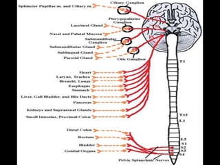
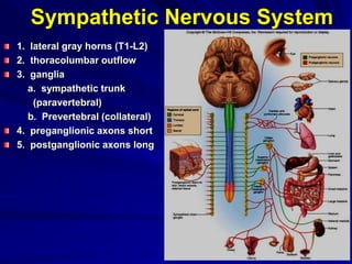
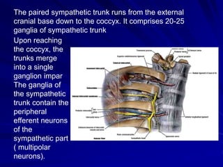
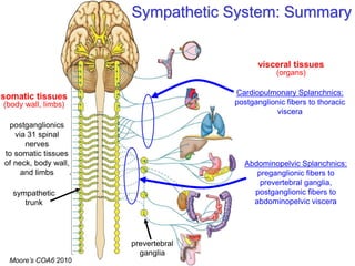
The sympathetic nervous system has three parts:
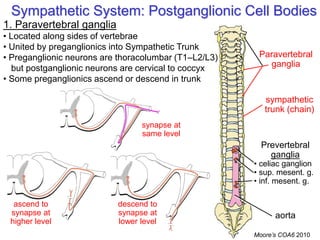
1. Preganglionic cell bodies located in the lateral gray horns of the spinal cord from T1 to L2/L3.
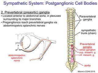
2. Ganglia located along the sympathetic trunk and around the abdominal aorta.
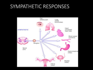
3. Postganglionic fibers innervating organs and blood vessels.


![Visceral Afferents and Referred Pain
Somatic sensation:
• conscious, sharp, well-localized
• touch, pain, temperature, pressure, proprioception
Visceral sensation:
• often unconscious; if conscious: dull, poorly-localized
• distension, blood gas, blood pressure, cramping, irritants
dorsal root ganglion
Visceral sensory nerves [GVA]
• run with sympathetic &
parasympathetic nerves
• cell bodies in dorsal root ganglion
• nerve ending in viscera](https://image.slidesharecdn.com/2020thesympatetnervoussystemnew-201119173908/85/The-Sympathetic-Nervous-System-27-320.jpg)