LEARNING
OBJECTIVE
(LO)
AT THEEND OF THE TOPIC 1-BHMS
STUDENT MUST BE ABLE TO ABIDE
BY THE AUTONOMIC NERVOUS
SYSTEM AND ITS APPLIED
ANATOMY.
3.
SPECIFIC LEARNING
OBJECTIVES (SLO)
•Definethe Autonomic Nervous System.
•Describe the divisions of ANS – sympathetic and
parasympathetic.
•Differentiate between sympathetic and
parasympathetic nervous systems.
•Explain the basic anatomy and organization of ANS.
•Name the neurotransmitters and receptors of ANS.
•Describe the effects of ANS on major organs.
•State the clinical significance of ANS.
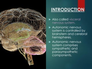
INTRODUCTION
Also calledvisceral
nervous system.
Autonomic nervous
system is controlled by
brainstem and cerebral
hemispheres.
Autonomic nervous
system comprises
sympathetic and
parasympathetic
components.
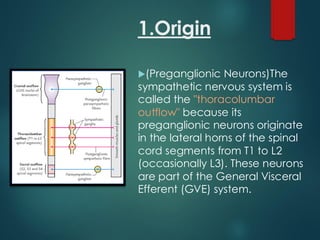
1.Origin
(Preganglionic Neurons)The
sympathetic nervoussystem is
called the "thoracolumbar
outflow" because its
preganglionic neurons originate
in the lateral horns of the spinal
cord segments from T1 to L2
(occasionally L3). These neurons
are part of the General Visceral
Efferent (GVE) system.
8.
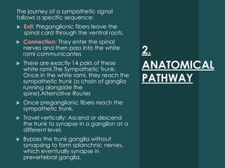
2.
ANATOMICAL
PATHWAY
The journey ofa sympathetic signal
follows a specific sequence:
Exit: Preganglionic fibers leave the
spinal cord through the ventral roots.
Connection: They enter the spinal
nerves and then pass into the white
rami communicantes
There are exactly 14 pairs of these
white rami.The Sympathetic Trunk:
Once in the white rami, they reach the
sympathetic trunk (a chain of ganglia
running alongside the
spine).Alternative Routes
Once preganglionic fibers reach the
sympathetic trunk.
Travel vertically: Ascend or descend
the trunk to synapse in a ganglion at a
different level.
Bypass the trunk ganglia without
synapsing to form splanchnic nerves,
which eventually synapse in
prevertebral ganglia.
9.
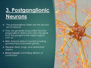
3. Postganglionic
Neurons
Thepostganglionic fibers are the second
set of neurons.
They are generally long (unlike the short
preganglionic fibers) and carry the signal
from the ganglia to the target organs,
such as :
Skin: Arrector pilorum muscles (causing
goosebumps) and sweat glands.
Viscera: Heart, lungs, and abdominal
organs.
Blood Vessels: Controlling dilation or
constriction.
10.
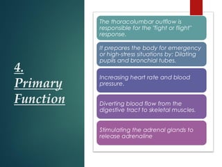
4.
Primary
Function
The thoracolumbar outflowis
responsible for the "fight or flight"
response.
It prepares the body for emergency
or high-stress situations by: Dilating
pupils and bronchial tubes.
Increasing heart rate and blood
pressure.
Diverting blood flow from the
digestive tract to skeletal muscles.
Stimulating the adrenal glands to
release adrenaline
11.
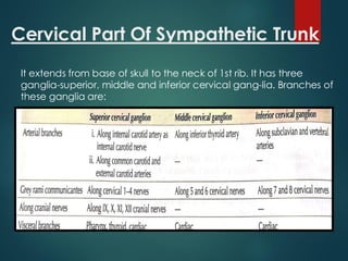
Cervical Part OfSympathetic Trunk
It extends from base of skull to the neck of 1st rib. It has three
ganglia-superior, middle and inferior cervical gang-lia. Branches of
these ganglia are:
12.
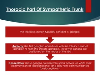
Thoracic Part OfSympathetic Trunk
Connections: These ganglia are linked to spinal nerves via white rami
communicantes (preganglionic) and grey rami communicantes
(postganglionic).
Anatomy:The first ganglion often fuses with the inferior cervical
ganglion to form the stellate ganglion. The lower ganglia are
positioned on the heads of the ribs.
The thoracic section typically contains 11 ganglia.
13.
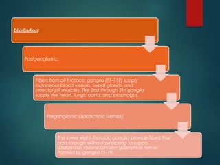
Distribution:
Postganglionic:
Fibers from allthoracic ganglia (T1–T12) supply
cutaneous blood vessels, sweat glands, and
arrector pili muscles. The 2nd through 5th ganglia
supply the heart, lungs, aorta, and esophagus.
Preganglionic (Splanchnic Nerves):
The lower eight thoracic ganglia provide fibers that
pass through without synapsing to supply
abdominal viscera:Greater splanchnic nerve:
Formed by ganglia T5–T9.
14.
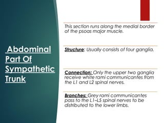
Abdominal
Part Of
Sympathetic
Trunk
This sectionruns along the medial border
of the psoas major muscle.
Structure: Usually consists of four ganglia.
Connection: Only the upper two ganglia
receive white rami communicantes from
the L1 and L2 spinal nerves.
Branches: Grey rami communicantes
pass to the L1–L5 spinal nerves to be
distributed to the lower limbs.
15.
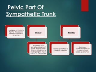
Pelvic Part Of
SympatheticTrunk
The pelvic part runs in
front of the sacrum,
medial to the sacral
foramina.
Structure:
It contains four
ganglia. At its lowest
point, the two trunks
unite and fuse into a
single ganglion impar
in front of the coccyx.
Branches:
Visceral branches: To
the pelvic plexuses.
Grey rami
communicantes: To
the sacral and
coccygeal nerves.
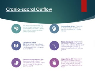
Cranio-sacral Outflow
This divisionis responsible for
conserving energy and "rest
and digest" functions. It arises
from cranial nerves and the
sacral spinal cord.
Preganglionic fibers: Originate
in the brainstem and travel
through four specific cranial
nerves:
Oculomotor Nerve
(III):Originates in the Edinger-
Westphal nucleus; relays in the
ciliary ganglion to control the
pupil and lens.
Facial Nerve (VII):Originates in
the superior salivatory nucleus;
relays in the submandibular and
pterygopalatine ganglia to
supply tear glands and salivary
glands.
Glossopharyngeal Nerve (IX):
Originates in the inferior
salivatory nucleus; relays in the
otic ganglion to supply the
parotid gland.
Vagus Nerve (X): Originates in
the dorsal nucleus of vagus ;
provides extensive innervation
to the heart, lungs, and the
majority of the gastrointestinal
tract.
18.
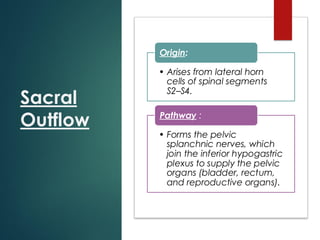
Sacral
Outflow
• Arises fromlateral horn
cells of spinal segments
S2–S4.
Origin:
• Forms the pelvic
splanchnic nerves, which
join the inferior hypogastric
plexus to supply the pelvic
organs (bladder, rectum,
and reproductive organs).
Pathway :
19.
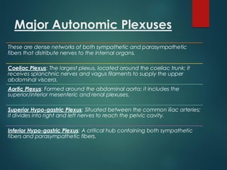
Major Autonomic Plexuses
Theseare dense networks of both sympathetic and parasympathetic
fibers that distribute nerves to the internal organs.
Coeliac Plexus: The largest plexus, located around the coeliac trunk; it
receives splanchnic nerves and vagus filaments to supply the upper
abdominal viscera.
Aortic Plexus: Formed around the abdominal aorta; it includes the
superior/inferior mesenteric and renal plexuses.
Superior Hypo-gastric Plexus: Situated between the common iliac arteries;
it divides into right and left nerves to reach the pelvic cavity.
Inferior Hypo-gastric Plexus: A critical hub containing both sympathetic
fibers and parasympathetic fibers.
20.
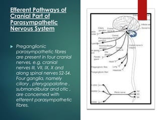
Efferent Pathways of
CranialPart of
Parasympathetic
Nervous System
Preganglionic
parasympathetic fibres
are present in four cranial
nerves, e.g. cranial
nerves III, VII, IX, X and
along spinal nerves S2-S4.
Four ganglia, namely
ciliary , pterygopalatine ,
submandibular and otic ,
are concerned with
efferent parasympathetic
fibres.
21.
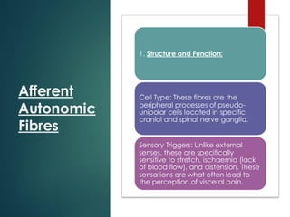
Afferent
Autonomic
Fibres
1. Structure andFunction:
Cell Type: These fibres are the
peripheral processes of pseudo-
unipolar cells located in specific
cranial and spinal nerve ganglia.
Sensory Triggers: Unlike external
senses, these are specifically
sensitive to stretch, ischaemia (lack
of blood flow), and distension. These
sensations are what often lead to
the perception of visceral pain.
22.
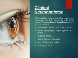
Clinical
Neuroanatomy
1.Removal of stellateganglion improves
the blood supply to the upper limb. But its
removal causes Horner's syndrome which
is comprised of :
Anhidrosis of the same side of face
Partial drooping of upper eyelid, i.e
ptosis
Enophthalmos
Constriction of the pupil
Loss of ciliospinal reflex
Flushing of face
23.
2. Arteries ofthe upper limb are
innervated by sympa thetic
fibres. Preganglionic fibres
originate from the cell bodies of
T2-T5 spinal segments. Fibres
ascend in the sympathetic trunk
and synapse with middle and
inferior cervical ganglia.
Postganglionic fibres join the
nerves which constitute the
brachial plexus. get distributed to
the arteries of the upper limbi in
each region.
These fibres may be cut to relieve
the symptoms of Raynaud's
disease.
24.
3. Arteries oflower limb are
supplied by lower three
thoracic and upper two
lumbar segments of the
spinal cord. Femoral artery
is supplied by sympathetic
fibres from femoral and
obturator nerves.
01
Posterior tibial artery
receives the
postganglionic fibres from
common peroneal and
tibial nerves. Buerger's
disease may be treated
by lumbar
sympathectomy.
02
Postganglionic fibres join
the nerves which
constitute the brachial
plexus. get distributed to
the arteries of the upper
limbi in each region.
03
25.
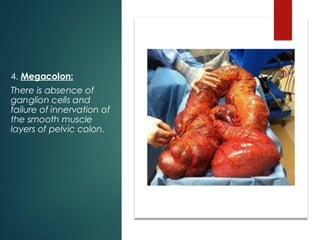
4. Megacolon:
There isabsence of
ganglion cells and
failure of innervation of
the smooth muscle
layers of pelvic colon.