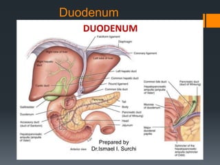
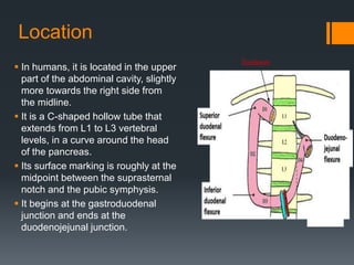
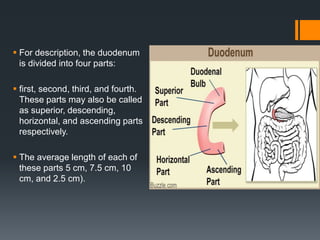
SlideShare a Scribd company logo
Duodenum
Prepared by
Dr.Ismael I. Surchi
Objectives
Anatomy and Physiological of duodenum.
Histology of duodenum.
Blood supply and lymphatic drainage of
duodenum.
Neurological innervation of the duodenum.
Physiological functions of the duodenum.
Common conditions of duodenum.
 The duodenum It is the initial
portion of the small intestine,
connecting the distal end (i.e.
the latter part) of the stomach
to the proximal end (i.e. the
upper part) of the jejunum.
 It receives partially digested
food (chyme) with high acidity
from the stomach, and
involves in neutralizing the
acidity of the food and
propelling it through the rest of
the small intestine for further
digestion and absorption.
Location
 In humans, it is located in the upper
part of the abdominal cavity, slightly
more towards the right side from
the midline.
 It is a C-shaped hollow tube that
extends from L1 to L3 vertebral
levels, in a curve around the head
of the pancreas.
 Its surface marking is roughly at the
midpoint between the suprasternal
notch and the pubic symphysis.
 It begins at the gastroduodenal
junction and ends at the
duodenojejunal junction.
 For description, the duodenum
is divided into four parts:
 first, second, third, and fourth.
These parts may also be called
as superior, descending,
horizontal, and ascending parts
respectively.
 The average length of each of
these parts 5 cm, 7.5 cm, 10
cm, and 2.5 cm).
Parts of duodenum
The first part of the duodenum begins at the pylorus and
runs upward and backward on the transpyloric plane at
the level of the 1st lumbar vertebra.
Relations:
Anteriorly: The quadrate lobe of the liver and the
gallbladder.
Posteriorly: The lesser sac (first inch only), the
gastroduodenal artery, the bile duct and the portal vein,
and the inferior vena cava.
Superiorly: The entrance into the lesser sac (the
epiploic foramen) • Inferiorly: The head of the pancreas
The second part of the duodenum runs vertically
downward in front of the hilum of the right kidney on
the right side of the 2nd and 3rd lumbar vertebrae.
Relations
Anteriorly: The fundus of the gallbladder and the right
lobe of the liver, the transverse colon, and the coils of
the small intestine .
Posteriorly: The hilum of the right kidney and the right
ureter.
Laterally: The ascending colon, the right colic flexure,
and the right lobe of the liver.
Medially: The head of the pancreas, the bile duct, and
themain pancreatic duct .
Third Part Of Duodenum
Runs horizontally to the left on the subcostal plane,
passing in front of the vertebral column and following
the lower margin of the head of the pancreas.
• Relations:
• Anteriorly: The root of the mesentery of the small
intestine, the superior mesenteric vessels contained
within it, and coils of jejunum.
Posteriorly: The right ureter, the right psoas muscle,
the inferior vena cava, and the aorta.
Superiorly: The head of the pancreas.
Inferiorly: Coils of jejunum
Fourth Part Of Duodenum
Runs upward and to the left to the duodenojejunal
flexure. The flexure is held in position by a
peritoneal fold, the ligament of Treitz, which is
attached to the right crus of the diaphragm.
Relations:
Anteriorly: The beginning of the root of the
mesentery and coils of jejunum.
Posteriorly: The left margin of the aorta and the
medial border of the left psoas muscle .
HISTOLOGY
 Histologically the duodenum is
similar to all the other hollow organs
of the gastrointestinal tract: mucosa,
submucosa and muscularis.
 The mucosa consists of simple
columnar epithelium (lamina
epithelialis), a connective
tissue layer (lamina propria) and
a smooth muscle layer (lamina
muscularis). The intestinal epithelial
cells (enterocytes) are overlaid by a
layer of glycoproteins and mucin.
 The submucosa comprises loose
connective tissue, numerous blood
vessels and the Meissner's plexus.
 The muscularis consists of an inner
circular and an outer
longitudinal musculature between
which the Auerbach’s plexus lies.
 The mucosa forms fingerlike projections called villi that extend into the
intestinal lumen. These are epithelial folds lined by two types of cells,
enterocytes and goblet cells.
 Enterocytes are simple columnar cells with basal elongated nuclei and
an apical brush border.
 The brush border is the microscopic representation of small protrusions
of the cell membrane, microvilli, which greatly increase the surface area
of the cell enhancing absorptive capacity.
 The mucous membrane of the duodenum is thick in the first part of the
duodenum, it is smooth in the remainder of the duodenum, it is thrown
into numerous circular folds called the plicae circulares.
 At the site where the bile duct and the main pancreatic duct pierce the
medial wall of the second part is a small, rounded elevation called the
major duodenal papilla .
 The accessory pancreatic duct, if present, opens into the duodenum on
a smaller papilla about 0.75 in. (1.9 cm) above the major duodenal
papilla.
 The other cell type is mucus secreting goblet cells that can be
recognized by the presence of an apical mucous cup. The core of the
villus is part of the lamina propria. The most numerous cells in the
lamina propria are immune cells, most of which are lymphocytes.
Because villi are the site of absorption of nutrition they have a rich
blood supply, each villus is supplied by central arterioles and drained
by central venules and a central lymph vessel.
 Underlying the villi are the intestinal glands, also called the crypts of
Lieberkühn. These glands are lined with numerous relatively
undifferentiated columnar cells that usually undergo two rounds of
mitosis before differentiating into either absorptive cells or goblet cells.
Enterocytes, goblet cells, paneth cells that secrete antibacterial
enzymes (recognized by eosinophilic granules in their apical
cytoplasm) and enteroendocrine cells also line the crypt. A thin layer of
smooth muscle marks the end of the mucosa, the muscularis mucosae.
 In the submucosa numerous pale stained glands are present, namely
the Brunner’s glands. These are branched tubular or alveo-tubular
glands lined with columnar secretory epithelium. Brunner’s glands
secrete large amounts of alkaline mucous that neutralize the acidic
contents from the stomach.
Blood Supply and Lymphatics
 The blood supply of the C-shaped duodenum is shared with the head
of the pancreas.
 The proximal segment of the duodenum is supplied by the
gastroduodenal artery and its branches which include the superior
pancreaticoduodenal artery.
 The distal segment of the duodenum is supplied by the superior
mesenteric artery and the inferior pancreaticoduodenal artery.
 The venous drainage follows the arteries and ultimately drains into the
portal system.
Lymphatic drainage
 The duodenum also has
lymphatic vessels which drain
into the pancreaticoduodenal
lymph nodes located along the
pancreaticoduodenal vessels
and the superior mesenteric
lymph nodes.
 Efferent vessels of these
nodes drain into coeliac and
sup.mesen.group of pre aortic
lymph nodes.
 Some vesseles drain into
hepatic nodes directly.
Innervation
Innervation
 Duodenum receives both sympathetic and parasympathetic
nerves from the celiac and superior mesenteric plexuses.
 The nerves of the duodenum travel throughout the
submucosal layer of the duodenum.
 The duodenum is richly innervated by the parasympathetic
nervous system which includes branches of the anterior and
posterior vagus trunks.
 These parasympathetic nerves pass through the celiac
plexuses and follow the celiac trunk toward the duodenum.
The nerves then synapse in ganglia in the gut plexuses in the
duodenum and reach their final targets through short
postsynaptic fibers.
 The sympathetic nerves are branches of the celiac plexus
which originate from T5 through T9. These sympathetic
nerves pass through the sympathetic chain and travel
through the greater splanchnic nerve and synapse in the
celiac ganglia. The postsynaptic sympathetic follow the
branches of the celiac trunk toward the duodenum.
Physiological function
 duodenum manifests three fundamental physiological phenomena:
motility, secretion, and absorption.
 The duodenum receives chyme (partially digested food mixed with
acid, mucus and pepsin) from the stomach, and acts as an
intermediate for the further digestion and motility of this food.
 The endocrine cells found in the duodenal epithelium secrete the
hormones secretin and cholecystokinin (CCK) in response to the
acids and fats present in chyme. These hormones act locally on the
pyloric sphincter of the stomach, and thereby regulate the movement
of food out of the pylorus (i.e. regulate gastric emptying).
 Secretin stimulates the release of bicarbonate and mucus from the
duodenal epithelium, and hence helps bring the high acidity (or very
low pH) of chyme back to normal, making it easier to be digested
further. These hormones also stimulate the secretion of bile from the
liver and gallbladder, and the secretion of digestive enzymes such as
trypsin, lipase and amylase from the pancreas.
 These secretions are received by the duodenum via the main and
accessory pancreatic ducts, and the food is mixed with these
secretions and prepared for further chemical digestion.
 Bile and the digestive enzymes initiate and continue the chemical
digestion of food inside the duodenum by breaking down all nutrient
macro-molecules into their simpler monomeric forms.
 The duodenum is the main part where calcium and iron absorption
takes place in the small intestine.
 The muscle layers of the duodenum contract and relax in a regulated
manner by way of peristalsis and segmentation movements to transfer
the food in an orderly way to the rest of the small intestine where
further digestion and absorption takes place.
Common conditions of duodenum.
Duodenal ulcers are breaks in the mucosa that reach down to
the level of the muscularis mucosa. In the duodenum, ulcers occur
most commonly in its first part. Duodenal ulcers are more common
than gastric ulcers, and are mostly caused by the action of the
bacterium Helicobacter pylori, or due to long term NSAID use.
 Other less common causes include autoimmune conditions,
Crohn’s disease, bile reflux, some viral infections, ingestion of
corrosive substances, excessive cigarette smoking etc
 These ulcers may heal with scar formation, or may cause further
complications such as bleeding, perforation, or gastric outlet
obstruction (due to edema surrounding the inflamed ulcer).
Duodenitis means inflammation of the duodenum, which can be
either acute (short-term) or chronic (long-term). It can be caused by
various reasons, and the most common ones are – infection with
Helicobacter pylori, and long term NSAID use.
Duodenal obstruction is usually caused by cancers, the most
common cause being pancreatic cancer. Metastatic deposits from
other cancers such as gastric or colorectal cancer may also cause
duodenal obstruction. Primary duodenal cancer is a less common
cause when compared with the aforementioned causes.
Duodenal cancers are less common when compared to other
gastrointestinal cancers such as gastric cancer and colorectal
cancer. However, it is the most common site for cancers occurring
in the small intestine. The condition named familial adenomatous
polyposis (FAP) is an important risk factor for duodenal cancer.
THANK YOU

More Related Content

What's hot

Anatomy of duodenum, duodenum structure, PPT of duodenum, power point present...
Anatomy of duodenum, duodenum structure, PPT of duodenum, power point present...Anatomy of duodenum, duodenum structure, PPT of duodenum, power point present...
Anatomy of duodenum, duodenum structure, PPT of duodenum, power point present...
drasarma1947
 
The jejunum and ileum
The jejunum and ileumThe jejunum and ileum
The jejunum and ileum
Idris Siddiqui
 
urinary Bladder.ppt
urinary Bladder.ppturinary Bladder.ppt
urinary Bladder.ppt
Dr. sana yaseen
 
Anatomy of stomach
Anatomy of stomachAnatomy of stomach
Anatomy of stomach
DINESH KUMAR D
 
SMALL INTESTINE AND LARGE INTESTINE
SMALL INTESTINE AND LARGE INTESTINESMALL INTESTINE AND LARGE INTESTINE
SMALL INTESTINE AND LARGE INTESTINE
Dr Nilesh Kate
 
Male and female urethra
Male and female urethraMale and female urethra
Male and female urethra
Devlop Shrestha
 
Anatomy Of Urinary Bladder
Anatomy Of Urinary BladderAnatomy Of Urinary Bladder
Anatomy Of Urinary Bladder
Umair Majeed
 
Stomach
StomachStomach
Histology of stomach
Histology of stomachHistology of stomach
Histology of stomach
Tehseen Anwar
 
peritoneum and peritoneal cavity
peritoneum and peritoneal  cavityperitoneum and peritoneal  cavity
peritoneum and peritoneal cavity
Jay Patel
 
Small intestine
Small intestine Small intestine
Small intestine
Brisso Mathew Arackal
 
Anatomy of stomach
Anatomy of stomachAnatomy of stomach
Anatomy of stomach
Sachin Patne
 
Extrahepatic Biliary Apparatus
Extrahepatic Biliary ApparatusExtrahepatic Biliary Apparatus
Extrahepatic Biliary Apparatus
DrUroojRehman
 
Small intestine
Small intestineSmall intestine
Small intestine
Madeleine Si
 
Urinary bladder and urethra
Urinary bladder  and urethraUrinary bladder  and urethra
Urinary bladder and urethraLawrence James
 
ANATOMY OF DUODENUM
ANATOMY OF DUODENUMANATOMY OF DUODENUM
ANATOMY OF DUODENUM
Dr. sana yaseen
 
Peritoneum i
Peritoneum iPeritoneum i
Peritoneum i
Dr Laxman Khanal
 
Anatomy of duodenum and pancreas
Anatomy of duodenum and pancreas Anatomy of duodenum and pancreas
Anatomy of duodenum and pancreas
Dr. Mohammad Mahmoud
 
Liver anatomy
Liver anatomyLiver anatomy
Liver anatomy
satha guru
 

What's hot (20)

Anatomy of duodenum, duodenum structure, PPT of duodenum, power point present...
Anatomy of duodenum, duodenum structure, PPT of duodenum, power point present...Anatomy of duodenum, duodenum structure, PPT of duodenum, power point present...
Anatomy of duodenum, duodenum structure, PPT of duodenum, power point present...
 
The jejunum and ileum
The jejunum and ileumThe jejunum and ileum
The jejunum and ileum
 
urinary Bladder.ppt
urinary Bladder.ppturinary Bladder.ppt
urinary Bladder.ppt
 
Anatomy of stomach
Anatomy of stomachAnatomy of stomach
Anatomy of stomach
 
SMALL INTESTINE AND LARGE INTESTINE
SMALL INTESTINE AND LARGE INTESTINESMALL INTESTINE AND LARGE INTESTINE
SMALL INTESTINE AND LARGE INTESTINE
 
Male and female urethra
Male and female urethraMale and female urethra
Male and female urethra
 
Anatomy Of Urinary Bladder
Anatomy Of Urinary BladderAnatomy Of Urinary Bladder
Anatomy Of Urinary Bladder
 
Stomach
StomachStomach
Stomach
 
Histology of stomach
Histology of stomachHistology of stomach
Histology of stomach
 
Blood supply of git
Blood supply of gitBlood supply of git
Blood supply of git
 
peritoneum and peritoneal cavity
peritoneum and peritoneal  cavityperitoneum and peritoneal  cavity
peritoneum and peritoneal cavity
 
Small intestine
Small intestine Small intestine
Small intestine
 
Anatomy of stomach
Anatomy of stomachAnatomy of stomach
Anatomy of stomach
 
Extrahepatic Biliary Apparatus
Extrahepatic Biliary ApparatusExtrahepatic Biliary Apparatus
Extrahepatic Biliary Apparatus
 
Small intestine
Small intestineSmall intestine
Small intestine
 
Urinary bladder and urethra
Urinary bladder  and urethraUrinary bladder  and urethra
Urinary bladder and urethra
 
ANATOMY OF DUODENUM
ANATOMY OF DUODENUMANATOMY OF DUODENUM
ANATOMY OF DUODENUM
 
Peritoneum i
Peritoneum iPeritoneum i
Peritoneum i
 
Anatomy of duodenum and pancreas
Anatomy of duodenum and pancreas Anatomy of duodenum and pancreas
Anatomy of duodenum and pancreas
 
Liver anatomy
Liver anatomyLiver anatomy
Liver anatomy
 

Similar to Duodenum by Ismail Surchi

digestive system of anatomypowerpoint pres
digestive system of anatomypowerpoint presdigestive system of anatomypowerpoint pres
digestive system of anatomypowerpoint pres
HgjUy7
 
Absorption rohit bharti
Absorption  rohit bhartiAbsorption  rohit bharti
ELIMINATION
ELIMINATIONELIMINATION
ELIMINATION
RAVINDRA MARKAD
 
Digestive system
Digestive systemDigestive system
Digestive system
Soneeshah
 
Thomson and shaffer first principles of gastroenterology - 6 - the small in...
Thomson and shaffer   first principles of gastroenterology - 6 - the small in...Thomson and shaffer   first principles of gastroenterology - 6 - the small in...
Thomson and shaffer first principles of gastroenterology - 6 - the small in...
Cristiane Medeiros
 
Digestive system
Digestive systemDigestive system
Digestive system
Santhosh Lingabathula
 
Digestive system
Digestive systemDigestive system
Digestive system
muthulakshmi623285
 
Anatomy of the esophagus.pptx
Anatomy of the esophagus.pptxAnatomy of the esophagus.pptx
Anatomy of the esophagus.pptx
EthelBwendo
 
Digestion anatomy
Digestion anatomyDigestion anatomy
Digestion anatomy
Amir rezagholizadeh
 
Peritoneum , Dr. Anudeep singh
Peritoneum , Dr. Anudeep singhPeritoneum , Dr. Anudeep singh
Peritoneum , Dr. Anudeep singh
ANUDEEP SINGH
 
HAP 4 SEMESTER 2 BPHARMACY AS PER PCI SYLLABUS
HAP 4 SEMESTER 2 BPHARMACY AS PER PCI SYLLABUSHAP 4 SEMESTER 2 BPHARMACY AS PER PCI SYLLABUS
HAP 4 SEMESTER 2 BPHARMACY AS PER PCI SYLLABUS
christinajohn24
 
Small intestine surgery
Small intestine surgerySmall intestine surgery
Small intestine surgery
garvsuthar
 
Digestive System.pptx
Digestive System.pptxDigestive System.pptx
Digestive System.pptx
nikhilgendre1
 
Large Intestine.pptxjdhdhdjdjdjdjdjehdhdh
Large Intestine.pptxjdhdhdjdjdjdjdjehdhdhLarge Intestine.pptxjdhdhdjdjdjdjdjehdhdh
Large Intestine.pptxjdhdhdjdjdjdjdjehdhdh
KennyjrLMunisi
 
1. INTROduction TO GIT - I (1) (3) - 1.pptx
1. INTROduction TO GIT - I (1) (3) - 1.pptx1. INTROduction TO GIT - I (1) (3) - 1.pptx
1. INTROduction TO GIT - I (1) (3) - 1.pptx
muneebrajaraja979
 
Digestive system
Digestive systemDigestive system
Digestive system
Chanukya Vanam . Dr
 
Combined 02 clinical training--anatomy
Combined 02 clinical training--anatomyCombined 02 clinical training--anatomy
Combined 02 clinical training--anatomyIknifem
 

Similar to Duodenum by Ismail Surchi (20)

Anatomy of digestive system-III.pptx
Anatomy of digestive system-III.pptxAnatomy of digestive system-III.pptx
Anatomy of digestive system-III.pptx
 
digestive system of anatomypowerpoint pres
digestive system of anatomypowerpoint presdigestive system of anatomypowerpoint pres
digestive system of anatomypowerpoint pres
 
Absorption rohit bharti
Absorption  rohit bhartiAbsorption  rohit bharti
Absorption rohit bharti
 
Digestive system-I.pptx
Digestive system-I.pptxDigestive system-I.pptx
Digestive system-I.pptx
 
ELIMINATION
ELIMINATIONELIMINATION
ELIMINATION
 
Digestive system
Digestive systemDigestive system
Digestive system
 
Thomson and shaffer first principles of gastroenterology - 6 - the small in...
Thomson and shaffer   first principles of gastroenterology - 6 - the small in...Thomson and shaffer   first principles of gastroenterology - 6 - the small in...
Thomson and shaffer first principles of gastroenterology - 6 - the small in...
 
Digestive system
Digestive systemDigestive system
Digestive system
 
Digestive system
Digestive systemDigestive system
Digestive system
 
Anatomy of the esophagus.pptx
Anatomy of the esophagus.pptxAnatomy of the esophagus.pptx
Anatomy of the esophagus.pptx
 
Digestion anatomy
Digestion anatomyDigestion anatomy
Digestion anatomy
 
Peritoneum , Dr. Anudeep singh
Peritoneum , Dr. Anudeep singhPeritoneum , Dr. Anudeep singh
Peritoneum , Dr. Anudeep singh
 
Digestive system
Digestive systemDigestive system
Digestive system
 
HAP 4 SEMESTER 2 BPHARMACY AS PER PCI SYLLABUS
HAP 4 SEMESTER 2 BPHARMACY AS PER PCI SYLLABUSHAP 4 SEMESTER 2 BPHARMACY AS PER PCI SYLLABUS
HAP 4 SEMESTER 2 BPHARMACY AS PER PCI SYLLABUS
 
Small intestine surgery
Small intestine surgerySmall intestine surgery
Small intestine surgery
 
Digestive System.pptx
Digestive System.pptxDigestive System.pptx
Digestive System.pptx
 
Large Intestine.pptxjdhdhdjdjdjdjdjehdhdh
Large Intestine.pptxjdhdhdjdjdjdjdjehdhdhLarge Intestine.pptxjdhdhdjdjdjdjdjehdhdh
Large Intestine.pptxjdhdhdjdjdjdjdjehdhdh
 
1. INTROduction TO GIT - I (1) (3) - 1.pptx
1. INTROduction TO GIT - I (1) (3) - 1.pptx1. INTROduction TO GIT - I (1) (3) - 1.pptx
1. INTROduction TO GIT - I (1) (3) - 1.pptx
 
Digestive system
Digestive systemDigestive system
Digestive system
 
Combined 02 clinical training--anatomy
Combined 02 clinical training--anatomyCombined 02 clinical training--anatomy
Combined 02 clinical training--anatomy
 

More from Ismail Surchi

Anxiolytics and antidepressants.
Anxiolytics and antidepressants.Anxiolytics and antidepressants.
Anxiolytics and antidepressants.
Ismail Surchi
 
diagnosis and management of ischemic cerebrovascular disease by Ismail Surchi
diagnosis and management of ischemic cerebrovascular disease by Ismail Surchidiagnosis and management of ischemic cerebrovascular disease by Ismail Surchi
diagnosis and management of ischemic cerebrovascular disease by Ismail Surchi
Ismail Surchi
 
Gynaecological history by Ismail Surchi
Gynaecological history by Ismail SurchiGynaecological history by Ismail Surchi
Gynaecological history by Ismail Surchi
Ismail Surchi
 
Ekg or ECG (clectrocariography)
Ekg or ECG (clectrocariography)Ekg or ECG (clectrocariography)
Ekg or ECG (clectrocariography)
Ismail Surchi
 
Urinary system by ismail surchi
Urinary system by ismail surchiUrinary system by ismail surchi
Urinary system by ismail surchi
Ismail Surchi
 
clostridium difficile by Ismail surchi
clostridium difficile by Ismail surchi clostridium difficile by Ismail surchi
clostridium difficile by Ismail surchi
Ismail Surchi
 
Kidney by ismail Surchi
Kidney by ismail SurchiKidney by ismail Surchi
Kidney by ismail Surchi
Ismail Surchi
 
Thyroid gland by ismail surchi
Thyroid gland by ismail surchiThyroid gland by ismail surchi
Thyroid gland by ismail surchi
Ismail Surchi
 

More from Ismail Surchi (8)

Anxiolytics and antidepressants.
Anxiolytics and antidepressants.Anxiolytics and antidepressants.
Anxiolytics and antidepressants.
 
diagnosis and management of ischemic cerebrovascular disease by Ismail Surchi
diagnosis and management of ischemic cerebrovascular disease by Ismail Surchidiagnosis and management of ischemic cerebrovascular disease by Ismail Surchi
diagnosis and management of ischemic cerebrovascular disease by Ismail Surchi
 
Gynaecological history by Ismail Surchi
Gynaecological history by Ismail SurchiGynaecological history by Ismail Surchi
Gynaecological history by Ismail Surchi
 
Ekg or ECG (clectrocariography)
Ekg or ECG (clectrocariography)Ekg or ECG (clectrocariography)
Ekg or ECG (clectrocariography)
 
Urinary system by ismail surchi
Urinary system by ismail surchiUrinary system by ismail surchi
Urinary system by ismail surchi
 
clostridium difficile by Ismail surchi
clostridium difficile by Ismail surchi clostridium difficile by Ismail surchi
clostridium difficile by Ismail surchi
 
Kidney by ismail Surchi
Kidney by ismail SurchiKidney by ismail Surchi
Kidney by ismail Surchi
 
Thyroid gland by ismail surchi
Thyroid gland by ismail surchiThyroid gland by ismail surchi
Thyroid gland by ismail surchi
 

Recently uploaded

ESC Beyond Borders _From EU to You_ InfoPack general.pdf
ESC Beyond Borders _From EU to You_ InfoPack general.pdfESC Beyond Borders _From EU to You_ InfoPack general.pdf
ESC Beyond Borders _From EU to You_ InfoPack general.pdf
Fundacja Rozwoju Społeczeństwa Przedsiębiorczego
 
The Roman Empire A Historical Colossus.pdf
The Roman Empire A Historical Colossus.pdfThe Roman Empire A Historical Colossus.pdf
The Roman Empire A Historical Colossus.pdf
kaushalkr1407
 
Mule 4.6 & Java 17 Upgrade | MuleSoft Mysore Meetup #46
Mule 4.6 & Java 17 Upgrade | MuleSoft Mysore Meetup #46Mule 4.6 & Java 17 Upgrade | MuleSoft Mysore Meetup #46
Mule 4.6 & Java 17 Upgrade | MuleSoft Mysore Meetup #46
MysoreMuleSoftMeetup
 
Basic phrases for greeting and assisting costumers
Basic phrases for greeting and assisting costumersBasic phrases for greeting and assisting costumers
Basic phrases for greeting and assisting costumers
PedroFerreira53928
 
GIÁO ÁN DẠY THÊM (KẾ HOẠCH BÀI BUỔI 2) - TIẾNG ANH 8 GLOBAL SUCCESS (2 CỘT) N...
GIÁO ÁN DẠY THÊM (KẾ HOẠCH BÀI BUỔI 2) - TIẾNG ANH 8 GLOBAL SUCCESS (2 CỘT) N...GIÁO ÁN DẠY THÊM (KẾ HOẠCH BÀI BUỔI 2) - TIẾNG ANH 8 GLOBAL SUCCESS (2 CỘT) N...
GIÁO ÁN DẠY THÊM (KẾ HOẠCH BÀI BUỔI 2) - TIẾNG ANH 8 GLOBAL SUCCESS (2 CỘT) N...
Nguyen Thanh Tu Collection
 
Operation Blue Star - Saka Neela Tara
Operation Blue Star   -  Saka Neela TaraOperation Blue Star   -  Saka Neela Tara
Operation Blue Star - Saka Neela Tara
Balvir Singh
 
How to Create Map Views in the Odoo 17 ERP
How to Create Map Views in the Odoo 17 ERPHow to Create Map Views in the Odoo 17 ERP
How to Create Map Views in the Odoo 17 ERP
Celine George
 
Chapter 3 - Islamic Banking Products and Services.pptx
Chapter 3 - Islamic Banking Products and Services.pptxChapter 3 - Islamic Banking Products and Services.pptx
Chapter 3 - Islamic Banking Products and Services.pptx
Mohd Adib Abd Muin, Senior Lecturer at Universiti Utara Malaysia
 
special B.ed 2nd year old paper_20240531.pdf
special B.ed 2nd year old paper_20240531.pdfspecial B.ed 2nd year old paper_20240531.pdf
special B.ed 2nd year old paper_20240531.pdf
Special education needs
 
CLASS 11 CBSE B.St Project AIDS TO TRADE - INSURANCE
CLASS 11 CBSE B.St Project AIDS TO TRADE - INSURANCECLASS 11 CBSE B.St Project AIDS TO TRADE - INSURANCE
CLASS 11 CBSE B.St Project AIDS TO TRADE - INSURANCE
BhavyaRajput3
 
aaaaaaaaaaaaaaaaaaaaaaaaaaaaaaaaaaaaaaaaaaaaaaaaaaaaaaa
aaaaaaaaaaaaaaaaaaaaaaaaaaaaaaaaaaaaaaaaaaaaaaaaaaaaaaaaaaaaaaaaaaaaaaaaaaaaaaaaaaaaaaaaaaaaaaaaaaaaaaaaaaaaaa
aaaaaaaaaaaaaaaaaaaaaaaaaaaaaaaaaaaaaaaaaaaaaaaaaaaaaaa
siemaillard
 
Instructions for Submissions thorugh G- Classroom.pptx
Instructions for Submissions thorugh G- Classroom.pptxInstructions for Submissions thorugh G- Classroom.pptx
Instructions for Submissions thorugh G- Classroom.pptx
Jheel Barad
 
Thesis Statement for students diagnonsed withADHD.ppt
Thesis Statement for students diagnonsed withADHD.pptThesis Statement for students diagnonsed withADHD.ppt
Thesis Statement for students diagnonsed withADHD.ppt
EverAndrsGuerraGuerr
 
Introduction to Quality Improvement Essentials
Introduction to Quality Improvement EssentialsIntroduction to Quality Improvement Essentials
Introduction to Quality Improvement Essentials
Excellence Foundation for South Sudan
 
PART A. Introduction to Costumer Service
PART A. Introduction to Costumer ServicePART A. Introduction to Costumer Service
PART A. Introduction to Costumer Service
PedroFerreira53928
 
The geography of Taylor Swift - some ideas
The geography of Taylor Swift - some ideasThe geography of Taylor Swift - some ideas
The geography of Taylor Swift - some ideas
GeoBlogs
 
Phrasal Verbs.XXXXXXXXXXXXXXXXXXXXXXXXXX
Phrasal Verbs.XXXXXXXXXXXXXXXXXXXXXXXXXXPhrasal Verbs.XXXXXXXXXXXXXXXXXXXXXXXXXX
Phrasal Verbs.XXXXXXXXXXXXXXXXXXXXXXXXXX
MIRIAMSALINAS13
 
Model Attribute Check Company Auto Property
Model Attribute  Check Company Auto PropertyModel Attribute  Check Company Auto Property
Model Attribute Check Company Auto Property
Celine George
 
Polish students' mobility in the Czech Republic
Polish students' mobility in the Czech RepublicPolish students' mobility in the Czech Republic
Polish students' mobility in the Czech Republic
Anna Sz.
 
The approach at University of Liverpool.pptx
The approach at University of Liverpool.pptxThe approach at University of Liverpool.pptx
The approach at University of Liverpool.pptx
Jisc
 

Recently uploaded (20)

ESC Beyond Borders _From EU to You_ InfoPack general.pdf
ESC Beyond Borders _From EU to You_ InfoPack general.pdfESC Beyond Borders _From EU to You_ InfoPack general.pdf
ESC Beyond Borders _From EU to You_ InfoPack general.pdf
 
The Roman Empire A Historical Colossus.pdf
The Roman Empire A Historical Colossus.pdfThe Roman Empire A Historical Colossus.pdf
The Roman Empire A Historical Colossus.pdf
 
Mule 4.6 & Java 17 Upgrade | MuleSoft Mysore Meetup #46
Mule 4.6 & Java 17 Upgrade | MuleSoft Mysore Meetup #46Mule 4.6 & Java 17 Upgrade | MuleSoft Mysore Meetup #46
Mule 4.6 & Java 17 Upgrade | MuleSoft Mysore Meetup #46
 
Basic phrases for greeting and assisting costumers
Basic phrases for greeting and assisting costumersBasic phrases for greeting and assisting costumers
Basic phrases for greeting and assisting costumers
 
GIÁO ÁN DẠY THÊM (KẾ HOẠCH BÀI BUỔI 2) - TIẾNG ANH 8 GLOBAL SUCCESS (2 CỘT) N...
GIÁO ÁN DẠY THÊM (KẾ HOẠCH BÀI BUỔI 2) - TIẾNG ANH 8 GLOBAL SUCCESS (2 CỘT) N...GIÁO ÁN DẠY THÊM (KẾ HOẠCH BÀI BUỔI 2) - TIẾNG ANH 8 GLOBAL SUCCESS (2 CỘT) N...
GIÁO ÁN DẠY THÊM (KẾ HOẠCH BÀI BUỔI 2) - TIẾNG ANH 8 GLOBAL SUCCESS (2 CỘT) N...
 
Operation Blue Star - Saka Neela Tara
Operation Blue Star   -  Saka Neela TaraOperation Blue Star   -  Saka Neela Tara
Operation Blue Star - Saka Neela Tara
 
How to Create Map Views in the Odoo 17 ERP
How to Create Map Views in the Odoo 17 ERPHow to Create Map Views in the Odoo 17 ERP
How to Create Map Views in the Odoo 17 ERP
 
Chapter 3 - Islamic Banking Products and Services.pptx
Chapter 3 - Islamic Banking Products and Services.pptxChapter 3 - Islamic Banking Products and Services.pptx
Chapter 3 - Islamic Banking Products and Services.pptx
 
special B.ed 2nd year old paper_20240531.pdf
special B.ed 2nd year old paper_20240531.pdfspecial B.ed 2nd year old paper_20240531.pdf
special B.ed 2nd year old paper_20240531.pdf
 
CLASS 11 CBSE B.St Project AIDS TO TRADE - INSURANCE
CLASS 11 CBSE B.St Project AIDS TO TRADE - INSURANCECLASS 11 CBSE B.St Project AIDS TO TRADE - INSURANCE
CLASS 11 CBSE B.St Project AIDS TO TRADE - INSURANCE
 
aaaaaaaaaaaaaaaaaaaaaaaaaaaaaaaaaaaaaaaaaaaaaaaaaaaaaaa
aaaaaaaaaaaaaaaaaaaaaaaaaaaaaaaaaaaaaaaaaaaaaaaaaaaaaaaaaaaaaaaaaaaaaaaaaaaaaaaaaaaaaaaaaaaaaaaaaaaaaaaaaaaaaa
aaaaaaaaaaaaaaaaaaaaaaaaaaaaaaaaaaaaaaaaaaaaaaaaaaaaaaa
 
Instructions for Submissions thorugh G- Classroom.pptx
Instructions for Submissions thorugh G- Classroom.pptxInstructions for Submissions thorugh G- Classroom.pptx
Instructions for Submissions thorugh G- Classroom.pptx
 
Thesis Statement for students diagnonsed withADHD.ppt
Thesis Statement for students diagnonsed withADHD.pptThesis Statement for students diagnonsed withADHD.ppt
Thesis Statement for students diagnonsed withADHD.ppt
 
Introduction to Quality Improvement Essentials
Introduction to Quality Improvement EssentialsIntroduction to Quality Improvement Essentials
Introduction to Quality Improvement Essentials
 
PART A. Introduction to Costumer Service
PART A. Introduction to Costumer ServicePART A. Introduction to Costumer Service
PART A. Introduction to Costumer Service
 
The geography of Taylor Swift - some ideas
The geography of Taylor Swift - some ideasThe geography of Taylor Swift - some ideas
The geography of Taylor Swift - some ideas
 
Phrasal Verbs.XXXXXXXXXXXXXXXXXXXXXXXXXX
Phrasal Verbs.XXXXXXXXXXXXXXXXXXXXXXXXXXPhrasal Verbs.XXXXXXXXXXXXXXXXXXXXXXXXXX
Phrasal Verbs.XXXXXXXXXXXXXXXXXXXXXXXXXX
 
Model Attribute Check Company Auto Property
Model Attribute  Check Company Auto PropertyModel Attribute  Check Company Auto Property
Model Attribute Check Company Auto Property
 
Polish students' mobility in the Czech Republic
Polish students' mobility in the Czech RepublicPolish students' mobility in the Czech Republic
Polish students' mobility in the Czech Republic
 
The approach at University of Liverpool.pptx
The approach at University of Liverpool.pptxThe approach at University of Liverpool.pptx
The approach at University of Liverpool.pptx
 

Duodenum by Ismail Surchi

  • 2. Objectives Anatomy and Physiological of duodenum. Histology of duodenum. Blood supply and lymphatic drainage of duodenum. Neurological innervation of the duodenum. Physiological functions of the duodenum. Common conditions of duodenum.
  • 3.  The duodenum It is the initial portion of the small intestine, connecting the distal end (i.e. the latter part) of the stomach to the proximal end (i.e. the upper part) of the jejunum.  It receives partially digested food (chyme) with high acidity from the stomach, and involves in neutralizing the acidity of the food and propelling it through the rest of the small intestine for further digestion and absorption.
  • 4. Location  In humans, it is located in the upper part of the abdominal cavity, slightly more towards the right side from the midline.  It is a C-shaped hollow tube that extends from L1 to L3 vertebral levels, in a curve around the head of the pancreas.  Its surface marking is roughly at the midpoint between the suprasternal notch and the pubic symphysis.  It begins at the gastroduodenal junction and ends at the duodenojejunal junction.
  • 5.  For description, the duodenum is divided into four parts:  first, second, third, and fourth. These parts may also be called as superior, descending, horizontal, and ascending parts respectively.  The average length of each of these parts 5 cm, 7.5 cm, 10 cm, and 2.5 cm).
  • 6. Parts of duodenum The first part of the duodenum begins at the pylorus and runs upward and backward on the transpyloric plane at the level of the 1st lumbar vertebra. Relations: Anteriorly: The quadrate lobe of the liver and the gallbladder. Posteriorly: The lesser sac (first inch only), the gastroduodenal artery, the bile duct and the portal vein, and the inferior vena cava. Superiorly: The entrance into the lesser sac (the epiploic foramen) • Inferiorly: The head of the pancreas
  • 7. The second part of the duodenum runs vertically downward in front of the hilum of the right kidney on the right side of the 2nd and 3rd lumbar vertebrae. Relations Anteriorly: The fundus of the gallbladder and the right lobe of the liver, the transverse colon, and the coils of the small intestine . Posteriorly: The hilum of the right kidney and the right ureter. Laterally: The ascending colon, the right colic flexure, and the right lobe of the liver. Medially: The head of the pancreas, the bile duct, and themain pancreatic duct .
  • 8. Third Part Of Duodenum Runs horizontally to the left on the subcostal plane, passing in front of the vertebral column and following the lower margin of the head of the pancreas. • Relations: • Anteriorly: The root of the mesentery of the small intestine, the superior mesenteric vessels contained within it, and coils of jejunum. Posteriorly: The right ureter, the right psoas muscle, the inferior vena cava, and the aorta. Superiorly: The head of the pancreas. Inferiorly: Coils of jejunum
  • 9. Fourth Part Of Duodenum Runs upward and to the left to the duodenojejunal flexure. The flexure is held in position by a peritoneal fold, the ligament of Treitz, which is attached to the right crus of the diaphragm. Relations: Anteriorly: The beginning of the root of the mesentery and coils of jejunum. Posteriorly: The left margin of the aorta and the medial border of the left psoas muscle .
  • 10. HISTOLOGY  Histologically the duodenum is similar to all the other hollow organs of the gastrointestinal tract: mucosa, submucosa and muscularis.  The mucosa consists of simple columnar epithelium (lamina epithelialis), a connective tissue layer (lamina propria) and a smooth muscle layer (lamina muscularis). The intestinal epithelial cells (enterocytes) are overlaid by a layer of glycoproteins and mucin.  The submucosa comprises loose connective tissue, numerous blood vessels and the Meissner's plexus.  The muscularis consists of an inner circular and an outer longitudinal musculature between which the Auerbach’s plexus lies.
  • 11.
  • 12.  The mucosa forms fingerlike projections called villi that extend into the intestinal lumen. These are epithelial folds lined by two types of cells, enterocytes and goblet cells.  Enterocytes are simple columnar cells with basal elongated nuclei and an apical brush border.  The brush border is the microscopic representation of small protrusions of the cell membrane, microvilli, which greatly increase the surface area of the cell enhancing absorptive capacity.  The mucous membrane of the duodenum is thick in the first part of the duodenum, it is smooth in the remainder of the duodenum, it is thrown into numerous circular folds called the plicae circulares.  At the site where the bile duct and the main pancreatic duct pierce the medial wall of the second part is a small, rounded elevation called the major duodenal papilla .  The accessory pancreatic duct, if present, opens into the duodenum on a smaller papilla about 0.75 in. (1.9 cm) above the major duodenal papilla.
  • 13.  The other cell type is mucus secreting goblet cells that can be recognized by the presence of an apical mucous cup. The core of the villus is part of the lamina propria. The most numerous cells in the lamina propria are immune cells, most of which are lymphocytes. Because villi are the site of absorption of nutrition they have a rich blood supply, each villus is supplied by central arterioles and drained by central venules and a central lymph vessel.  Underlying the villi are the intestinal glands, also called the crypts of Lieberkühn. These glands are lined with numerous relatively undifferentiated columnar cells that usually undergo two rounds of mitosis before differentiating into either absorptive cells or goblet cells. Enterocytes, goblet cells, paneth cells that secrete antibacterial enzymes (recognized by eosinophilic granules in their apical cytoplasm) and enteroendocrine cells also line the crypt. A thin layer of smooth muscle marks the end of the mucosa, the muscularis mucosae.  In the submucosa numerous pale stained glands are present, namely the Brunner’s glands. These are branched tubular or alveo-tubular glands lined with columnar secretory epithelium. Brunner’s glands secrete large amounts of alkaline mucous that neutralize the acidic contents from the stomach.
  • 14. Blood Supply and Lymphatics  The blood supply of the C-shaped duodenum is shared with the head of the pancreas.  The proximal segment of the duodenum is supplied by the gastroduodenal artery and its branches which include the superior pancreaticoduodenal artery.  The distal segment of the duodenum is supplied by the superior mesenteric artery and the inferior pancreaticoduodenal artery.  The venous drainage follows the arteries and ultimately drains into the portal system.
  • 15. Lymphatic drainage  The duodenum also has lymphatic vessels which drain into the pancreaticoduodenal lymph nodes located along the pancreaticoduodenal vessels and the superior mesenteric lymph nodes.  Efferent vessels of these nodes drain into coeliac and sup.mesen.group of pre aortic lymph nodes.  Some vesseles drain into hepatic nodes directly.
  • 17. Innervation  Duodenum receives both sympathetic and parasympathetic nerves from the celiac and superior mesenteric plexuses.  The nerves of the duodenum travel throughout the submucosal layer of the duodenum.  The duodenum is richly innervated by the parasympathetic nervous system which includes branches of the anterior and posterior vagus trunks.  These parasympathetic nerves pass through the celiac plexuses and follow the celiac trunk toward the duodenum. The nerves then synapse in ganglia in the gut plexuses in the duodenum and reach their final targets through short postsynaptic fibers.  The sympathetic nerves are branches of the celiac plexus which originate from T5 through T9. These sympathetic nerves pass through the sympathetic chain and travel through the greater splanchnic nerve and synapse in the celiac ganglia. The postsynaptic sympathetic follow the branches of the celiac trunk toward the duodenum.
  • 18. Physiological function  duodenum manifests three fundamental physiological phenomena: motility, secretion, and absorption.  The duodenum receives chyme (partially digested food mixed with acid, mucus and pepsin) from the stomach, and acts as an intermediate for the further digestion and motility of this food.  The endocrine cells found in the duodenal epithelium secrete the hormones secretin and cholecystokinin (CCK) in response to the acids and fats present in chyme. These hormones act locally on the pyloric sphincter of the stomach, and thereby regulate the movement of food out of the pylorus (i.e. regulate gastric emptying).  Secretin stimulates the release of bicarbonate and mucus from the duodenal epithelium, and hence helps bring the high acidity (or very low pH) of chyme back to normal, making it easier to be digested further. These hormones also stimulate the secretion of bile from the liver and gallbladder, and the secretion of digestive enzymes such as trypsin, lipase and amylase from the pancreas.
  • 19.  These secretions are received by the duodenum via the main and accessory pancreatic ducts, and the food is mixed with these secretions and prepared for further chemical digestion.  Bile and the digestive enzymes initiate and continue the chemical digestion of food inside the duodenum by breaking down all nutrient macro-molecules into their simpler monomeric forms.  The duodenum is the main part where calcium and iron absorption takes place in the small intestine.  The muscle layers of the duodenum contract and relax in a regulated manner by way of peristalsis and segmentation movements to transfer the food in an orderly way to the rest of the small intestine where further digestion and absorption takes place.
  • 20. Common conditions of duodenum. Duodenal ulcers are breaks in the mucosa that reach down to the level of the muscularis mucosa. In the duodenum, ulcers occur most commonly in its first part. Duodenal ulcers are more common than gastric ulcers, and are mostly caused by the action of the bacterium Helicobacter pylori, or due to long term NSAID use.  Other less common causes include autoimmune conditions, Crohn’s disease, bile reflux, some viral infections, ingestion of corrosive substances, excessive cigarette smoking etc  These ulcers may heal with scar formation, or may cause further complications such as bleeding, perforation, or gastric outlet obstruction (due to edema surrounding the inflamed ulcer).
  • 21. Duodenitis means inflammation of the duodenum, which can be either acute (short-term) or chronic (long-term). It can be caused by various reasons, and the most common ones are – infection with Helicobacter pylori, and long term NSAID use. Duodenal obstruction is usually caused by cancers, the most common cause being pancreatic cancer. Metastatic deposits from other cancers such as gastric or colorectal cancer may also cause duodenal obstruction. Primary duodenal cancer is a less common cause when compared with the aforementioned causes. Duodenal cancers are less common when compared to other gastrointestinal cancers such as gastric cancer and colorectal cancer. However, it is the most common site for cancers occurring in the small intestine. The condition named familial adenomatous polyposis (FAP) is an important risk factor for duodenal cancer.