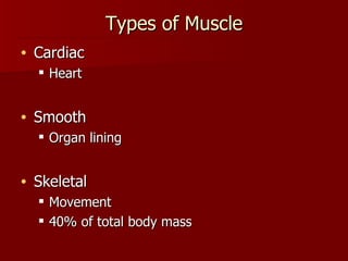
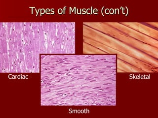
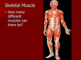
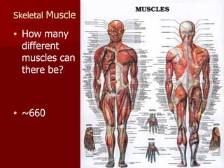
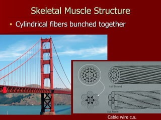
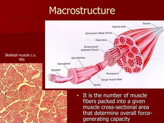
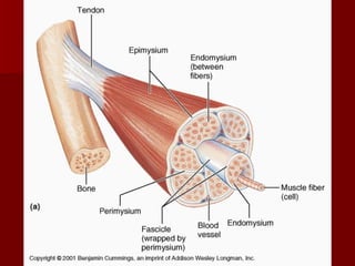
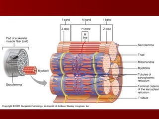
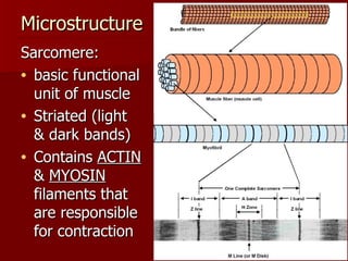
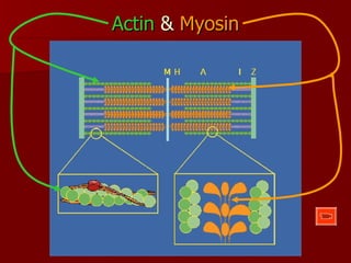
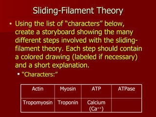
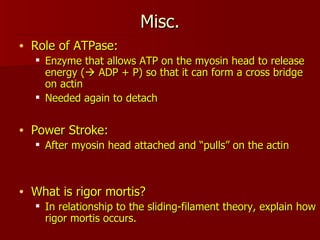
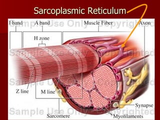
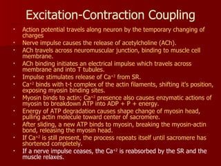
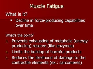
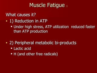
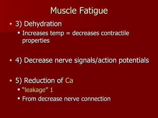
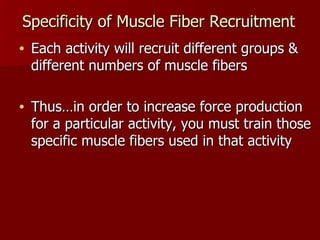
Applied Exercise Physiology Section 5: Body Systems Topic 1: Muscular System Prepared by Mr. Cerny Niagara Wheatfield Senior High School
Types of Muscle Cardiac Heart Smooth Organ lining Skeletal Movement 40% of total body mass
Types of Muscle (con’t) Cardiac Smooth Skeletal
Skeletal Muscle  How many different muscles can there be?
Skeletal  Muscle  How many different muscles can there be?  ~660
Skeletal Muscle Structure Cylindrical fibers bunched together Cable wire c.s.
 
It is the number of muscle fibers packed into a given muscle cross-sectional area that determine overall force-generating capacity Macrostructure Skeletal muscle c.s.  40x
 
 
Microstructure Sarcomere: basic functional unit of muscle Striated (light & dark bands) Contains  ACTIN  &  MYOSIN  filaments that are responsible for contraction
Actin  &  Myosin
Sliding-Filament Theory Using the list of “characters” below, create a storyboard showing the many different steps involved with the sliding-filament theory. Each step should contain a colored drawing (labeled if necessary) and a short explanation. “ Characters:” Calcium (Ca ++ ) Troponin Tropomyosin ATPase ATP Myosin Actin
Misc. Role of ATPase: Enzyme that allows ATP on the myosin head to release energy (   ADP + P) so that it can form a cross bridge on actin Needed again to detach Power Stroke: After myosin head attached and “pulls” on the actin What is rigor mortis? In relationship to the sliding-filament theory, explain how rigor mortis occurs.
Where did the Ca ++  come from? Ca ++  is stored and released from the sarcoplasmic reticulum (SR) SR is a network of tubules within the muscle fiber
Sarcoplasmic Reticulum
Nerves & Muscles An action potential (signal traveling along a neuron) causing contraction is termed  Excitation-Contraction Coupling
Excitation-Contraction Coupling Action potential travels along neuron by the temporary changing of charges Nerve impulse causes the release of acetylcholine (ACh). ACh travels across neuromuscular junction, binding to muscle cell membrane. ACh binding initiates an electrical impulse which travels across membrane and into T tubules. Impulse stimulates release of Ca +2  from SR. Ca +2  binds with t-t complex of the actin filaments, shifting it's position, exposing myosin binding sites. Myosin binds to actin; Ca +2  presence also causes enzymatic actions of myosin to breakdown ATP into ADP + P + energy. Energy of ATP degradation causes shape change of myosin head, pulling actin molecule toward center of sacromere. After sliding, a new ATP binds to myosin, breaking the myosin-actin bond, releasing the myosin head. If Ca +2  is still present, the process repeats itself until sacromere has shortened completely. If a nerve impulse ceases, the Ca +2  is reabsorbed by the SR and the muscle relaxes.
Muscle Fatigue What is it? Decline in force-producing capabilities over time What’s the point? Prevents exhausting of metabolic (energy-producing) reserve (like enzymes) Limits the buildup of harmful products Reduces the likelihood of damage to the contractile elements (ex.: sarcomeres)
Muscle Fatigue  1 What causes it? 1) Reduction in ATP Under high stress, ATP utilization  reduced faster than ATP production 2) Peripheral metabolic bi-products Lactic acid H (and other free radicals)
Muscle Fatigue 3) Dehydration Increases temp = decreases contractile properties 4) Decrease nerve signals/action potentials 5) Reduction of  Ca   “ leakage ”  1 From decrease nerve connection
Specificity of Muscle Fiber Recruitment Each activity will recruit different groups & different numbers of muscle fibers Thus…in order to increase force production for a particular activity, you must train those specific muscle fibers used in that activity
Specificity of Muscle Fiber Recruitment What does this mean for  training ? In order for optimum adaptation to occur during training, muscles should be trained in a manner similar to the way they are expected to perform during competition What does this mean for  rehabilitation ? Return to work in a timely manner requires you to look at exactly what that worker will be doing
Muscle Types Difference dependant on contraction time (rate of cross-bridge cycling)  NOT amount of force produced “Twitch”: response of skeletal muscle fiber to a single electrical stimulus Muscle Types: Slow twitch (Type I) Fast twitch (Type II)
Muscle Types nonoxidative Few Sprinting /bursts No Few Fast Same (slightly more) Fast twitch (II) Oxidative Many Endurance Yes Many Slow Same (slightly less) Slow twitch (I) Oxidative / nonoxidative # capillaries Activity Resistance to fatigue # mit. Speed Force
Muscle  Injury DOMS “ Delayed onset muscle soreness” Damage to sarcomere properties Actual protein molecules in sarcomere Titin Nebulin Costamere Exacerbated by dehydration Recovering faster with  steroids
 
Muscle Strength “ Bigger” muscles: Usually NOT increase in muscle cells  (hyperplasia) Usually an increase in existing tissue size (hypertrophy) Factors contributing: Genetics Exercise Nutrition Endocrine Environmental Nervous

Muscular System

  • 1.
    Applied Exercise PhysiologySection 5: Body Systems Topic 1: Muscular System Prepared by Mr. Cerny Niagara Wheatfield Senior High School
  • 2.
    Types of MuscleCardiac Heart Smooth Organ lining Skeletal Movement 40% of total body mass
  • 3.
    Types of Muscle(con’t) Cardiac Smooth Skeletal
  • 4.
    Skeletal Muscle How many different muscles can there be?
  • 5.
    Skeletal Muscle How many different muscles can there be? ~660
  • 6.
    Skeletal Muscle StructureCylindrical fibers bunched together Cable wire c.s.
  • 7.
  • 8.
    It is thenumber of muscle fibers packed into a given muscle cross-sectional area that determine overall force-generating capacity Macrostructure Skeletal muscle c.s. 40x
  • 9.
  • 10.
  • 11.
    Microstructure Sarcomere: basicfunctional unit of muscle Striated (light & dark bands) Contains ACTIN & MYOSIN filaments that are responsible for contraction
  • 12.
    Actin & Myosin
  • 13.
    Sliding-Filament Theory Usingthe list of “characters” below, create a storyboard showing the many different steps involved with the sliding-filament theory. Each step should contain a colored drawing (labeled if necessary) and a short explanation. “ Characters:” Calcium (Ca ++ ) Troponin Tropomyosin ATPase ATP Myosin Actin
  • 14.
    Misc. Role ofATPase: Enzyme that allows ATP on the myosin head to release energy (  ADP + P) so that it can form a cross bridge on actin Needed again to detach Power Stroke: After myosin head attached and “pulls” on the actin What is rigor mortis? In relationship to the sliding-filament theory, explain how rigor mortis occurs.
  • 15.
    Where did theCa ++ come from? Ca ++ is stored and released from the sarcoplasmic reticulum (SR) SR is a network of tubules within the muscle fiber
  • 16.
  • 17.
    Nerves & MusclesAn action potential (signal traveling along a neuron) causing contraction is termed Excitation-Contraction Coupling
  • 18.
    Excitation-Contraction Coupling Actionpotential travels along neuron by the temporary changing of charges Nerve impulse causes the release of acetylcholine (ACh). ACh travels across neuromuscular junction, binding to muscle cell membrane. ACh binding initiates an electrical impulse which travels across membrane and into T tubules. Impulse stimulates release of Ca +2 from SR. Ca +2 binds with t-t complex of the actin filaments, shifting it's position, exposing myosin binding sites. Myosin binds to actin; Ca +2 presence also causes enzymatic actions of myosin to breakdown ATP into ADP + P + energy. Energy of ATP degradation causes shape change of myosin head, pulling actin molecule toward center of sacromere. After sliding, a new ATP binds to myosin, breaking the myosin-actin bond, releasing the myosin head. If Ca +2 is still present, the process repeats itself until sacromere has shortened completely. If a nerve impulse ceases, the Ca +2 is reabsorbed by the SR and the muscle relaxes.
  • 19.
    Muscle Fatigue Whatis it? Decline in force-producing capabilities over time What’s the point? Prevents exhausting of metabolic (energy-producing) reserve (like enzymes) Limits the buildup of harmful products Reduces the likelihood of damage to the contractile elements (ex.: sarcomeres)
  • 20.
    Muscle Fatigue 1 What causes it? 1) Reduction in ATP Under high stress, ATP utilization reduced faster than ATP production 2) Peripheral metabolic bi-products Lactic acid H (and other free radicals)
  • 21.
    Muscle Fatigue 3)Dehydration Increases temp = decreases contractile properties 4) Decrease nerve signals/action potentials 5) Reduction of Ca “ leakage ” 1 From decrease nerve connection
  • 22.
    Specificity of MuscleFiber Recruitment Each activity will recruit different groups & different numbers of muscle fibers Thus…in order to increase force production for a particular activity, you must train those specific muscle fibers used in that activity
  • 23.
    Specificity of MuscleFiber Recruitment What does this mean for training ? In order for optimum adaptation to occur during training, muscles should be trained in a manner similar to the way they are expected to perform during competition What does this mean for rehabilitation ? Return to work in a timely manner requires you to look at exactly what that worker will be doing
  • 24.
    Muscle Types Differencedependant on contraction time (rate of cross-bridge cycling) NOT amount of force produced “Twitch”: response of skeletal muscle fiber to a single electrical stimulus Muscle Types: Slow twitch (Type I) Fast twitch (Type II)
  • 25.
    Muscle Types nonoxidativeFew Sprinting /bursts No Few Fast Same (slightly more) Fast twitch (II) Oxidative Many Endurance Yes Many Slow Same (slightly less) Slow twitch (I) Oxidative / nonoxidative # capillaries Activity Resistance to fatigue # mit. Speed Force
  • 26.
    Muscle InjuryDOMS “ Delayed onset muscle soreness” Damage to sarcomere properties Actual protein molecules in sarcomere Titin Nebulin Costamere Exacerbated by dehydration Recovering faster with steroids
  • 27.
  • 28.
    Muscle Strength “Bigger” muscles: Usually NOT increase in muscle cells (hyperplasia) Usually an increase in existing tissue size (hypertrophy) Factors contributing: Genetics Exercise Nutrition Endocrine Environmental Nervous