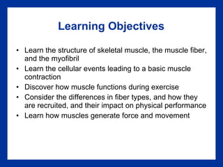
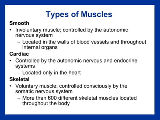
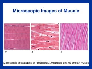
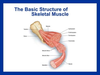
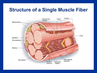
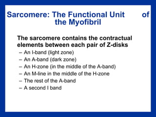
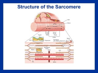
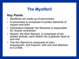
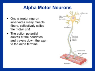
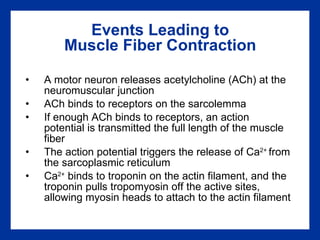
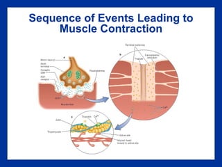
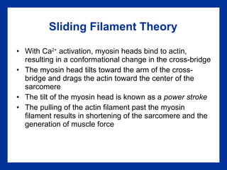
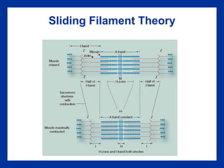
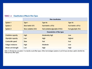
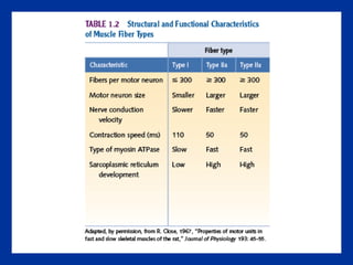
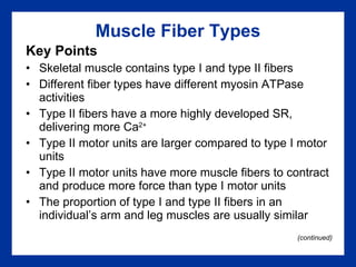
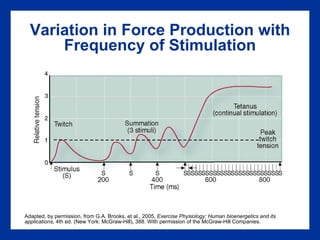
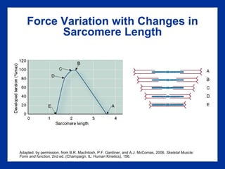
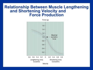
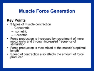
1 Structure and Function of Exercising Muscle chapter
Learning Objectives Learn the structure of skeletal muscle, the muscle fiber, and the myofibril Learn the cellular events leading to a basic muscle contraction Discover how muscle functions during exercise Consider the differences in fiber types, and how they are recruited, and their impact on physical performance Learn how muscles generate force and movement
Types of Muscles Smooth Involuntary muscle; controlled by the autonomic nervous system Located in the walls of blood vessels and throughout internal organs Cardiac Controlled by the autonomic nervous and endocrine systems Located only in the heart Skeletal Voluntary muscle; controlled consciously by the somatic nervous system More than 600 different skeletal muscles located throughout the body
Microscopic Images of Muscle Microscopic photographs of  (a)  skeletal,  (b)  cardiac, and  (c)  smooth muscle a b c
The Basic Structure of  Skeletal Muscle
Structure of a Single Muscle Fiber
Skeletal Muscle Fiber Structure Key Points An individual muscle cell is called a muscle fiber A muscle fiber is enclosed by a plasma membrane called the sarcolemma The cytoplasm of a muscle fiber is called a sarcoplasm Within the sarcoplasm, the extensive T-tubules allow transport of substances throughout the muscle fiber  The sarcoplasmic reticulum (SR) stores calcium
An Electron Micrograph of Myofibrils © Custom Medical Stock Photo Myofibrils
Sarcomere: The Functional Unit  of the Myofibril The sarcomere contains the contractual elements between each pair of Z-disks An I-band (light zone) An A-band (dark zone) An H-zone (in the middle of the A-band) An M-line in the middle of the H-zone The rest of the A-band A second I band
Structure of the Sarcomere
The Myofibril Key Points Myofibrils are made up of sarcomeres A sarcomere is composed of protein filaments of myosin and actin Interactions between the filaments is responsible for muscle contraction Myosin, the thick filament, is composed of two protein strands, each folded into a globular head at one end The thin filament is composed of actin, tropomyosin, and troponin, with one end attached to a Z-disk
Alpha Motor Neurons One   -motor neuron innervates many muscle fibers, collectively called the  motor unit The action potential arrives at the dendrites and travels down the axon to the axon terminal
Events Leading to Muscle Fiber Contraction A motor neuron releases acetylcholine (ACh) at the neuromuscular junction ACh binds to receptors on the sarcolemma If enough ACh binds to receptors, an action potential is transmitted the full length of the muscle fiber The action potential triggers the release of Ca 2+  from the sarcoplasmic reticulum Ca 2+  binds to troponin on the actin filament, and the troponin pulls tropomyosin off the active sites, allowing myosin heads to attach to the actin filament
Sequence of Events Leading to Muscle Contraction
Sliding Filament Theory With Ca 2+  activation, myosin heads bind to actin, resulting in a conformational change in the cross-bridge The myosin head tilts toward the arm of the cross-bridge and drags the actin toward the center of the sarcomere The tilt of the myosin head is known as a  power stroke The pulling of the actin filament past the myosin filament results in shortening of the sarcomere and the generation of muscle force
Sliding Filament Theory
Molecular Events of Cross-Bridge Cycling in Skeletal Muscle Fig. 12.9, p. 405 from HUMAN PHYSIOLOGY, 4th ed. By Dee Unglaub Silverthorn. Copyright © 2007 by Pearson Education, Inc. Adapted by permission.
Muscle Fiber Contraction Key Points Muscle contraction is initiated by an   -motor neuron action potential The motor neuron releases ACh, which opens up ion gates in the muscle cell membrane Sodium enters the muscle cell depolarizing the plasmalemma  The action potential travels throughout the plasmalemma and through the T-tubules, which releases stored Ca 2+  ions from the sarcoplasmic reticulum (continued)
Muscle Fiber Contraction  (continued) Key Points Ca 2+  ions bind with troponin, lifting the tropomyosin molecules off the active sites on the actin filament  The myosin head binds to the active actin site The myosin head binds ATP, and ATPase on the myosin heads splits ATP into ADP and P i , releasing energy to fuel the muscle contraction  The myosin head tilts, pulling the thin filament past the thick filament — power stroke Muscle contraction ends when Ca 2+  is actively pumped out of the sarcoplasm back to the sarcoplasmic reticulum
Muscle Biopsy The muscle biopsy allows us to study muscle fibers and the effects of acute exercise and chronic training on muscle fiber composition The skin is first anesthetized and then a small incision is made through the skin  A hollow Bergstrom needle is inserted into the muscle belly to take the sample The sample is mounted, frozen, thinly sliced, and examined under a microscope
Muscle Biopsy
Muscle Fiber Types Slow-twitch fibers, Type I (~50%), oxidative Fast-twitch fibers, Type II Type IIa (25%), fast oxidative/glycolytic (FOG) Type IIx in humans (~25%) ~ IIb in animals,  fast glycolytic (FG) Type IIc (1-3%) The percentage of each fiber type is variable among muscles, among individuals, and with exercise training
A Photomicrograph Showing Type I, Type IIa, and Type IIx Muscle Fibers Type I (black), type IIa (white), and type IIx (gray) muscle fibers
Determination of Myosin Isoforms Myosin isoforms are determined by electrophoretic separation of the proteins and by staining. Determining the myosin isoform helps to identify the fiber type.
 
 
Single Muscle Fiber Physiology Peak power is different between muscle fiber types All fiber types tend to reach their peak power at ~20% peak force
Muscle Fiber Types Key Points Skeletal muscle contains type I and type II fibers Different fiber types have different myosin ATPase activities Type II fibers have a more highly developed SR, delivering more Ca 2+ Type II motor units are larger compared to type I motor units Type II motor units have more muscle fibers to contract and produce more force than type I motor units The proportion of type I and type II fibers in an individual’s arm and leg muscles are usually similar (continued)
Muscle Fiber Types  (continued) Key Points Type I fibers have higher aerobic endurance and are well suited to low-intensity endurance activities Type II fibers are better suited for anaerobic activity Type IIa fibers play a major role in high intensity exercise Type IIx fibers are activated when the force demanded of a muscle is high
Determination of Fiber Type Fiber type is genetically determined (twin studies) Fiber type is determined by the   -motor neuron that innervates the muscle fibers Endurance training, strength training, and muscular inactivity may cause a shift in myosin isoforms Exercise training  ↓  type IIx and  ↑  type IIa Aging may shift the relative distribution of type I and type II fibers  ↓  type II and ↑  type I
Motor Unit Recruitment Principle of orderly recruitment Motor units are activated on the basis of a fixed order type I  ->  type IIa  ->  type IIx
Size Principle Size principle The order of motor unit recruitment is directly related to the motor neuron size
Motor Unit Recruitment Key Points Motor units give an all-or-none response Activating more motor units and thus more muscle fibers produces more force Motor units are recruited in an orderly way to generate force or for long duration events type I  ->  type IIa  ->  type IIx
 
Athletes and Fiber Type Key Points Muscle fiber composition differs in athletes by sport and event Speed and strength events are characterized by a higher percentage of type II fibers Endurance events are characterized by a higher percentage of type I fibers
Types of Muscle Contraction Concentric contraction: Force is developed while the muscle is shortening Isometric contraction: Force is generated but the length of the muscle is unchanged Eccentric contraction: Force is generated while the muscle is lengthening
Variation in Force Production with Frequency of Stimulation Adapted, by permission, from G.A. Brooks, et al., 2005,  Exercise Physiology: Human bioenergetics and its applications,  4th ed. (New York: McGraw-Hill), 388. With permission of the McGraw-Hill Companies.
Force Variation with Changes in Sarcomere Length Adapted, by permission, from B.R. MacIntosh, P.F. Gardiner, and A.J. McComas, 2006,  Skeletal Muscle: Form and function,  2nd ed. (Champaign, IL: Human Kinetics), 156.
Relationship Between Muscle Lengthening and Shortening Velocity and  Force Production
Muscle Force Generation Key Points 3 types of muscle contraction Concentric Isometric Eccentric Force production is increased by recruitment of more motor units and through increased frequency of stimulation Force production is maximized at the muscle’s optimal length Speed of contraction also affects the amount of force produced

Chapter 01 Power Points

  • 1.
    1 Structure andFunction of Exercising Muscle chapter
  • 2.
    Learning Objectives Learnthe structure of skeletal muscle, the muscle fiber, and the myofibril Learn the cellular events leading to a basic muscle contraction Discover how muscle functions during exercise Consider the differences in fiber types, and how they are recruited, and their impact on physical performance Learn how muscles generate force and movement
  • 3.
    Types of MusclesSmooth Involuntary muscle; controlled by the autonomic nervous system Located in the walls of blood vessels and throughout internal organs Cardiac Controlled by the autonomic nervous and endocrine systems Located only in the heart Skeletal Voluntary muscle; controlled consciously by the somatic nervous system More than 600 different skeletal muscles located throughout the body
  • 4.
    Microscopic Images ofMuscle Microscopic photographs of (a) skeletal, (b) cardiac, and (c) smooth muscle a b c
  • 5.
    The Basic Structureof Skeletal Muscle
  • 6.
    Structure of aSingle Muscle Fiber
  • 7.
    Skeletal Muscle FiberStructure Key Points An individual muscle cell is called a muscle fiber A muscle fiber is enclosed by a plasma membrane called the sarcolemma The cytoplasm of a muscle fiber is called a sarcoplasm Within the sarcoplasm, the extensive T-tubules allow transport of substances throughout the muscle fiber The sarcoplasmic reticulum (SR) stores calcium
  • 8.
    An Electron Micrographof Myofibrils © Custom Medical Stock Photo Myofibrils
  • 9.
    Sarcomere: The FunctionalUnit of the Myofibril The sarcomere contains the contractual elements between each pair of Z-disks An I-band (light zone) An A-band (dark zone) An H-zone (in the middle of the A-band) An M-line in the middle of the H-zone The rest of the A-band A second I band
  • 10.
  • 11.
    The Myofibril KeyPoints Myofibrils are made up of sarcomeres A sarcomere is composed of protein filaments of myosin and actin Interactions between the filaments is responsible for muscle contraction Myosin, the thick filament, is composed of two protein strands, each folded into a globular head at one end The thin filament is composed of actin, tropomyosin, and troponin, with one end attached to a Z-disk
  • 12.
    Alpha Motor NeuronsOne  -motor neuron innervates many muscle fibers, collectively called the motor unit The action potential arrives at the dendrites and travels down the axon to the axon terminal
  • 13.
    Events Leading toMuscle Fiber Contraction A motor neuron releases acetylcholine (ACh) at the neuromuscular junction ACh binds to receptors on the sarcolemma If enough ACh binds to receptors, an action potential is transmitted the full length of the muscle fiber The action potential triggers the release of Ca 2+ from the sarcoplasmic reticulum Ca 2+ binds to troponin on the actin filament, and the troponin pulls tropomyosin off the active sites, allowing myosin heads to attach to the actin filament
  • 14.
    Sequence of EventsLeading to Muscle Contraction
  • 15.
    Sliding Filament TheoryWith Ca 2+ activation, myosin heads bind to actin, resulting in a conformational change in the cross-bridge The myosin head tilts toward the arm of the cross-bridge and drags the actin toward the center of the sarcomere The tilt of the myosin head is known as a power stroke The pulling of the actin filament past the myosin filament results in shortening of the sarcomere and the generation of muscle force
  • 16.
  • 17.
    Molecular Events ofCross-Bridge Cycling in Skeletal Muscle Fig. 12.9, p. 405 from HUMAN PHYSIOLOGY, 4th ed. By Dee Unglaub Silverthorn. Copyright © 2007 by Pearson Education, Inc. Adapted by permission.
  • 18.
    Muscle Fiber ContractionKey Points Muscle contraction is initiated by an  -motor neuron action potential The motor neuron releases ACh, which opens up ion gates in the muscle cell membrane Sodium enters the muscle cell depolarizing the plasmalemma The action potential travels throughout the plasmalemma and through the T-tubules, which releases stored Ca 2+ ions from the sarcoplasmic reticulum (continued)
  • 19.
    Muscle Fiber Contraction (continued) Key Points Ca 2+ ions bind with troponin, lifting the tropomyosin molecules off the active sites on the actin filament The myosin head binds to the active actin site The myosin head binds ATP, and ATPase on the myosin heads splits ATP into ADP and P i , releasing energy to fuel the muscle contraction The myosin head tilts, pulling the thin filament past the thick filament — power stroke Muscle contraction ends when Ca 2+ is actively pumped out of the sarcoplasm back to the sarcoplasmic reticulum
  • 20.
    Muscle Biopsy Themuscle biopsy allows us to study muscle fibers and the effects of acute exercise and chronic training on muscle fiber composition The skin is first anesthetized and then a small incision is made through the skin A hollow Bergstrom needle is inserted into the muscle belly to take the sample The sample is mounted, frozen, thinly sliced, and examined under a microscope
  • 21.
  • 22.
    Muscle Fiber TypesSlow-twitch fibers, Type I (~50%), oxidative Fast-twitch fibers, Type II Type IIa (25%), fast oxidative/glycolytic (FOG) Type IIx in humans (~25%) ~ IIb in animals, fast glycolytic (FG) Type IIc (1-3%) The percentage of each fiber type is variable among muscles, among individuals, and with exercise training
  • 23.
    A Photomicrograph ShowingType I, Type IIa, and Type IIx Muscle Fibers Type I (black), type IIa (white), and type IIx (gray) muscle fibers
  • 24.
    Determination of MyosinIsoforms Myosin isoforms are determined by electrophoretic separation of the proteins and by staining. Determining the myosin isoform helps to identify the fiber type.
  • 25.
  • 26.
  • 27.
    Single Muscle FiberPhysiology Peak power is different between muscle fiber types All fiber types tend to reach their peak power at ~20% peak force
  • 28.
    Muscle Fiber TypesKey Points Skeletal muscle contains type I and type II fibers Different fiber types have different myosin ATPase activities Type II fibers have a more highly developed SR, delivering more Ca 2+ Type II motor units are larger compared to type I motor units Type II motor units have more muscle fibers to contract and produce more force than type I motor units The proportion of type I and type II fibers in an individual’s arm and leg muscles are usually similar (continued)
  • 29.
    Muscle Fiber Types (continued) Key Points Type I fibers have higher aerobic endurance and are well suited to low-intensity endurance activities Type II fibers are better suited for anaerobic activity Type IIa fibers play a major role in high intensity exercise Type IIx fibers are activated when the force demanded of a muscle is high
  • 30.
    Determination of FiberType Fiber type is genetically determined (twin studies) Fiber type is determined by the  -motor neuron that innervates the muscle fibers Endurance training, strength training, and muscular inactivity may cause a shift in myosin isoforms Exercise training ↓ type IIx and ↑ type IIa Aging may shift the relative distribution of type I and type II fibers ↓ type II and ↑ type I
  • 31.
    Motor Unit RecruitmentPrinciple of orderly recruitment Motor units are activated on the basis of a fixed order type I -> type IIa -> type IIx
  • 32.
    Size Principle Sizeprinciple The order of motor unit recruitment is directly related to the motor neuron size
  • 33.
    Motor Unit RecruitmentKey Points Motor units give an all-or-none response Activating more motor units and thus more muscle fibers produces more force Motor units are recruited in an orderly way to generate force or for long duration events type I -> type IIa -> type IIx
  • 34.
  • 35.
    Athletes and FiberType Key Points Muscle fiber composition differs in athletes by sport and event Speed and strength events are characterized by a higher percentage of type II fibers Endurance events are characterized by a higher percentage of type I fibers
  • 36.
    Types of MuscleContraction Concentric contraction: Force is developed while the muscle is shortening Isometric contraction: Force is generated but the length of the muscle is unchanged Eccentric contraction: Force is generated while the muscle is lengthening
  • 37.
    Variation in ForceProduction with Frequency of Stimulation Adapted, by permission, from G.A. Brooks, et al., 2005, Exercise Physiology: Human bioenergetics and its applications, 4th ed. (New York: McGraw-Hill), 388. With permission of the McGraw-Hill Companies.
  • 38.
    Force Variation withChanges in Sarcomere Length Adapted, by permission, from B.R. MacIntosh, P.F. Gardiner, and A.J. McComas, 2006, Skeletal Muscle: Form and function, 2nd ed. (Champaign, IL: Human Kinetics), 156.
  • 39.
    Relationship Between MuscleLengthening and Shortening Velocity and Force Production
  • 40.
    Muscle Force GenerationKey Points 3 types of muscle contraction Concentric Isometric Eccentric Force production is increased by recruitment of more motor units and through increased frequency of stimulation Force production is maximized at the muscle’s optimal length Speed of contraction also affects the amount of force produced